

Assessment of anatomic variations of the brachial artery bifurcation using vascular Doppler ultrasound: a cross-sectional study
- PMID: 38659618
- PMCID: PMC11042541
- DOI: 10.1590/1677-5449.202301172
Assessment of anatomic variations of the brachial artery bifurcation using vascular Doppler ultrasound: a cross-sectional study
Abstract
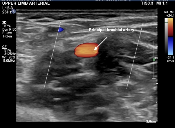
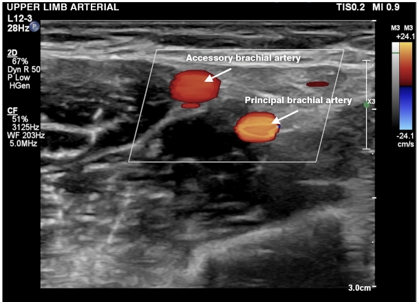
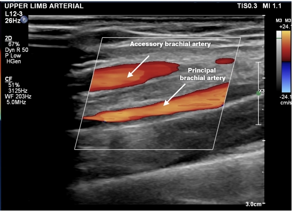
Background: Anatomical variations in arteries of the upper limb, such as presence of an accessory brachial artery, are common and widely described in the literature, mainly in cadaveric studies, but it is now possible to diagnose them using vascular Doppler ultrasound.
Objectives: To identify the incidence of accessory brachial artery using vascular Doppler ultrasound and compare the findings with cadaveric studies.
Methods: This was a prospective study that examined 500 upper limbs of 250 volunteers assessed with vascular Doppler ultrasound using the Sonosite Titan portable ultrasound machine.
Results: 15.6% of the participants in our study had the accessory brachial artery anatomical variation. Our percentage is in line with the average rates found in cadaveric studies, which ranged from 0.2% to 22%. Being aware of this variation is fundamental in procedures such as peripheral venipuncture, arteriovenous fistula creation, catheterization, forearm flaps, emergency surgeries on the limb and even correction of fractures by cast.
Conclusions: The accessory brachial artery is a frequent variant in the upper limb. The percentage of individuals with an accessory brachial artery in our study was 15.6%, which agrees with data from the literature on cadaveric studies.
Contexto: Variações anatômicas em artérias do membro superior, como a presença da artéria braquial acessória, são comuns e amplamente descritas na literatura, principalmente por estudos em cadáveres. No entanto, atualmente, é possível realizar o diagnóstico através do eco-Doppler vascular.
Objetivos: Identificar a incidência da artéria braquial acessória pelo eco-Doppler e comparar os achados com estudos cadavéricos.
Métodos: Tratou-se de um estudo prospectivo em 500 membros superiores de 250 voluntários avaliados pelo eco-Doppler com o aparelho portátil de ultrassom Sonosite Titan.
Resultados: Dos participantes do nosso estudo, 15,6% apresentaram a variação anatômica da artéria braquial acessória. A porcentagem está dentro da média encontrada em estudos cadavéricos, que varia de 0,2 até 22%. Ter conhecimento dessa variação é fundamental em procedimentos como punção venosa periférica, fístula arteriovenosa, cateterismo, retalhos de antebraço, cirurgias de emergência no membro superior e até mesmo correção de fraturas por gesso.
Conclusões: A artéria braquial acessória é uma variante frequente no membro superior. O percentual de indivíduos com a artéria braquial acessória em nosso estudo foi de 15,6% e coincide com os dados da literatura de estudos cadavéricos.
Keywords: Doppler ultrasound; anatomical variation; brachial artery.
Copyright© 2024 The authors.
Conflict of interest statement
Conflicts of interest: No conflicts of interest declared concerning the publication of this article.
Figures


References
-
- Rodriguez-Niedenführ M, Vázquez T, Nearn L, Ferreira B, Parkin I, Sañudo JR. Variations of the arterial pattern in the upper limb revised: a morphological and statistical study, with a review of the literature. J Anat. 2001;199(5):547–566. doi: 10.1046/j.1469-7580.2001.19950547.x. - DOI - PMC - PubMed
-
- Coleman SS, Anson BJ. Arterial patterns in the hand based upon a study of 650 specimens. Surg Gynecol Obstet. 1961;113(4):409–424. - PubMed
-
- Green PH. An account of the varieties in the arterial system of the human body. Dublin: James Marshal Leckie; 1830. pp. 16–20.
-
- Rodriguez-Niedenführ M, Vázquez T, Parkin I, Sañudo JR. Arterial patterns of the human upper limb: update of anatomical variations and embryological development. Eur J Anat. 2003;7(1):21–28.
LinkOut - more resources
Full Text Sources
Miscellaneous