[Preprint]. 2024 Apr 20:2024.04.15.589650.
doi: 10.1101/2024.04.15.589650.

Functional cardiac consequences of β-adrenergic stress-induced injury in the mdx mouse model of Duchenne muscular dystrophy

Affiliations

Functional cardiac consequences of β-adrenergic stress-induced injury in the mdx mouse model of Duchenne muscular dystrophy

Conner C Earl et al. bioRxiv. .

Update in

Abstract

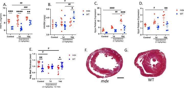
Cardiomyopathy is the leading cause of death in Duchenne muscular dystrophy (DMD), however, in the mdx mouse model of DMD, the cardiac phenotype differs from that seen in DMD-associated cardiomyopathy. Although some have used pharmacologic stress to enhance the cardiac phenotype in the mdx model, many methods lead to high mortality, variable cardiac outcomes, and do not recapitulate the structural and functional cardiac changes seen in human disease. Here, we describe a simple and effective method to enhance the cardiac phenotype model in mdx mice using advanced 2D and 4D high-frequency ultrasound to monitor cardiac dysfunction progression in vivo. For our study, mdx and wild-type (WT) mice received daily low-dose (2 mg/kg/day) isoproterenol injections for 10 days. Histopathologic assessment showed that isoproterenol treatment increased myocyte injury, elevated serum cardiac troponin I levels, and enhanced fibrosis in mdx mice. Ultrasound revealed reduced ventricular function, decreased wall thickness, increased volumes, and diminished cardiac reserve in mdx mice compared to wild-type. Our findings highlight the utility of low-dose isoproterenol in mdx mice as a valuable model for exploring therapies targeting DMD-associated cardiac complications.

Keywords: 4DUS; Duchenne muscular dystrophy; cardiac strain; isoproterenol; mdx; mouse model.


Conflict of interest statement

Declaration of Interest CJG is a paid consultant of FUJIFILM VisualSonics Inc. Declaration of generative AI and AI-assisted technologies in the writing process During the preparation of this work, the authors used ChatGPT(OpenAI, San Francisco, California) in some minor instances to rephrase and/or summarize text in order to improve readability. After using this tool, the authors reviewed and edited the content as needed and take full responsibility for the content of the publication.

Figures

Figure 1:
Figure 1:
Study Overview. (A) Schematic representing subcutaneous injection of 2 mg/kg/day isoproterenol to induce β-adrenergic cardiac stimulation and injury. (B) Histopathologic study timeline. (C) Imaging study timeline.
Figure 2:
Figure 2:
Isoproterenol promotes sarcolemmal injury in dystrophin-deficient cardiac myocytes. The acute and chronic effects of isoproterenol (2 mg/kg/day) were assessed in mdx and wild-type (WT) mice treated for 1- (n=10 per group) or 10 days (n=5 per group). Control mice were injected with an equal volume of saline for 10 days (n=5 mdx and n=4 WT). (A-D) To assess sarcolemmal damage, mid-chamber coronal sections of ventricles were immunolabeled with anti-IgM (red) and counterstained with wheat germ agglutinin (WGA; green) to visualize tissue morphology. (A) Cardiac injury was detected transiently in mdx mice after isoproterenol challenge with IgM+ cardiac myocytes occupying ~8.5% of the myocardial area after a single isoproterenol treatment. Cardiac myocyte injury was scarcely detected in mdx mice after chronic isoproterenol administration or in WT mice across all conditions. (B-C) Representative mdx (B) and WT (C) whole ventricle cross-section and high magnification images show prominent areas of injury (red) after acute isoproterenol treatment in mdx mice. Bar=500 µm. (C) High-magnification images confirm that the IgM+ signal is localized to cardiac myocytes in mdx mice. (D) Serum cardiac troponin I (cTnI) levels were measured by ELISA as an independent assay of cardiac injury. Serum cTnI levels were also transiently elevated in mdx mice after acute isoproterenol treatment (n=5 per group). Data are presented as mean ± SEM. All p values are based on ANOVA with Tukey’s multiple comparison test. **p<0.01 and ***p<0.001 versus WT within a treatment condition. ## indicates p<0.01 between treatment groups within a genotype.
Figure 3:
Figure 3:
Distinct cardiac growth and stress responses to β-adrenergic stimulation in dystrophic hearts. Heart weight normalized to tibia length (HW/TL) and heart weight normalized to body mass (HW/BM) were used as indices of cardiac growth in mdx and WT mice treated for 1- (n=10 per group) or 10-days (n=5 per group), and control mice (n=5 mdx and n=4 WT). (A) HW/TL ratio was higher in mdx mice compared to WT in all conditions. Chronic isoproterenol treatment resulted in an increased HW/TL ratio in WT mice only. (B) HW/BM ratio increased with chronic isoproterenol treatment in WT mice relative to control and mdx mice. (C-D) QPCR analyses of mdx and WT ventricles expressed as mRNA levels relative to WT control. (C) Nppa and (B) Nppb mRNA expression increased in mdx mice with chronic isoproterenol challenge and relative to WT. (E) Ventricular wall thickness was measured at key time points by ultrasound (n=10 per genotype). At baseline, wall thickness was greater in mdx hearts relative to WT. However, in response to isoproterenol treatment wall thickness decreased in mdx mice becoming less thick than WT at day 14. Mid-chamber coronal sections of mdx (F) and WT (G) ventricles stained with Masson’s trichrome show characteristic fibrosis and thinning of the free wall in mdx mice at day 14. Bar=1 mm. Data are presented as mean±SEM. All p values are based on 2-way ANOVA with Tukey’s multiple comparison test. *p<0.05, **p<0.01, ***p<0.001 and ***p<0.0001 versus WT within a treatment condition. #p<0.05, ##p<0.01, ###p<0.001 and ####p<0.0001 between treatment groups within a genotype.
Figure 4:
Figure 4:
Isoproterenol promotes fibrotic replacement of the dystrophic myocardium. (A-G) Fibrotic area was measured on mid-chamber cross-sections stained with Sirius Red Fast Green stain (SRFG) in control (n=5 mdx and n=4 WT) and chronic isoproterenol treated (n=5 per genotype) mice. (A) The area of the myocardium occupied by collagen (red stain) is increased in (B) mdx mice after chronic isoproterenol challenge relative to control and (C) iso-treated WT mice. Bar=500 µm. (D-E) Collagen density was visualized by polarized light microscopy in iso-treated (D) mdx and (E) WT mice. (D-E) Fibrotic lesions of iso-treated mdx mice contained prominent birefringent areas indicating the presence of densely bundled collagen fibers (red and orange) in addition to the overall increase in collagen area. Representative overlay images of brightfield and polarized light (F) mdx and (G) WT ventricles. Areas of birefringence are pseudo-colored yellow to enhance image contrast in image overlays (F-G). (H-K) QPCR analyses of transcripts encoding markers of (H-I) activated fibroblasts and (J-K) connective tissue proteins. (H) Acta2 mRNA levels were elevated in mdx mice relative to WT at baseline and with isoproterenol stimulation. (I) Induction of Postn transcripts was greater with isoproterenol stimulation in mdx relative to WT. Genes encoding extracellular matrix components (J) Col1a1 and (K) Fn1 were further increased in mdx mice relative to WT with iso-treatment. Data are expressed as mRNA levels relative to WT control group and presented as mean±SEM. All p values are based on ANOVA with Tukey’s multiple comparison test. *p<0.05, **p<0.01, and ***p<0.001 versus WT within a treatment condition. #p<0.05, ##p<0.01, ###p<0.001 and ####p<0.0001 between treatment groups within a genotype.
Figure 5:
Figure 5:
Cardiac function decreases with isoproterenol injury in dystrophic hearts. (A) LVEF in mdx mice was significantly reduced by day 7, and further reduced at day 14. (B) The left ventricular end-diastolic volume and (C) end-systolic volume in mdx mice were significantly greater than WT at day 14 signifying left ventricular dilation. (D) There were no significant changes in the cardiac output or (E) heart rate in either WT or mdx groups. (F) 2D ultrasound parasternal long-axis images at peak systole for both mdx and WT mice at day 0, day 7, and day 14 with left ventricular ejection fraction (EF) label. These findings suggest that the mdx mice exhibited dilated cardiomyopathy by day 14 when given an isoproterenol challenge. Data presented as mean±SEM. All p values are based on ANOVA with Tukey’s multiple comparison test. *p<0.05, **p<0.01, and ***p<0.001 versus WT at a specified timepoint. #p<0.05, ##p<0.01, and ###p<0.001 mdx difference between timepoints.
Figure 6:
Figure 6:
Cardiac response to isoproterenol challenge decreases over time in dystrophic hearts. (A,B) The initial compensatory response to an isoproterenol administration measured at exactly 1 minute before and 1 minute after the injection was apparent in the increase in heart rate and LVEF in both mdx and WT mice. (C) In the 2D ultrasound images of the left ventricle, this compensation can be seen as endocardial walls coming close together during systole. (C,D) After 7 days of exposure to isoproterenol injury, this compensatory response is impaired in the mdx mice, where the ∆heart rate and ∆LVEF are reduced in comparison to WT. (E) There is little to no qualitative change in the left ventricular chamber in the mdx mice immediately after isoproterenol injection compared to WT. After prolonged exposure to an isoproterenol challenge, mdx mice’s ability to compensate for a single injection is impaired in comparison to the robust compensation of WT mice. Data presented as mean±SEM. All p values are based on ANOVA with Tukey’s multiple comparison test. **p<0.01, and ****p<0.0001 versus WT. #p<0.05, ##p<0.01, ###p<0.001, and ####p<0.0001 difference between pre and post injection.
Figure 7:
Figure 7:
4D strain magnitude decreases in dystrophic hearts with isoproterenol injury. Global circumferential, longitudinal, radial, and surface area strain were calculated with 4DUS measurements (A-D). In the mdx mice with isoproterenol injury, the magnitude of strain was significantly lowered by day 7 (E-F). There was a slight recovery in ventricular strain by day 14, yet the strain magnitude remains significantly reduced. 4D strain decreases in dystrophic hearts when given an isoproterenol challenge. After the completion of the isoproterenol challenge, there is some functional recovery, but irreversible damage remains. Data presented as mean±SEM. All p values are based on ANOVA with Tukey’s multiple comparison test. *p<0.05, **p<0.01, ***p<0.001 and ****p<0.0001 versus WT at a specified timepoint. ##p<0.01, ###p<0.001, and ####p<0.0001 mdx difference between timepoints.

References

    1. Romitti PA, Zhu Y, Puzhankara S, James KA, Nabukera SK, Zamba GK, Ciafaloni E, Cunniff C, Druschel CM, Mathews KD, Matthews DJ, Meaney FJ, Andrews JG, Conway KM, Fox DJ, Street N, Adams MM, Bolen J. Prevalence of Duchenne and Becker muscular dystrophies in the United States. Pediatrics. 2015;135(3):513–21. doi: 10.1542/peds.2014-2044. - DOI - PMC - PubMed
    1. Hoffman EP, Brown RH, Kunkel LM. Dystrophin: The protein product of the duchenne muscular dystrophy locus. Cell. 1987;51(6):919–28. - PubMed
    1. Petrof BJ, Shrager JB, Stedman HH, Kelly AM, Sweeney HL. Dystrophin protects the sarcolemma from stresses developed during muscle contraction. Proc Natl Acad Sci USA. 1993;90(8):3710–4. - PMC - PubMed
    1. Tidball JG, Welc SS, Wehling-Henricks M. Immunobiology of inherited muscular dystrophies. Compr Physiol. 2018;8(4):1313–56. doi: 10.1002/cphy.c170052. - DOI - PMC - PubMed
    1. Birnkrant DJ, Bushby K, Bann CM, Alman BA, Apkon SD, Blackwell A, Case LE, Cripe L, Hadjiyannakis S, Olson AK, Sheehan DW, Bolen J, Weber DR, Ward LM. Diagnosis and management of Duchenne muscular dystrophy, part 2: respiratory, cardiac, bone health, and orthopaedic management. Lancet Neurol. 2018;17(4):347–61. doi: 10.1016/s1474-4422(18)30025-5. - DOI - PMC - PubMed

Publication types