[Preprint]. 2024 Apr 18:2024.04.17.588758.
doi: 10.1101/2024.04.17.588758.

Polycystins recruit cargo to distinct ciliary extracellular vesicle subtypes

Affiliations

Polycystins recruit cargo to distinct ciliary extracellular vesicle subtypes

Inna A Nikonorova et al. bioRxiv. .

Update in

Abstract

Therapeutic use of tiny extracellular vesicles (EVs) requires understanding cargo loading mechanisms. Here, we used a modular proximity label approach to identify EV cargo associated with the transient potential channel (TRP) polycystin PKD-2 of C. elegans. Polycystins are conserved receptor-TRP channel proteins affecting cilium function; dysfunction causes polycystic kidney disease in humans and mating deficits in C. elegans. Polycystin-2 EV localization is conserved from algae to humans, hinting at an ancient and unknown function. We discovered that polycystins associate with and direct specific cargo to EVs: channel-like PACL-1, dorsal and ventral membrane C-type lectins PAMLs, and conserved tumor necrosis-associated factor (TRAF) signaling adaptors TRF-1 and TRF-2. Loading of these components relied on polycystin-1 LOV-1. Our modular EV-TurboID approach can be applied in both cell- and tissue-specific manners to define the composition of distinct EV subtypes, addressing a major challenge of the EV field.


Conflict of interest statement

Declaration of Interests The authors declare no competing interests.

Figures

Extended Data Fig. 1.
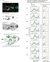
Extended Data Fig. 1.. Targeting TurboID module to PKD-2::GFP (related to Fig. 1).
a, anti-GFP nanobody::mScarlet::TurboID is uniformly abundant in cell bodies, neurites, and cilia in an untargeted state in the absence of any GFP-labeled protein. b, Coexpression of the anti-GFP nanobody::mScarlet::TurboID and pkd-2::GFP results in specific targeting of the TurboID module to PKD-2::GFP subcellular locations, including cilia and ciliary EVs. Male tails in panels a and b are oriented in different positions (dorsal on a, ventral on b), so cell bodies are not captured in panel b.
Extended Data Fig. 2.
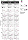
Extended Data Fig. 2.. LOV-1 is required for loading TRFs to ciliary PKD-2 EVs (related to Fig. 2).
a-b, Average fluorescence profiles through cilia of RnB neurons of WT and lov-1 null males for the pair of PKD-2::GFP and TRF-1::mScarlet (a) and for PKD-2::mScarlet and TRF-2::GFP (b). c, Scheme of molecular mechanism loading TRFs to polycystin EVs. Summary of findings.
Extended Data Fig. 3.
Extended Data Fig. 3.. TRFs are not required for polycystin EV release.
a, Flattened z-stacks showing colocalization of PKD-2::GFP and LOV-1::mScarlet in the cilium and on EVs in WT, trf-1 null, and trf-2 null animals. a’, Average fluorescence profiles through cilia of RnB neurons. b, Summary of findings.
Extended Data Fig. 4.
Extended Data Fig. 4.. TRFs require each other for their loading to ciliary EVs.
a, Flattened z-stacks showing average fluorescence profiles of PKD-2::GFP and TRF-1::mScarlet along the cilium of trf-2 null animals. TRF-1 is depleted form the ciliary tip and is not loaded to PKD-2::GFP EVs. b, Summary of findings. c, Flattened z-stacks (uniformly adjusted) showing representative images of TRF-2::GFP and PKD-2::mScarlet levels in the cell bodies of RnB neurons. In the trf-1 null mutant, TRF-2::GFP levels are reduced three times.
Extended Data Fig. 5.
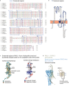
Extended Data Fig. 5.. PACL-1 predicted 3D structure exhibits channel-like properties.
a, Alignment of PCAL-1 homologs within the Caenorhabditis genus. Regions of the high similarity are boxed and numbered. b, Diagram of PACL-1 amino acid sequence, regions of the highest similarity from panel a are shown as blue-shaded amino acid stretches. c, Surface hydrophobicity of PACL-1 (arrow points to the predicted hydrophilic tunnel) and charge distribution (positively-charged cluster on the cytoplasmic part is in blue).d, Structure overlay of the bacterial sodium channel (four subunits, blue) and PACL-1 AlphaFold predicted structure (a single subunit, yellow). Our search in the Protein Data Bank for structural folds akin to the AlphaFold-predicted structural model of PACL-1 yielded a bacterial sodium channel (PDB ID: 4lto) as a candidate (a, c). The structural similarity of PACL-1 and the bacterial sodium channel is confined to transmembrane helices 3 and 4, connected by an extracellular loop (a, c); all are regions less conserved among Caenorhabditis species (b, c). These helices potentially might stabilize the conserved amphipathic helix. Thus, specific amino acid composition within these regions may be less critical for protein function. Examination of the evolutionary conservation of PACL-1 across Caenorhabditis species revealed almost invariable conservation within specific segments, notably an amphipathic helix (a putative pore – region 5 on panels a and b forms hydrophilic tunnel on panel c) and a positively charged cluster located on the cytoplasmic side of the predicted pore entrance (region 7 and 10 on panels a and b, blue cluster on panel c) that might serve as an "anion sink." Among the sequence homologs of PACL-1 outside of the Caenorhabditis genus were a four-pass transmembrane protein A0A016SLD6_9BILA (Acey_s0209.g2096 gene) of Ancylostoma ceylanicum (intestinal hookworm) that distantly resembles TWiK potassium channels (Tandem of P-domains in a Weakly Inward rectifying K+ channel). Taken together, these observations suggest a role for PACL-1 as a channel. Whether PACL-1 functions as an anion or cation channel necessitates experimental validation beyond the scope of this study.
Extended Data Fig. 6.
Extended Data Fig. 6.. PACL-1::GFP colocalizes with PKD-2::mScarlet in cilia and EVs.
a, Fluorescence profiles through cilia of RnB neurons. a’, Colocalization plots show a high level of correlation between the PACL-1:GFP and PKD-2::mScarlet fluorescence intensities in the ciliary shaft (blue) and ciliary tip (yellow). b, Summary of molecular mechanism. PACL-1 associates with the polycystin complex and is released in EVs.
Extended Data Fig. 7.
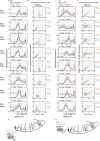
Extended Data Fig. 7.. PAML-1 and PAML-2 are polycystin-interacting transmembrane C-type lectins with dorso-ventral specialization.
a, Fluorescence profiles through cilia of RnB neurons for PAML-1::GFP. a’, Colocalization plots showing a correlation between the PAML-1::GFP and PKD-2::mScarlet fluorescence intensities in ciliary shaft (blue) and ciliary tip (yellow). b, Summary cartoon for PAML-1 presence in cilia and EVs. c, Fluorescence profiles through cilia of RnB neurons for PAML-2::mScarlet. c’, Colocalization plots showing a correlation between the PAML-2::mScarlet and PKD-2::GFP fluorescence intensities in ciliary shaft (blue) and ciliary tip (yellow). d, Summary cartoon for PAML-2 presence in cilia and EVs. Fluorescence values are normalized to the average of minimum and maximum values for each cilium.
Extended Data Fig. 8.
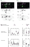
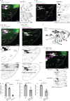
Extended Data Fig. 8.. Cell-specific expression of PAML-1 in ventral rays 2, 3, 4, and 8 inhibits expression of PAML-2, resulting in PAML-2 ciliary localization only in dorsal rays 1, 5, 7, and 9.
a, All PKD-2::GFP-expressing neurons express PAML-2::mScarlet. Image taken at the L4 molt stage (the last molting stage at which most genes required for sexual maturity start their expression). b, Expression patterns of PAML-1::GFP and PAML-2::mScarlet at the L4 molt stage. While PAML-2 is present in all neurons, PAML-1 expression is restricted to a ventral subset of the neurons. At this stage, ray 2, 4, and HOB cell bodies are PAML-1-positive. c, The paml-1 null mutation results in ectopic ciliary localization of PAML-2 in ventral rays 2, 3, 4, 8 (upper left panel). The ciliary presence coincides with increased paml-2::mScarlet presence in cell bodies of those neurons (upper right panel). We measured the cell bodies of neurons 2 and 8 because they are easily identifiable by location; the proximity of neuronal cell bodies 2 and 4 to neurons 5 and 7 made their identification ambiguous, and thus, we did not take these measurements. In dorsal neurons (lower panels), levels of PAML-2 in cilia and cell bodies were unaffected by the paml-1 null mutation. *Mann-Whitney U Test p < .00001 We propose that the mechanism of selective transport of PAML-1 to the cilium of ventral neurons stems from the different affinities of PAML-1 and PAML-2 for the polycystin complex. In ventral neurons that express both paml-1 and paml-2, only PAML-1 is recruited to the cilium, presumably due to its strong binding to the polycystin complex. Conversely, PAML-2 likely has lower affinity to polycystins and thus binds the polycystin complex solely in the absence of PAML-1, as observed in dorsal neurons. This mechanism produces two distinct types of polycystin-carrying ciliary EVs, originating from dorsal and ventral neurons, carrying either PAML-1 or PAML-2 cell-specific markers.
Extended Data Fig. 9.
Extended Data Fig. 9.. CWP-5 expression pattern in PKD-2 expressing neurons displays ray-specificity.
a, Flattened z-stack showing PKD-2::mScarlet and CWP-5::GFP expression. Note the absence of CWP-5::GFP in the cell body of ray 2. b, CWP-5::GFP summarized expression pattern. c, Normalized fluorescence profiles through cilia of RnB neurons for CWP-5::GFP and PKD-2::mScarlet. Note the shouldering effect of CWP::GFP profile compared to PKD-2::mScarlet in the transition zone and the area proximal to the ciliary tip. c’, Colocalization plots show less correlation between PKD-2::mScaret and CWP-5::GFP compared to PACL-1::GFP (Extended Data Fig.5a’), suggesting that CWP-5 has ciliary functions independent of the polycystins.
Extended Data Fig. 10.
CWP-5::GFP ciliary localization in ray 2 and 8 is observed in 50% cases. a, Representative image showing CWP-5::GFP presence in cilia or R2B and R8B neurons (circled). b, Summary of PKD-2 interactor expression patterns. Within the R2B and R8B neurons, CWP-5::GFP showed weak and inconsistent ciliary presence (observed at low levels in 50% cases: 19 out of 26 R2B cilia were CWP-5::GFP positive despite no detectable fluorescence in the R2B cell body, and 28 out of 50 R8B cilia were weakly CWP-5::GFP-positive despite high levels of CWP-5::GFP in the R8B cell body). These results suggest the existence of molecular mechanisms inhibiting transport of CWP-5 from the R8B cell body to the cilium. The source of CWP-5::GFP in the R2B cilium was unclear because CWP-5::GFP was not present in the R2B cell body. We propose three explanations for the absence of CWP-5 in the cell body and its presence in the cilium of the R2B neuron. First, R2B might express CWP-5 at exceedingly low levels, evading detection by our imaging technique. However, CWP-5 relocation to the cilium might be so efficient that even minute amounts in the cell body would lead to detectable accumulation within the cilium. Second, synthesis of the CWP-5 protein might occur exclusively at the ciliary base of R2B, resulting in its exclusive presence in the cilium and not the cell body. Third, the accumulation of ciliary CWP-5 in R2B cilia might arise from the fusion of CWP-5-carrying environmental EVs with the cilium of the R2B neuron. The latter scenario suggests that the R2B neuron might function as a recipient cell for EVs released from other rays of the same or different animal.
Fig. 1.
Fig. 1.. Proximity labeling within PKD-2 EVs identified candidate interactors.
a, Scheme of EV harvest and enrichment, followed by pulldown of candidate interactors biotinylated by TurboID targeted to PKD-2 EVs. b, Scheme of male tail anatomical structures that release PKD-2 EVs. Each sensory ray of the male tail harbors a ciliated dendritic ending protruding into the environment and releasing PKD-2 EVs from the tip of its sensory cilium. c, Visualization of PKD-2::GFP EVs carrying TurboID. d, Identified top candidate interactors.
Fig. 2.
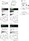
Fig. 2.. LOV-1 is required for loading TRFs to ciliary PKD-2 EVs.
a, Scheme of fluorescent profiling along cilia. b-e, Flattened z-stacks show the ciliary presence of endogenous FP-tagged PKD-2 with TRF-1::mScarlet (b) and TRF-2::GFP (c) in the wild-type cilium and lov-1 mutant (d, e). Representative average fluorescent profiles for each case are shown (b’, c’, d’, e’). Fluorescence values are normalized to the average of minimum and maximum values for each cilium. f-f’, Scheme of the molecular mechanism for loading TRFs to ciliary EVs. In the lov-1 mutant, cilia produce EVs without TRFs (f), whereas in the pkd-2 mutant, ciliary EVs contain neither polycystins nor TRFs (f’).
Fig. 3.
Fig. 3.. TRFs require each other for their loading to ciliary EVs.
a, Flattened z-stack shows that disruption of trf-2 abrogates loading of TRF-1::mScarlet to PKD-2::GFP EVs. TRF-1::mScarlet stays in the cilium and does not reach the ciliary tip, as shown by fluorescent profiling (a’). Fluorescence values are normalized to the average of minimum and maximum values for each cilium. b, Flattened z-stack shows that disruption of trf-1 abrogates ciliary localization of TRF-2::GFP, and thus, no TRF-2::GFP is loaded to PKD-2::mScarlet EVs. b’, Quantification of total fluorescence within the cilium shows that TRF-2::GFP ciliary levels in the trf-1 mutant are reduced tenfold compared to WT cilia, whereas PKD-2::mScarlet total levels are not affected (WT n = 17 animals and 73 cilia, trf-1 null n = 36 animals and 112 cilia).. c, Flattened z-stack through cell bodies of RnB neurons showing reduced cytoplasmic levels of TRF-2::GFP, with quantification shown on panel c’ (WT n = 11 animals and 96 cilia, trf-1 null n = 15 animals and 147 cilia). d, Homology of C. elegans TRFs to human TRAFs. e, Scheme of the molecular mechanism for loading TRFs to ciliary EVs. In the trf-2 mutant, cilia produce EVs without TRF-1, whereas the trf-1 mutation causes reduced levels of TRF-2 and abrogates TRF-2 ciliary localization. *Mann-Whitney U Test p < .00001.
Fig. 4.
Fig. 4.. The polycystin complex associates with and recruits a channel-like protein PACL-1 and to cilia and EVs.
a-b, Flattened z-stacks showing colocalization of PKD-2::mScarlet with PACL-1::GFP in WT (a) and lov-1 mutant (b) animals. c, Scheme of molecular mechanism showing that loading of PACL-1::GFP to ciliary EVs requires LOV-1. d, Assessment of male mating behavior shows that the pacl-1 mutant is deficient in responding to hermaphrodite contact. *Studenťs t test p<0.05.
Fig. 5.
Fig. 5.. The polycystin complex associates with transmembrane C-type lectins that specify dorsal and ventral populations of polycystin cilia and EVs.
a-b, Flattened z-stacks showing colocalization of PKD-2 reporters with PAML-1::GFP (a) and PAML-2::mScarlet (b) in WT. c-d, Disruption of lov-1 abrogates ciliary localization of PAML-1::GFP (c) and PAML-2::mScarlet (d). e, Scheme of PAML-1::GFP and PAML-2::mScarlet localization in RnB neuronal cell bodies and cilia. f, Flattened z-stack showing that disruption of pacl-1 results in PAML-2::mScarlet ciliary localization in ventral neurons. g-h, Assessment of male mating behavior shows that the paml-1; paml-2 double mutant is deficient in responding to hermaphrodite contact (g), and location of vulva (h).* *Studenťs t test p<0.05.
Fig. 6.
Fig. 6.. The polycystin complex is not required for shedding of ciliary EVs as evidenced by tracking cargo CWP-5.
a, Flattened z-stacks showing colocalization of PKD-2::mScarlet with CWP-5::GFP with a zoomed-in inset. a’, Representative average fluorescent profile of CWP-5::GFP and PKD-2::mScarlet along the cilium. Note the prominent presence of CWP-5::GFP in the areas depleted of PKD-2::mScarlet – in the transition zone (TZ) and the neck of the cilium, b-c, Flattened z-stacks showing that disruption of lov-1 (b) and pkd-2 (c) does not alter CWP-5::GFP ciliary and EV localization. d, Scheme of molecular mechanism showing that loading CWP-5::GFP to cilia and ciliary EVs is independent of the polycystins.
Fig. 7.
Fig. 7.. Summary of cargo loading to polycystin ciliary EVs of C. elegans.
A single genetic perturbation leads to a drastic change in EV cargo composition. Compare WT polycystin EVs (a) with EVs of the lov-1 and pkd-2 mutants (b).

References

    1. Hilgendorf K. I., Myers B. R. & Reiter J. F. Emerging mechanistic understanding of cilia function in cellular signalling. Nat Rev Mol Cell Biol 1–19 (2024). - PMC - PubMed
    1. Hogan M. C. et al. Characterization of PKD protein-positive exosome-like vesicles. J Am Soc Nephrol 20, 278–288 (2009). - PMC - PubMed
    1. Wyart C., Carbo-Tano M., Cantaut-Belarif Y., Orts-Del’Immagine A. & Böhm U. L. Cerebrospinal fluid-contacting neurons: multimodal cells with diverse roles in the CNS. Nat Rev Neurosci 24, 540–556 (2023). - PubMed
    1. Orts-Del’Immagine A. et al. Sensory Neurons Contacting the Cerebrospinal Fluid Require the Reissner Fiber to Detect Spinal Curvature In Vivo. Current Biology 30, 827–839.e4 (2020). - PubMed
    1. Djenoune L. et al. The dual developmental origin of spinal cerebrospinal fluid-contacting neurons gives rise to distinct functional subtypes. Sci Rep 7, 719 (2017). - PMC - PubMed

Publication types