[Preprint]. 2024 Apr 15:2024.04.12.589218.
doi: 10.1101/2024.04.12.589218.

Influenza virus antibodies inhibit antigen-specific de novo B cell responses in mice

Affiliations

Influenza virus antibodies inhibit antigen-specific de novo B cell responses in mice

Eileen Goodwin et al. bioRxiv. .

Update in

Abstract

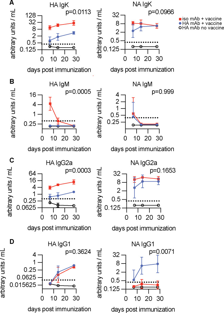
Antibody responses to influenza vaccines tend to be focused on epitopes encountered during prior influenza exposures, with little production of de novo responses to novel epitopes. To examine the contribution of circulating antibody to this phenomenon, we passively transferred a hemagglutinin (HA)-specific monoclonal antibody (mAb) into mice before immunizing with whole inactivated virions. The HA mAb inhibited de novo HA-specific antibodies, plasmablasts, germinal center B cells, and memory B cells, while responses to a second antigen in the vaccine, neuraminidase (NA), were uninhibited. The HA mAb potently inhibited de novo antibody responses against epitopes near the HA mAb binding site. The HA mAb also promoted IgG1 class switching, an effect that, unlike the inhibition of HA responses, relied on signaling through Fc-gamma receptors. These studies suggest that circulating antibodies inhibit de novo B cell responses in an antigen-specific manner, which likely contributes to differences in antibody specificities elicited during primary and secondary influenza virus exposures.


Conflict of interest statement

Declaration of Interests S.E.H. is a co-inventors on patents that describe the use of nucleoside-modified mRNA as a vaccine platform. S.E.H reports receiving consulting fees from Sanofi, Pfizer, Lumen, Novavax, and Merck.

Figures

Fig. 1:
Fig. 1:. HA-specific antibody potently inhibits antibody responses to HA but not NA.
BALB/c mice received either 1μg H28-D14 mAb (blue circles) or 1μg isotype control (red squares) prior to intramuscular immunization with 2,000 hemagglutinating units (HAU) of BPL-inactivated H1N1. Open circles represent serum from unimmunized mice that received 1μg H28-D14 mAb. IgK (A), IgM (B), IgG2a (C), and IgG1 (D) titers against HA and NA protein following immunization are shown. Dashed lines indicate the limit of detection. Data represents three independent experiments with n=3–5 mice per group. Geometric mean titer +/− 95% CI are plotted. Area under the curve (AUC) was calculated for individual mice and groups were compared with Welch’s ANOVA (A, C, HA D) with Dunnett’s T3 multiple comparisons or Kruskal-Wallis test (B, NA D) with Dunn’s multiple comparisons. Reported p-values for comparison between H28-D14 IgG2a (blue) and isotype control IgG2a (red) groups.
Figure 2:
Figure 2:. HA-specific antibody inhibits HA-specific B cells from becoming plasmablasts, germinal center B cells, and memory B cells.
BALB/c mice received either 1μg H28-D14 mAb (blue circles) or 1μg isotype control (red squares) prior to intramuscular immunization with 2,000 HAU BPL-inactivated H1N1. Open circles represent unimmunized mice. (A) Plasmablasts (CD3F4/80Gr1Ter119B220+CD138+) and (B) HA probe-binding plasmablasts in draining lymph nodes of mice measured by flow cytometry at days 4, 7, 10, 14, 21, and 28 post-immunization. (C) Example flow plots of the plasmablast compartment at day 7 binding HA probe. (D) IgM-secreting cells in draining lymph nodes at day 7 post-immunization enumerated by ELISpot. (E) Germinal center B cells (CD3F4/80Gr1Ter119B220+GL7+Fas+) and (F) HA probe-binding germinal center B cells in draining lymph nodes of mice measured by flow cytometry at days 4, 7, 10, 14, 21, and 28 post-immunization. (G) Example flow plots of the GC B cell compartment at day 14 binding HA probe. (H) Memory B cells (CD3F4/80Gr1Ter119B220+GL7CD138CD38+) and HA probe-binding memory B cells in the spleen measured by flow cytometry at >1 year post-immunization. Data represents four independent experiments with n=3–8 mice per group. Arithmetic mean +/− standard deviation are plotted. Dots (D, H) represent individual mice. Significant differences between H28-D14 IgG2a (blue circles) and ctrl IgG2a (red squares) groups by two-way ANOVA with Tukey’s multiple comparisons test (A, B, D-F) or unpaired T test (H) are reported. *p<0.05, ** p<0.01, *** p<0.001, **** p<0.0001, ns=not significant.
Figure 3:
Figure 3:. FcγR interactions mediate IgG1 increase but are dispensable for the inhibitory effects of HA antibody.
BALB/c mice received either 1μg H28-D14 mAb (blue circles), 1μg H28-D14 with D265A mutation (purple triangles), or 1μg isotype control (red squares) prior to intramuscular immunization with 2,000 HAU BPL-inactivated H1N1. Open circles represent unimmunized mice that received 1μg H28-D14 mAb. Serum IgK (A), IgM (B), IgG2a (C), and IgG1 (D) titers against HA and NA protein following immunization are shown. Dashed lines indicate the limit of detection. Data represents three independent experiments with n=3–5 mice per group. Geometric mean titer +/− 95% CI are plotted. Area under the curve (AUC) was calculated for individual mice and groups were compared with Welch’s ANOVA (A, C, HA D) with Dunnett’s T3 multiple comparisons or Kruskal-Wallis test (B, NA D) with Dunn’s multiple comparisons. Reported p-values for comparison between H28-D14 IgG2a (blue circles) and H28-D14 D265A (purple triangles) groups.
Figure 4:
Figure 4:. Anti-Sb antibody most potently inhibits de novo antibody responses to Sb antigenic site and nearby neighboring antigenic sites
(A) Structure of PR8 H1N1 HA protein with residues contributing to each antigenic site indicated by color. (B-D) BALB/c mice received either 1μg H28-D14 mAb (blue circles) or 1μg isotype control (red squares) prior to intramuscular immunization with 2,000 HAU BPL-inactivated H1N1. Open circles represent serum from unimmunized mice that received 1μg H28-D14 mAb. Serum IgK (B), IgG2a (C), and IgG1 (D) titers against PR8 HA epitope panel at day 28 post-immunization are shown. Data points represent individual mice. Dashed lines indicate the limit of detection. Data represents three independent experiments with n=3–5 mice per group. Geometric mean +/− 95% CI are plotted. Significant differences between H28-D14 (blue circles) and control IgG2a (red squares) groups by unpaired t-test with Welch’s correction are reported.

Similar articles

References

    1. Anderson C. F., & Mosser D. M. (2002). Cutting Edge: Biasing Immune Responses by Directing Antigen to Macrophage Fcγ Receptors. The Journal of Immunology, 168(8), 3697–3701. 10.4049/jimmunol.168.8.3697 - DOI - PubMed
    1. Angeletti D., Gibbs J. S., Angel M., Kosik I., Hickman H. D., Frank G. M., Das S. R., Wheatley A. K., Prabhakaran M., Leggat D. J., McDermott A. B., & Yewdell J. W. (2017). Defining B cell immunodominance to viruses. Nature Immunology, 18(4), 456–463. 10.1038/ni.3680 - DOI - PMC - PubMed
    1. Arevalo P., McLean H. Q., Belongia E. A., & Cobey S. (2020). Earliest infections predict the age distribution of seasonal influenza a cases. ELife, 9, 1–30. 10.7554/eLife.50060 - DOI - PMC - PubMed
    1. Bandukwala H. S., Clay B. S., Tong J., Mody P. D., Cannon J. L., Shilling R. A., Verbeek J. S., Weinstock J. V., Solway J., & Sperling A. I. (2007). Signaling through FcγRIII is required for optimal T helper type (Th)2 responses and Th2-mediated airway inflammation. Journal of Experimental Medicine, 204(8), 1875–1889. 10.1084/jem.20061134 - DOI - PMC - PubMed
    1. Baudino L., Shinohara Y., Nimmerjahn F., Furukawa J.-I., Nakata M., Martínez-Soria E., Petry F., Ravetch J. V., Nishimura S.-I., & Izui S. (2008). Crucial Role of Aspartic Acid at Position 265 in the CH2 Domain for Murine IgG2a and IgG2b Fc-Associated Effector Functions. The Journal of Immunology, 181(9), 6664–6669. 10.4049/jimmunol.181.9.6664 - DOI - PubMed

Publication types