[Preprint]. 2024 Jun 4:2024.04.16.589830.
doi: 10.1101/2024.04.16.589830.

Neurophysiological and Autonomic Dynamics of Threat Processing During Sustained Social Fear Generalization

Affiliations

Neurophysiological and Autonomic Dynamics of Threat Processing During Sustained Social Fear Generalization

Jourdan J Pouliot et al. bioRxiv. .

Update in

Abstract

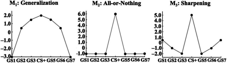
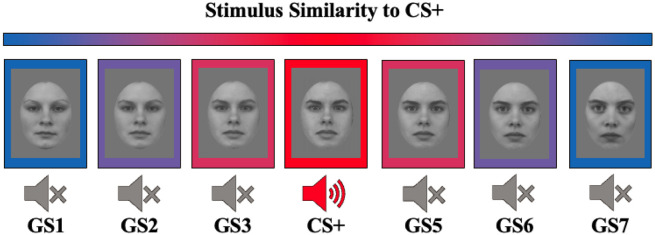
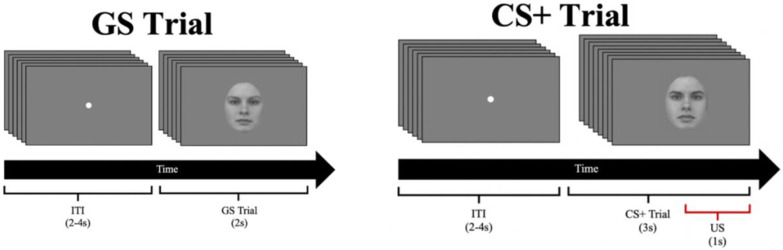
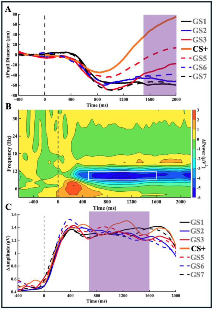
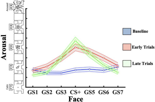
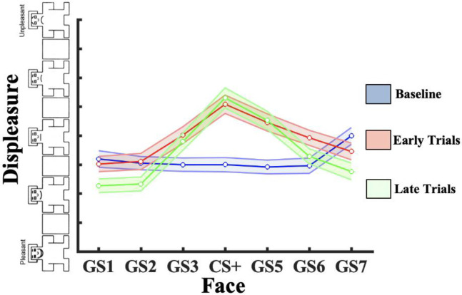
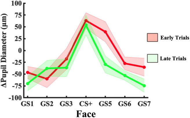
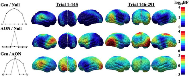
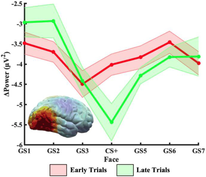
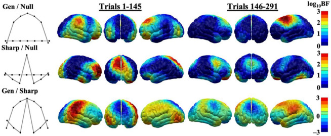
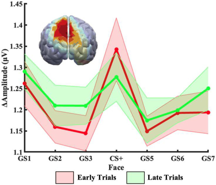
Survival in dynamic environments requires that organisms learn to predict danger from situational cues. One key facet of threat prediction is generalization from a predictive cue to similar cues, ensuring that a cue-outcome contingency is applied beyond the original learning environment. Generalization has been observed in laboratory studies of aversive conditioning: behavioral and physiological processes generalize responses from a stimulus paired with threat (the CS+) to unpaired stimuli, with response magnitudes varying with CS+ similarity. In contrast, work focusing on sensory responses in visual cortex has found a sharpening pattern, in which responses to stimuli closely resembling the CS+ are maximally suppressed, potentially reflecting lateral inhibitory interactions with the CS+ representation. Originally demonstrated with simple visual cues, changes in visuocortical tuning have also been observed in threat generalization learning across facial identities. It is unclear to what extent these visuocortical changes represent transient or sustained effects and if generalization learning requires prior conditioning to the CS+. The present study addressed these questions using EEG and pupillometry in an aversive generalization paradigm involving hundreds of trials using a gradient of facial identities. Visuocortical ssVEP sharpening occurred after dozens of trials of generalization learning without prior differential conditioning, but diminished as learning continued. By contrast, generalization of alpha power suppression, pupil dilation, and self-reported valence and arousal was seen throughout the experiment. Findings are consistent with threat processing models emphasizing the role of changing visucocortical and attentional dynamics when forming, curating, and shaping fear memories as observers continue learning about stimulus-outcome contingencies.


Figures

Figure 1.
Figure 1.
Hypothetical patterns emerging during generalization learning. Left: Quadratic trend showing a broad generalization pattern. Middle: discrimination of only the CS+. Right: Difference-of-Gaussians/sharpening pattern with suppression of responses to GSs with high similarity to the CS+.
Figure 2.
Figure 2.
Gradient of facial stimuli. Four faces were generated using MorphX software to create systematically differing facial morphs, used as gradient stimuli (GS). The GS1, the GS7, and the conditioned stimulus (CS+) were used as the basis of the facial morphs. The GS2 and GS3 are, respectively, 30:70 and 70:30 blends of GS1 and CS+. The GS5 and GS6 are, respectively, 30:70 and 70:30 blends of CS+ and GS7. The CS+ in the middle was paired with presentation of 92 dB(A) white noise.
Figure 3.
Figure 3.
Trial structure. Stimuli were presented following a fixation dot shown during an intertrial interval (ITI) ranging from 2-4 seconds along a uniform distribution. Gradient stimuli (GS1, GS2, GS3, GS5, GS6, GS7) were presented individually in the center of the screen for 2 seconds, flickering at a driving frequency of 15Hz. The conditioned stimulus (CS+) was presented in the center of the screen for 3 seconds, co-terminating with presentation of the unconditioned stimulus (US) during the final second.
Figure 4.
Figure 4.
Dependent variables derived from physiological measures. A) Baseline corrected pupil size time course, averaged by condition across all participants. The purple window indicates the averaged epoch used for statistical analysis (1500ms to 2000ms). B) Baseline corrected time-frequency spectrogram from channel Oz averaged across all participants and conditions, after convolution with a family of Morlet wavelets. The white window indicates where data were averaged across frequencies (10Hz to 12Hz) and time (500ms to 1600ms) prior to statistical analysis. C) Baseline corrected time-varying 15Hz amplitude from channel Oz averaged across all participants and conditions, alongside a time-averaged topography. The purple window indicates the averaged epoch used for statistical analysis (700ms to 1600ms).
Figure 5.
Figure 5.
Arousal Condition Means. Arousal ratings were taken during baseline before conditioning, during the first half of trials, and during the second half of trials. An increase along the y-axis indicates an increase in reported physiological arousal or emotional intensity. SEM bars included to indicate significant differences.
Figure 6.
Figure 6.
Valence Condition Means. Valence ratings were taken during baseline before conditioning, during the first half of trials, and during the second half of trials. An increase along the y-axis indicates an increase in reported negative valence or displeasure. SEM bars included to indicate significant differences.
Figure 7.
Figure 7.
Averaged Pupillary Response Condition Means. Pupil responses in each condition averaged across the analytic epoch (1500ms-2000ms) and across subjects. Shaded error bars depicting the within-subjects standard error of the mean (SEM) included to indicate condition differences.
Figure 8.
Figure 8.
Brain Mapping of Alpha Suppression Bayesian Model Fits. log10Bayes Factors were computed comparing fit of electrocortical responses to each hypothesized model to the null, alongside a direct comparison of model evidence between both models. This was done for all channels. Topographies map model fits at each chain to their corresponding cortical region. Results show widespread overlap of fit to both models, and general increase in model fit over time. Strongest model fit to both models occurs during later trials in right occipitotemporal and central regions.
Figure 9.
Figure 9.
Averaged Alpha Power Condition Means. Baseline-corrected alpha power was averaged across subjects and across the analytic epoch (500ms-1600ms). Additionally, power was averaged across a 5-channel right occipitotemporal cluster showing greatest model fit to the all-or-nothing model to demonstrate changes in cortical tuning with respect to the stimulus gradient. Shaded error bars depicting the within-subjects standard error of the mean (SEM) included to indicate condition differences.
Figure 10.
Figure 10.
Brain Mapping of ssVEP Bayesian Model Fits. log10Bayes Factors were computed comparing fit of electrocortical responses to each hypothesized model to the null, alongside a direct comparison of model evidence between both models. This was done for all channels. Topographies map model fits at each channel to their corresponding cortical region. Results show large differences in model fit between conditioning periods. Most notable is the occipitoparietal channels, where fit to the sharpening pattern is decisive during early trials but decreases to strong in later trials.
Figure 11.
Figure 11.
Averaged Steady-State Visual Evoked Potential (ssVEP) Condition Means. Baseline-corrected ssVEP amplitude was averaged across subjects and across the analytic epoch (700ms-1600ms). Additionally, amplitude was averaged across a 5-channel occipitoparietal cluster showing greatest model fit to the sharpening model to demonstrate changes in cortical tuning with respect to the stimulus gradient. Shaded error bars depicting the within-subjects standard error of the mean (SEM) included to indicate condition differences.

Similar articles

References

    1. Adrian E. D., & Matthews B. H. (1934). The Berger rhythm: potential changes from the occipital lobes in man. Brain, 57(4), 355–385. - PubMed
    1. Ahrens L. M., Pauli P., Reif A., Mühlberger A., Langs G., Aalderink T., & Wieser M. J. (2016). Fear conditioning and stimulus generalization in patients with social anxiety disorder. Journal of Anxiety Disorders, 44, 36–46. 10.1016/jjanxdis.2016.10.003 - DOI - PubMed
    1. Ala T. S., Graversen C., Wendt D., Alickovic E., Whitmer W. M., & Lunner T. (2020). An exploratory Study of EEG Alpha Oscillation and Pupil Dilation in Hearing-Aid Users During Effortful listening to Continuous Speech. PLOS ONE, 15(7), e0235782. 10.1371/journal.pone.0235782 - DOI - PMC - PubMed
    1. Andersen S. K., Müller M. M., & Hillyard S. A. (2009). Color-selective attention need not be mediated by spatial attention. Journal of Vision, 9(6), 2–2. 10.1167/9.6.2 - DOI - PubMed
    1. Antov M. I., Plog E., Bierwirth P., Keil A., & Stockhorst U. (2020). Visuocortical tuning to a threat-related feature persists after extinction and consolidation of conditioned fear. Scientific reports, 10(1), 3926. 10.1038/s41598-020-60597-z - DOI - PMC - PubMed

Publication types