[Preprint]. 2024 Apr 19:2024.04.17.589977.
doi: 10.1101/2024.04.17.589977.

Functional and antigenic landscape of the Nipah virus receptor binding protein

Affiliations

Functional and antigenic landscape of the Nipah virus receptor binding protein

Brendan B Larsen et al. bioRxiv. .

Update in

Abstract

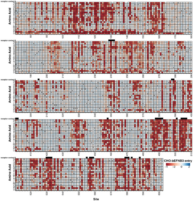
Nipah virus recurrently spills over to humans, causing fatal infections. The viral receptor-binding protein (RBP or G) attaches to host receptors and is a major target of neutralizing antibodies. Here we use deep mutational scanning to measure how all amino-acid mutations to the RBP affect cell entry, receptor binding, and escape from neutralizing antibodies. We identify functionally constrained regions of the RBP, including sites involved in oligomerization, along with mutations that differentially modulate RBP binding to its two ephrin receptors. We map escape mutations for six anti-RBP antibodies, and find that few antigenic mutations are present in natural Nipah strains. Our findings offer insights into the potential for functional and antigenic evolution of the RBP that can inform the development of antibody therapies and vaccines.


Figures

Figure 1.
Figure 1.. Effects of RBP mutations on entry into CHO-bEFNB3 cells.
For each mutation, the entry score reflects the cell entry efficiency of a pseudovirus with that RBP mutation relative to the unmutated RBP. Negative values (red) indicate impaired entry, zero (white) indicates no effect, and positive values (blue) indicate improved entry. The wildtype amino-acid in the Malaysia strain RBP at each site is indicated with a ‘X’. Mutations that were not measured with high confidence in our experiments are indicated with a light gray box. Residues that directly contact the EFNB3 receptor (<= 4 angstroms of the receptor calculated from PDB 3D12) are indicated with black boxes above the heatmap. An interactive version of this heatmap is available at https://dms-vep.org/Nipah_Malaysia_RBP_DMS/htmls/E3_entry_heatmap.html. See fig S9 for comparable data for cell entry into CHO-bEFNB2 cells.
Figure 2.
Figure 2.. Functional constraint on different regions of RBP for entry into CHO-bEFNB3 cells.
A) RBP tetrameric structure colored by the effect of mutations on cell entry (PDBs: 7TXZ, 7TY0, 3D12). Each site is colored by the mean effect of all amino-acid mutations at that site, with red indicating impaired cell entry, and white indicating entry comparable to unmutated RBP. B) Effects of mutations on cell entry projected on the RBP head oriented to visualize the RBP dimerization face. C) Boxplot showing the impacts of mutations on cell entry across different RBP regions. D) Average effects of mutations at each RBP site on cell entry. E) Average effect of mutations at each site on cell entry for the RBP neck viewed from the side or top. Each chain has a unique color scale as indicated in the color-scale bars, with darker colors indicating impaired entry. F) Effects of mutations on entry for the interface between the distal heads (chains A and B). G) Effects of mutations on entry at the RBP/EFNB3 interface. The same color scale is used as in (A). EFNB3 is shown as a gray cartoon. H) Correlation between effects of mutations on cell entry in CHO-bEFNB2 vs CHO-bEFNB3 cells. Each point is the average effect of mutations at a site. Points are colored by distance to the closest receptor residue (contact sites defined as < 4 Å to receptor, close > 4 and < 8 Å, and distant >10 Å). I) Validation assays showing the correlation between single mutant pseudovirus titers in CHO-bEFNB3 cells versus effects measured in DMS. Three independent measurements were made for each validation pseudovirus. Infectivity was quantified by luciferase signal (RLU/μL) at 48 hours after infection of CHO-bEFNB3 cells.
Figure 3.
Figure 3.. Effects of RBP mutations on binding to bEFNB2 or bEFNB3.
A) Neutralization of unmutated Nipah RBP/F pseudoviruses by monomeric or dimeric soluble bEFNB2 or bEFNB3. B) Correlation between biolayer interferometry (BLI) measurements of binding affinity with DMS measured effects of mutations on binding. The magnitude of binding was assessed by quantifying the change in total area under the curve (AUC) relative to unmutated RBP for binding of the indicated RBP head domains to immobilized dimeric bEFNB2 or bEFNB3 (see fig S18 for raw BLI sensorgrams). C) Tetrameric structure of RBP colored by the site-average effects of mutations on binding to bEFNB2. Sites that are missing binding measurements (typically because mutations are highly deleterious for cell entry) are colored black. D) Correlation between the effects of mutations on binding to bEFNB2 versus bEFNB3. Mutations with notable effects on binding are labeled. E) Location of sites on RBP’s head that have notable mutations on binding. For heatmaps showing effects of all mutations on binding as measured by DMS, see fig S14,15.
Figure 4.
Figure 4.. DMS maps of escape mutations from six monoclonal antibodies.
A) Key sites of escape for each antibody. The height of each letter is proportional to the escape caused by that amino-acid mutation, and letters are colored by the effect of that mutation on entry in CHO-bEFNB3 cells (dark green indicates well tolerated mutations, light yellow indicates impaired cell entry). These logo plots show the top escape sites for each antibody; for all sites see (https://dms-vep.org/Nipah_Malaysia_RBP_DMS/htmls/mab_plot_all.html). For panels A-C, only mutations that decrease in antibody neutralization are shown. B) Antibody escape mapped onto the structure of RBP’s head. The escape averaged across all mutations at each site is shown as a white-blue color scale, with sites where mutations cause strong escape in darker blue, and those not affecting neutralization in white. Dark gray sites have no escape measurements due to mutations strongly impairing cell entry. C) Variability of RBP among known Nipah virus sequences at sites of antibody escape mapped in this study. Sites are classified as conserved or polymorphic based on whether they show variation among at least two Nipah RBP sequences. D) Correlation between the IC50 measured for single mutant Nipah RBP pseudoviruses for antibody nAH1.3 in validation assays versus escape measured in DMS. The upper limit of IC50 values in the validation assays was 10 μg/mL (dashed line). See fig S26 for escape logo plots that show only mutations that are single-nucleotide accessible.

References

    1. Chua K. B., Bellini W. J., Rota P. A., Harcourt B. H., Tamin A., Lam S. K., Ksiazek T. G., Rollin P. E., Zaki S. R., Shieh W., Goldsmith C. S., Gubler D. J., Roehrig J. T., Eaton B., Gould A. R., Olson J., Field H., Daniels P., Ling A. E., Peters C. J., Anderson L. J., Mahy B. W., Nipah virus: a recently emergent deadly paramyxovirus. Science 288, 1432–1435 (2000). - PubMed
    1. Chua K. B., Koh C. L., Hooi P. S., Wee K. F., Khong J. H., Chua B. H., Chan Y. P., Lim M. E., Lam S. K., Isolation of Nipah virus from Malaysian Island flying-foxes. Microbes Infect. 4, 145–151 (2002). - PubMed
    1. Epstein J. H., Prakash V., Smith C. S., Daszak P., McLaughlin A. B., Meehan G., Field H. E., Cunningham A. A., Henipavirus infection in fruit bats (Pteropus giganteus), India. Emerg. Infect. Dis. 14, 1309–1311 (2008). - PMC - PubMed
    1. Epstein J. H., Anthony S. J., Islam A., Kilpatrick A. M., Ali Khan S., Balkey M. D., Ross N., Smith I., Zambrana-Torrelio C., Tao Y., Islam A., Quan P. L., Olival K. J., Khan M. S. U., Gurley E. S., Hossein M. J., Field H. E., Fielder M. D., Briese T., Rahman M., Broder C. C., Crameri G., Wang L.-F., Luby S. P., Lipkin W. I., Daszak P., Nipah virus dynamics in bats and implications for spillover to humans. Proc. Natl. Acad. Sci. U. S. A. 117, 29190–29201 (2020). - PMC - PubMed
    1. Epstein J. H., Field H. E., Luby S., Pulliam J. R. C., Daszak P. , Nipah virus: impact, origins, and causes of emergence. Curr. Infect. Dis. Rep. 8, 59–65 (2006). - PMC - PubMed

Publication types