. 2024 Apr 24;3(4):pgae141.
doi: 10.1093/pnasnexus/pgae141. eCollection 2024 Apr.

Alteration of mechanical stresses in the murine brain by age and hemorrhagic stroke

Affiliations

Alteration of mechanical stresses in the murine brain by age and hemorrhagic stroke

Siyi Zheng et al. PNAS Nexus. .

Abstract

Residual mechanical stresses, also known as solid stresses, emerge during rapid differential growth or remodeling of tissues, as observed in morphogenesis and tumor growth. While residual stresses typically dissipate in most healthy adult organs, as the growth rate decreases, high residual stresses have been reported in mature, healthy brains. However, the origins and consequences of residual mechanical stresses in the brain across health, aging, and disease remain poorly understood. Here, we utilized and validated a previously developed method to map residual mechanical stresses in the brains of mice across three age groups: 5-7 days, 8-12 weeks, and 22 months. We found that residual solid stress rapidly increases from 5-7 days to 8-12 weeks and remains high in mature 22 months mice brains. Three-dimensional mapping revealed unevenly distributed residual stresses from the anterior to posterior coronal brain sections. Since the brain is rich in negatively charged hyaluronic acid, we evaluated the contribution of charged extracellular matrix (ECM) constituents in maintaining solid stress levels. We found that lower ionic strength leads to elevated solid stresses, consistent with its unshielding effect and the subsequent expansion of charged ECM components. Lastly, we demonstrated that hemorrhagic stroke, accompanied by loss of cellular density, resulted in decreased residual stress in the murine brain. Our findings contribute to a better understanding of spatiotemporal alterations of residual solid stresses in healthy and diseased brains, a crucial step toward uncovering the biological and immunological consequences of this understudied mechanical phenotype in the brain.

Keywords: age-related alteration; hemorrhagic stroke; mouse brain; residual solid stress.


Figures

Fig. 1.
Fig. 1.
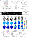
Residual solid stresses in murine brain are age-dependent. A) The fresh brain (graphic generated using BioRender.com) is embedded in 2% agarose, then sliced with a Compresstome to obtain a specific thickness of 250 μm. B) Slices are left in a PBS at room temperature for 20 min to deform as the solid stress is released in the tissue slice. C) The deformed slices are embedded in 1% agarose, then fixed with formalin overnight, and washed with PBS. D) The deformed slice is imaged via confocal microscopy. E) The normalized deformation, Dn, an index of residual solid stress, is defined as the average height difference from curved surface to the midline. F) The area ratio, Ra, defined as the ratio of surface area and projection area, is used as another index of solid stress. G) The curvature, k, defined as the reciprocal of the curvature radius, is used as the third index of solid stress. Mean curvature, Km, is the average of mean curvature on tissue slices. H) The orthogonal views of deformation of representative brain and kidney (negative control) slices from 5–7 days, 8–12 weeks, and 22 months mice. I) The projection images, J) deformation maps, and K) mean curvature maps of representative brain and kidney slices from 5–7 days, 8–12 weeks, and 22 months mice. L) Residual solid stresses are estimated in multiple ways by quantifying the normalized deformation, M) area ratio, and N) mean curvature. Brain slices have significantly higher normalized deformation, area ratio, and mean curvature than kidney slices in all age groups. Brain slices have increased normalized deformation, area ratio, and mean curvature from 5–7 days to 8–12weeks, and then keep plateau from 8–12 weeks to 22 months (mean ± SEM, n = 5 mice, two-tailed t test).
Fig. 2.
Fig. 2.
Viscoelastic properties in murine brain are age-dependent. A) Young's modulus and B) rate-dependent stiffening defined as instantaneous/equilibrium modulus ratio are age-dependent in brain (mean ± SEM, n = 5 mice, two-tailed t test). Brain from 8–12 weeks mice has the highest Young's modulus among all age groups. Instantaneous/equilibrium modulus ratio has a decreasing tendency in mice brains from 5–7 days to 22 months. C) The instantaneous/equilibrium modulus ratio is strain dependent (mean ± SEM, n = 5 mice, one-way ANOVA test). Brain tissue from 8–12 weeks and 22 months mice have decreasing tendency of instantaneous/equilibrium modulus ratio with strain. D) Fast relaxation time constant, and slow relaxation time constant, changes with age (mean ± SEM, n = 5 mice, two-tailed t test). Brain from 5–7 days mice has both the highest fast and slow relaxation time constants. The changing tendency of E) fast and F) slow relaxation time constant with strain (mean ± SEM, n = 5 mice, one-way ANOVA test).
Fig. 3.
Fig. 3.
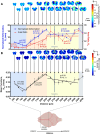
Residual solid stress distribution in the whole brain. A) The normalized deformation and area ratio of brain slices in the coronal direction (mean ± SEM, n = 3 mice, one-way ANOVA test) demonstrated a heterogenous distribution along the anterior–posterior axis. B) The mean curvature of brain slices in the coronal direction (mean ± SEM, n = 3 mice, one-way ANOVA test) demonstrated a heterogenous distribution along the anterior–posterior axis.
Fig. 4.
Fig. 4.
The effect of ionic strength on residual solid stresses. A) Projected microscopy images, B) normalized deformation maps, and C) mean curvature maps of representative brain slices from 8–12 weeks mice under different ionic strength. D) The normalized deformation, E) area ratio, and F) mean curvature show a decreasing trend with increasing ionic strength (mean ± SEM, n = 3–5 slices, two-tailed t test).
Fig. 5.
Fig. 5.
Hemorrhagic stroke results in lower residual solid stress in murine brains. A) The loss of cellular density within the lesion core and the depletion of NeuN-positive neurons on the periphery of the lesion core. B) The infiltrated Cd13-positive immune cells phagocytose cellular debris and in conjunction with locally recruited Gfap-positive astrocytes take part in the formation of the astroglial border. C) The projected image from microscopy, D) deformation maps, and E) mean curvature maps of representative brain sections from hemorrhagic stroke and healthy mice. F) The normalized deformation, G) area ratio, and H) mean curvature are lower in lobes with hemorrhagic stroke compared to normal brain sections (mean ± SEM, n = 3 mice, two-tailed t test). Healthy brain sections have higher normalized deformation, area ratio, and mean curvature than hemorrhagic stroke brain sections.

References

    1. Nia HT, Munn LL, Jain RK. 2020. Physical traits of cancer. Science. 370:eaaz0868. - PMC - PubMed
    1. Zhang S, et al. 2023. The peritumor microenvironment: physics and immunity. Trends Cancer. 9:609–623. - PMC - PubMed
    1. Nia HT, et al. 2016. Solid stress and elastic energy as measures of tumour mechanopathology. Nat Biomed Eng. 1:0004. - PMC - PubMed
    1. Kalli M, Stylianopoulos T. 2018. Defining the role of solid stress and matrix stiffness in cancer cell proliferation and metastasis. Front Oncol. 8:55. - PMC - PubMed
    1. Huycke TR, et al. 2019. Genetic and mechanical regulation of intestinal smooth muscle development. Cell. 179:90–105.e121. - PMC - PubMed