. 2024 Apr 10:17:1009404.
doi: 10.3389/fnmol.2024.1009404. eCollection 2024.

PLC-γ-Ca2+ pathway regulates axonal TrkB endocytosis and is required for long-distance propagation of BDNF signaling

Affiliations

PLC-γ-Ca2+ pathway regulates axonal TrkB endocytosis and is required for long-distance propagation of BDNF signaling

Guillermo Moya-Alvarado et al. Front Mol Neurosci. .

Abstract

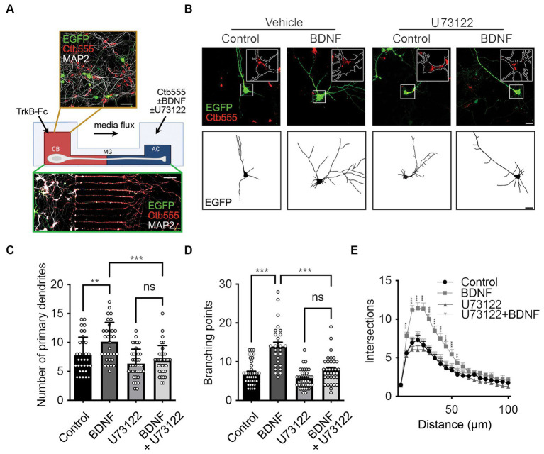
Brain-derived neurotrophic factor (BDNF) and its tropomyosin receptor kinase B (TrkB) are important signaling proteins that regulate dendritic growth and maintenance in the central nervous system (CNS). After binding of BDNF, TrkB is endocytosed into endosomes and continues signaling within the cell soma, dendrites, and axon. In previous studies, we showed that BDNF signaling initiated in axons triggers long-distance signaling, inducing dendritic arborization in a CREB-dependent manner in cell bodies, processes that depend on axonal dynein and TrkB activities. The binding of BDNF to TrkB triggers the activation of different signaling pathways, including the ERK, PLC-γ and PI3K-mTOR pathways, to induce dendritic growth and synaptic plasticity. How TrkB downstream pathways regulate long-distance signaling is unclear. Here, we studied the role of PLC-γ-Ca2+ in BDNF-induced long-distance signaling using compartmentalized microfluidic cultures. We found that dendritic branching and CREB phosphorylation induced by axonal BDNF stimulation require the activation of PLC-γ in the axons of cortical neurons. Locally, in axons, BDNF increases PLC-γ phosphorylation and induces intracellular Ca2+ waves in a PLC-γ-dependent manner. In parallel, we observed that BDNF-containing signaling endosomes transport to the cell body was dependent on PLC-γ activity and intracellular Ca2+ stores. Furthermore, the activity of PLC-γ is required for BDNF-dependent TrkB endocytosis, suggesting a role for the TrkB/PLC-γ signaling pathway in axonal signaling endosome formation.

Keywords: PLC-γ; TrkB; axonal transport; brain-derived neurotrophic factor; calcium; dendritic branching; endocytosis; signaling endosomes.


Conflict of interest statement

The authors declare that the research was conducted in the absence of any commercial or financial relationships that could be construed as a potential conflict of interest.

Figures

Figure 1
Figure 1
Axonal PLC-γ activity is required for dendritic arborization induced by BDNF in rat cortical neurons. (A) Schematics showing the experimental design and example of a lower and a higher magnification image of the cortical neurons in compartmentalized culture. Cortical neurons (DIV 6) were transfected with a plasmid expressing EGFP (green). The cell body compartment (CB) was incubated with TrkB-Fc (100 ng/ml). The axonal compartment (AC) was stimulated with BDNF (50 ng/ml) in addition to Ctb555 (red) in the presence or absence of the PLC-γ inhibitor U73122 (5 μM). Treatments were applied for 48 h. Finally, neurons were fixed, and immunofluorescence was performed against MAP2 (white). Upper panel, scale bar 50 μm. Lower panel, scale bar 50 μm. (B) Upper panels, representative images of the CB of compartmentalized rat cortical neurons whose axons were treated with DMSO (control), U73122, BDNF or BDNF following preincubation with U73122. Ctb555 is shown in red and EGFP in green. Insets are 2.5x-zoom images of the Ctb555 signal and the cell body contour is labeled with a white line. Lower panels, binary images obtained from EGFP-associated fluorescence was used to perform morphological analysis. Scale bar, 20 μm. (C–E) Quantification of primary dendrites (C) and branching points (D) and Sholl analysis (E) for neurons labeled with EGFP/MAP2/Ctb555 for each treatment. n = 27–38 neurons from 3 independent experiments. The results are expressed as the means ± SEMs. **p < 0.01, ***p < 0.001. Statistical analysis was performed by one-way ANOVA followed by the Bonferroni correction for multiple comparisons (C,D). The Sholl’s analysis results were statistically analyzed by two-way ANOVA followed by the Bonferroni correction for multiple comparisons (E).
Figure 2
Figure 2
Axonal PLC-γ activity is required for somatodendritic CREB phosphorylation in rat cortical neurons. (A) Schematic representation of the protocol used for stimulating neurons. DIV 5 cortical neurons were retrograde labeled with Ctb555 (in red) overnight. At DIV 6, the culture medium was changed to serum-free medium for 90 min in the presence or absence of U73122 (5 μM) in the AC and stimulated with BDNF (50 ng/ml) for 180 min in the AC in the presence or absence of U73122 with the flux toward the AC. Finally, the cultures were fixed, and phosphorylated CREB (pCREB, S133) immunofluorescence was analyzed in cell bodies. (B) Representative figures of nuclear pCREB in neurons with or without BDNF stimulation labeled with Ctb555 (in red) added to axons in the presence or absence of axonal U73122 as indicated in (A). Scale bar, 10 μm. (C) Quantification of pCREB in the nucleus of neurons labeled with Ctb555 (red) in each condition. n = 90–114 neurons from 3 independent experiments as shown in (B). (D) Schematic representation of the protocol used for stimulating neurons. DIV 5 cortical neurons were retrograde labeled with Ctb555 (in red) overnight. At 6 DIV, the culture medium was changed to serum-free medium for 90 min in the presence or absence of U73122 (5 μM) in the CB with the flux toward the CB, and then the AC was incubated with BDNF (50 ng/mL) for 180 min. Finally, the cultures were fixed, and pCREB was analyzed in cell bodies. (E) Representative figures of nuclear pCREB (in green) in neurons with or without BDNF stimulation labeled with Ctb555 (in red) added to axons in the presence or absence of U73122 in the CB. (F) Quantification of pCREB in the nucleus in neurons labeled with Ctb555 in each condition. n = 43–60 neurons from 2 independent experiments as indicated in (D). The results are expressed as the means ± SEMs. ns, non significant. *p < 0.05. **p < 0.01, ***p < 0.01. Statistical analysis was performed by one-way ANOVA followed by the Bonferroni correction for multiple comparisons.
Figure 3
Figure 3
BDNF in axons promotes axonal PLC-γ phosphorylation in rat cortical neurons. (A) Representative images of phosphorylated PLC-γ (pPLC-γ, pY783.28, green) in axons of compartmentalized rat cortical neurons left unstimulated (control), stimulated with 50 ng/ml BDNF for 20 min (BDNF) or stimulated with BDNF in the presence of 0.2 μM K252a (BDNF + K252a). Axons were labeled with Ctb555 overnight before treatment to assess correct compartmentalization of the culture. (B) Quantification of the immunofluorescence signal associated with pPLC-γ was performed in a rectangular ROI drawn in the border between the beginning of the microgroove and the AC and continuing for 30 μm toward the microgroove that was delimited by Ctb555 fluorescence, shown as a white rectangles in (A) and in Supplementary Figure S2A as distal microgroove. n = 5 chambers (the value in each chamber corresponds to the average of 5 different microgrooves) performed in five independent cultures. Statistical analysis was performed by one-way ANOVA followed by the Bonferroni correction for multiple comparisons. **p < 0.01. (C) Compartmentalized neurons were treated as described in (A) (but K252a was not used in this experiment). A representative image of total PLC-γ in the AC quantified as indicated in (B) used to assess pPLC-γ immunostaining is shown. (D) Quantification of the immunofluorescence signal associated with total PLC-γ. Statistical analysis was performed by Student’s t-test. No significant differences were found. Scale bar, 10 μm.
Figure 4
Figure 4
Axonal BDNF promotes an increase in intracellular Ca2+ in a PLC-γ-dependent manner in rat cortical neurons. (A) Evaluation of Ca2+ signaling induced by BDNF. The change in fluorescence intensity associated with Fluo4-AM (2 μM) was used to measure the concentration change in cytosolic Ca2+. Representative images of compartmentalized cultures loaded with Fluo4-AM in the AC treated with vehicle or BDNF (50 ng/ml) in the AC with or without U73122 (5 μM) pretreatment. Live-cell imaging of each axonal field in the AC recorded before BDNF treatment (0 s) and during 60 s of BDNF treatment. Scale bar, 10 μm. (B) Mean Fluo4-AM fluorescence intensity (±SEM) for each treatment at different snapshot times. Fab represents the average fluorescence of the baseline. We calculate the average of the calcium recordings for the vehicle or U17322 treatments, and subsequently standardize the data of the baseline, BDNF and BDNF + U17622 calcium recordings by these respective values. n = 8–12 axons from three independent compartmentalized cultures. Statistical analysis was performed by two-way ANOVA followed by the Bonferroni correction for multiple comparisons. **p < 0.01. (C) Quantification of the velocity of Ca2+ back-propagation of the fluorescence signal associated with Fluo4-AM under BDNF conditions.
Figure 5
Figure 5
Axonal PLC-γ activity and intracellular Ca2+ are required for axonal BDNF-containing signaling endosome generation and retrograde transport to cell bodies in mouse cortical neurons. (A) DIV 7 compartmentalized cortical neurons were retrograde labeled with Ctb555 (red) overnight. At DIV 8, the cell body compartment was treated with TrkB-Fc, and the AC was treated with DyLight 488-labeled streptavidin (f-streptavidin, green) alone or with biotinylated BDNF conjugated to DyLight 488-labelled streptavidin (f-BDNF, green), as indicated in the methodology section, for 6 h in the absence or presence of vehicle or 5 μM U73122 (U73122) or 20 μM BAPTA-AM (BAPTA). Then, the cells were fixed, mounted in Mowiol containing Hoechst (blue) and prepared for confocal microscopy. Scale bar, 5 μm (B) Quantification of green-labelled vesicles in cell bodies of neurons containing Ctb555. Only vesicles larger than 200 nm2 were considered for the analysis. Forty-five neurons from three independent compartmentalized cultures were considered. Statistical analysis was performed by one-way ANOVA followed by the Bonferroni correction for multiple comparisons. ****p < 0.0001. (C) Left panels, representative images of f-streptavidin or f-BDNF (green) associated fluorescence. White lines are selecting the region of the microgroove label with Ctb555 (red) shown in the right panels. White arrows indicate green-labelled vesicles. Right panels, Representative images of axons (labeled with Ctb555, red) in microgrooves of neurons treated as described in (A). Scale bar, 5 μm. (D) Quantification of green-labelled vesicles in microgrooves of axons containing Ctb555. Forty-five microgrooves from three independent compartmentalized cultures were considered for the analysis. Statistical analysis was performed by one-way ANOVA followed by the Bonferroni correction for multiple comparisons. ****p < 0.0001.
Figure 6
Figure 6
PLC-γ activity and intracellular calcium are required for TrkB internalization in mouse cortical neurons. Neurons (DIV 6) were transfected with a plasmid expressing Flag-TrkB. After 48 h, the neurons were incubated with an anti-Flag antibody. The neurons were treated with BDNF (50 ng/ml) in the presence or absence of U73122 (5 μM) or BAPTA-AM (5 μM) or ANA-12 (10 μM) or BDNF (50 ng/ml) in the presence or absence of U73343 (5 μM) for 20 min to induce endocytosis at 37°C. Finally, the neurons were fixed, and the Flag epitope was detected by immunostaining as indicated in Supplementary Figure S4A. The plasma membrane associated Flag antibody was recognized with an anti-mouse Alexa488 (mFlag, green) before permeabilizing cells, then, cells were permeabilized and the total Flag epitope was recognized with an anti-mouse Alexa555 (tFlag). (A) Representative images of the endocytosis of Flag-TrkB in control cells treated with BDNF, U73122, U73343, BDNF with U73122 or BDNF with U73343. The figure shows plasma membrane associated Flag-TrkB (in green), and total Flag-TrkB in red, blue is the nucleus labelled with Hoechst. The yellow line is limiting the outer border of the cell, the green line is limiting membrane versus cytosolic associated Flag-TrkB and the white one the nucleus. Scale bar, 5 μm. (B) Quantification of internalized Flag-TrkB was achieved by dividing the fluorescence associated with internalized Flag-TrkB (only red fluorescence between the green and white lines) by the fluorescence intensity associated with total Flag-TrkB. n = 14–18 neurons from 3 independent experiments. Scale bar, 5 μm. (C) Representative images of the endocytosis of Flag-TrkB in control cells treated with BDNF, BAPTA-AM, ANA-12, BDNF with BAPTA-AM or BDNF with ANA-12. The figure shows plasma membrane associated Flag-TrkB (mFlag, in green), and total Flag-TrkB (tFlag, in red), blue is the nucleus labelled with Hoechst. The green line is limiting membrane versus cytosolic associated Flag-TrkB and the white one the nucleus. Scale bar, 5 μm. (D) Quantification of internalized Flag-TrkB in neurons treated with BAPTA-AM and ANA-12. n = 15–23 neurons from 3 independent experiments. The results are expressed as the mean ± SEM. ***p < 0.001; ****p < 0.0001. Statistical analysis was performed by one-way ANOVA followed by Bonferroni’s post-test for multiple comparisons.
Figure 7
Figure 7
Model summarizing the role of known BDNF–TrkB signaling pathways on long-distance BDNF signaling. The findings from our study suggest that BDNF in the axon activates TrkB receptors and PLC-γ, leading to an increase in intracellular calcium concentration (Ca2+). This, in turn, promotes the endocytosis of the receptor and the formation of signaling endosomes. The first step in this process enables the retrograde transport of signaling endosomes to the cell body, contributing to an elevation in CREB phosphorylation and dendritic arborization. Notably, we observed that cell body PLC-γ activity was not essential for CREB phosphorylation induced by axonal BDNF. In previous research, we demonstrated that PI3K activity is not necessary for endocytosis or transport of signaling endosomes but plays a crucial role in mTOR-dependent translation of CREB target genes (Moya-Alvarado et al., 2023). This process was also needed for dendritic arborization induced by BDNF in the axons.

Similar articles

Cited by

References

    1. Alonso M., Vianna M. R., Depino A. M., Mello E Souza T., Pereira P., Szapiro G., et al. . (2002a). Bdnf-triggered events in the rat hippocampus are required for both short- and long-term memory formation. Hippocampus 12, 551–560. doi: 10.1002/hipo.10035 - DOI - PubMed
    1. Alonso M., Vianna M. R., Izquierdo I., Medina J. H. (2002b). Signaling mechanisms mediating Bdnf modulation of memory formation in vivo in the hippocampus. Cell. Mol. Neurobiol. 22, 663–674. doi: 10.1023/A:1021848706159 - DOI - PMC - PubMed
    1. Arimura N., Kimura T., Nakamuta S., Taya S., Funahashi Y., Hattori A., et al. . (2009). Anterograde transport of TrkB in axons is mediated by direct interaction with Slp1 and Rab27. Dev. Cell 16, 675–686. doi: 10.1016/j.devcel.2009.03.005, PMID: - DOI - PubMed
    1. Beattie E. C., Zhou J., Grimes M. L., Bunnett N. W., Howe C. L., Mobley W. C. (1996). A signaling endosome hypothesis to explain Ngf actions: potential implications for neurodegeneration. Cold Spring Harb. Symp. Quant. Biol. 61, 389–406. PMID: - PubMed
    1. Bleasdale J. E., Thakur N. R., Gremban R. S., Bundy G. L., Fitzpatrick F. A., Smith R. J., et al. . (1990). Selective inhibition of receptor-coupled phospholipase C-dependent processes in human platelets and polymorphonuclear neutrophils. J. Pharmacol. Exp. Ther. 255, 756–768. PMID: - PubMed