. 2024 Apr 10:14:1367359.
doi: 10.3389/fcimb.2024.1367359. eCollection 2024.

Unveiling Cryptosporidium parvum sporozoite-derived extracellular vesicles: profiling, origin, and protein composition

Affiliations

Unveiling Cryptosporidium parvum sporozoite-derived extracellular vesicles: profiling, origin, and protein composition

Lucia Bertuccini et al. Front Cell Infect Microbiol. .

Abstract

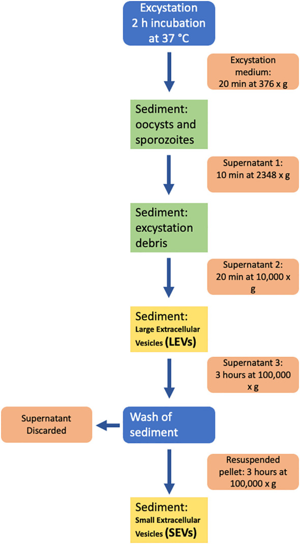
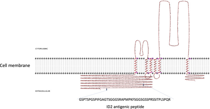
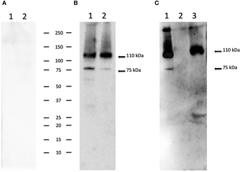
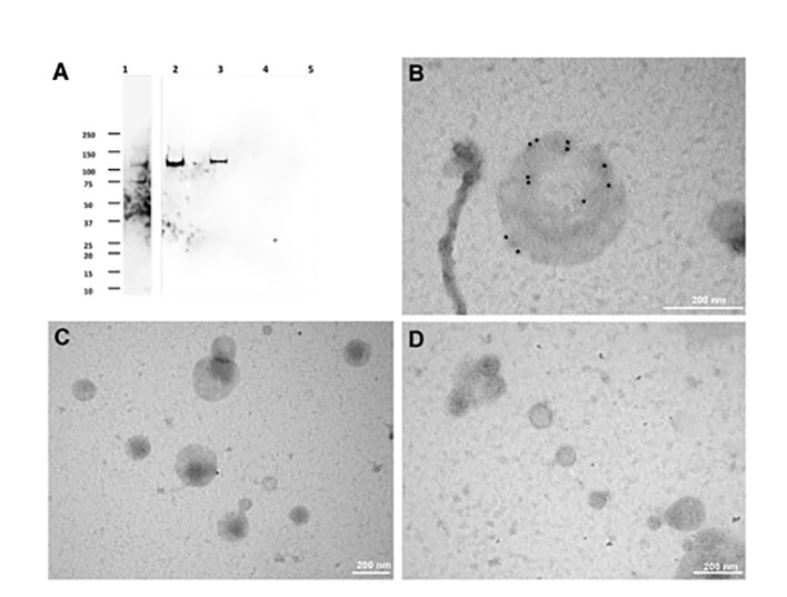
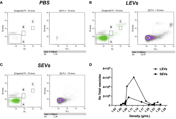
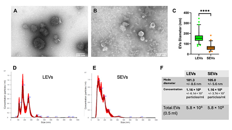
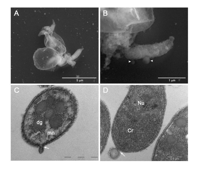
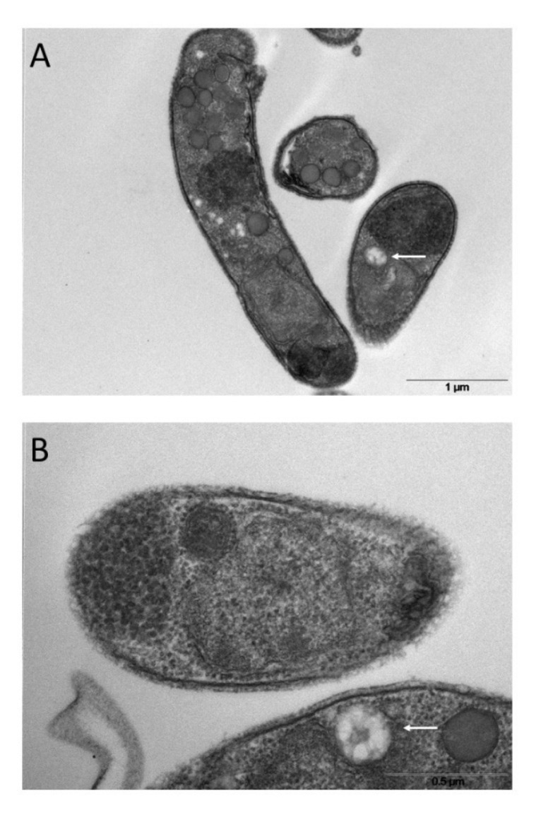
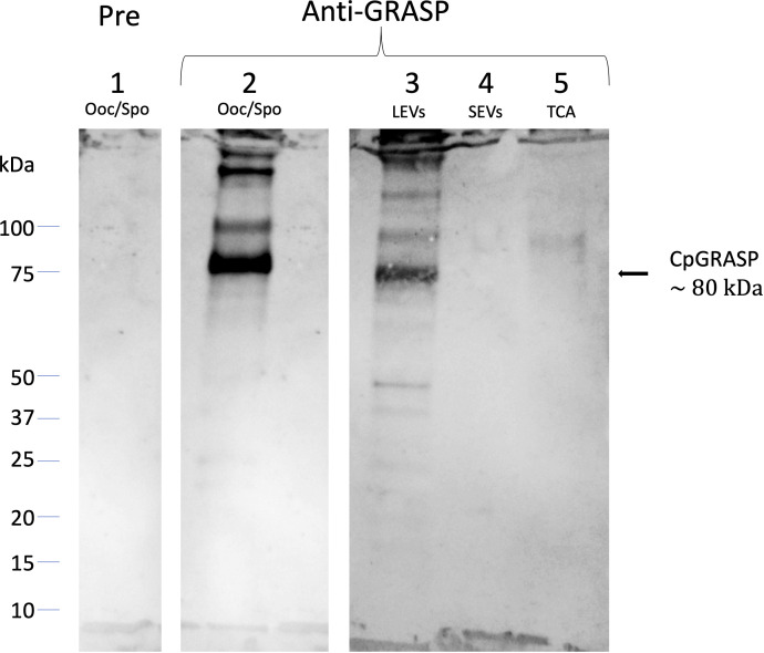
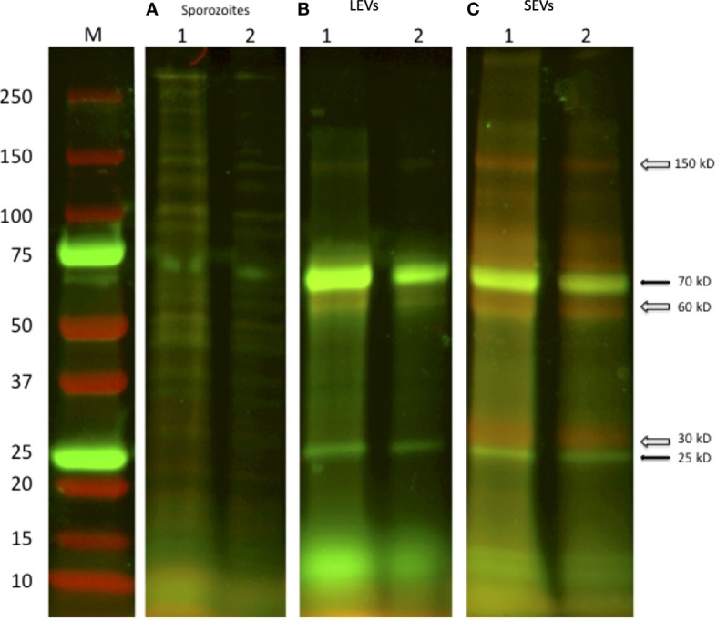
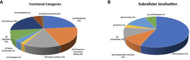
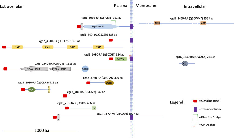
Cryptosporidium parvum is a common cause of a zoonotic disease and a main cause of diarrhea in newborns. Effective drugs or vaccines are still lacking. Oocyst is the infective form of the parasite; after its ingestion, the oocyst excysts and releases four sporozoites into the host intestine that rapidly attack the enterocytes. The membrane protein CpRom1 is a large rhomboid protease that is expressed by sporozoites and recognized as antigen by the host immune system. In this study, we observed the release of CpRom1 with extracellular vesicles (EVs) that was not previously described. To investigate this phenomenon, we isolated and resolved EVs from the excystation medium by differential ultracentrifugation. Fluorescence flow cytometry and transmission electron microscopy (TEM) experiments identified two types of sporozoite-derived vesicles: large extracellular vesicles (LEVs) and small extracellular vesicles (SEVs). Nanoparticle tracking analysis (NTA) revealed mode diameter of 181 nm for LEVs and 105 nm for SEVs, respectively. Immunodetection experiments proved the presence of CpRom1 and the Golgi protein CpGRASP in LEVs, while immune-electron microscopy trials demonstrated the localization of CpRom1 on the LEVs surface. TEM and scanning electron microscopy (SEM) showed that LEVs were generated by means of the budding of the outer membrane of sporozoites; conversely, the origin of SEVs remained uncertain. Distinct protein compositions were observed between LEVs and SEVs as evidenced by their corresponding electrophoretic profiles. Indeed, a dedicated proteomic analysis identified 5 and 16 proteins unique for LEVs and SEVs, respectively. Overall, 60 proteins were identified in the proteome of both types of vesicles and most of these proteins (48 in number) were already identified in the molecular cargo of extracellular vesicles from other organisms. Noteworthy, we identified 12 proteins unique to Cryptosporidium spp. and this last group included the immunodominant parasite antigen glycoprotein GP60, which is one of the most abundant proteins in both LEVs and SEVs.

Keywords: Cryptosporidium; GP60; aspartyl protease; excystation; exosome; extracellular vesicles; rhomboid; sporozoite.


Conflict of interest statement

The authors declare that the research was conducted in the absence of any commercial or financial relationships that could be construed as a potential conflict of interest.

Figures

Figure 1
Figure 1
Diagram of the procedure used for the isolation of extracellular vesicles from the excystation medium.
Figure 2
Figure 2
Two-dimensional model of CpRom1 in relation to the cell membrane. The diagram shows the protein six transmembrane portion, two cytoplasmic loops (above the membrane), the large N-terminal extracellular region including the ID2 antigen, and the small extracellular region at the C-terminus (above the membrane). The small arrows show the position of the ID2 peptide (see text), and the amino acid sequence of the ID2 is expanded below.
Figure 3
Figure 3
CpRom1 assesment in oocyst and sporozoite samples by Western blotting. (A) Western blotting on oocysts and sporozoites lysates probed with mouse pre-immune serum: 1, lysate from 5×10⁶ unexcysted oocysts; 2, lysate from 5×10⁶ oocysts after 30 min of excystation. (B) Western blotting on oocysts and sporozoites lysates probed with mouse anti-CpRom1 serum: 1, lysate from 5×10⁶ unexcysted oocysts; 2, lysate from 5×10⁶ oocysts after 30 min of excystation. (C) Western blotting on TCA-precipitated supernatant of excystation medium probed with mouse anti-CpRom1 serum: lysate from 1×10⁷ oocysts after 30 min of excystation, TCA-precipitated supernatant before the induction of excystation. Ladder shows molecular weights expressed in kDa.
Figure 4
Figure 4
Immunolocalization of CpRom1 by Western blotting on microvesicle extracts and immunoelectron microscopy of the corresponding extracellular vesicles. (A) Immunoblotting with anti-CpRom1 mouse serum: 1, unexcysted oocysts lysate; 2, excysted soporozoites; 3, LEVs extract; 4, SEVs extract; 5, TCA-precipitated supernatant of SEVs. (B) Negative staining immunoelectron microscopy of LEVs labelled with anti-CpRom1 rabbit serum. (C) Negative staining immunoelectron microscopy of LEVs labelled with pre-immune rabbit serum. (D) Negative staining immunoelectron microscopy of SEVs labelled with anti-CpRom1 rabbit serum.
Figure 5
Figure 5
Flow cytometry analysis of gradient fractions of LEVs and SEVs. (A) PBS buffer used to resuspend EVs as negative control. (B) Dot plot of LEVs fraction at 1.1 g/ml density. (C) Dot plot of SEVs at 1.1 g/ml density. (D) diagram comparing the distribution of LEVs and SEVs in the gradient fractions. Left panels indicate gate dimensions (A ≤ 200 nm, B=500 nm and C=1 µm). Right dot plot panels show vesicles distribution in terms of fluorescence.
Figure 6
Figure 6
Physical characterization of LEVs and SEVs by electron-microscopy and nanoparticle tracking analysis (NTA). (A) TEM negative staining of LEVs. (B) TEM negative staining of SEVs. (C) graph showing the vesicle size distribution in LEVs (green) and SEVs (orange). Statistical analysis was based on TEM micrographs; dots indicate maximum and minimum values (****=p-value<0.0001, Mann-Whitney test). (D) graph showing the size distribution of LEVs as dtermined by NTA. (E) graph showing the distribution of SEVs as determined by NTA. (F) table reporting the mode diameter, the concentration of LEVs and SEVs, the total number of EVs (LEVs or SEVs) in 0.5 ml as determined by NTA.
Figure 7
Figure 7
Electron microscopy images of EVs at their release from sporozoites. (A) Backscattered electron SEM micrograph of two excysted oocysts showing the release of vesicles during the sporozoites egress. (B) High magnification of an egressed sporozoite showing two budding vesicles (head arrows). (C) TEM micrograph showing the plasma membrane budding of a vesicle from the apical region of a sporozoite (white arrow). (D) TEM micrograph of a vesicle budding from the posterior region of a sporozoite. Dg: dense granules; mn: micronemes; Cr: crystalloid; Nu: nucleus.
Figure 8
Figure 8
Ultrathin sections of excysted sporozoites (A, B) showing possible MVB-like organelles inside the cells (white arrows).
Figure 9
Figure 9
Western blotting experiments on oocyst-sporozoite lysates and EVs probed with mouse anti-CpGRASP serum. 1, lysate oocyst-sporozoite probed with pre-immune serum; 2, lysate oocyst-sporozoite; 3, LEVs extract; 4, SEVs extract; 5, SEVs supernatant precipitated with TCA. 4-20% SDS-PAGE, lane 1 probed with 1:500 with mouse serum before the immunization; lane 2-5 probed with 1:500 mouse serum after the immunization with recombinant CpGRASP.
Figure 10
Figure 10
SDS-PAGE of two independent excystation experiments followed by labelling with the fluorescent dyes NHS-AF647 (red) and CFSE (green) of the different stages of centrifugation. (A) 1: lysate of 1×10⁷oocysts; 2: lysate of sporozoite of 5 × 10⁶ oocysts. (B) LEVs and (C) SEVs electrophoretic profiles, 1: sediment from 1×10⁷oocysts; 2: sediment from 5 × 10⁶ oocysts, respectively. Black arrows indicate common green bands; open arrows indicate red bands present only in the extract of SEVs.
Figure 11
Figure 11
Quantitative representation of the different categories of the proteins identified in the extracellular vesicles. (A) graphic of the EVs proteins distributed on the basis of their presumptive functions. The classification was based on the sequence homologies with characterized proteins. (B) graphic of the EVs proteins distributed based on their presumptive sub-cellular localization. Prediction was made with DeepLoc-1.0 (http://www.cbs.dtu.dk/services/DeepLoc/).
Figure 12
Figure 12
Cartoon showing some structural features of the Cryptosporidium-characteristic proteins (see text). UniProtKB accessions are reported in brackets. Proteins were divided into three categories according to the presence of hydrophobic motifs (i. e. presence/absence of a signal peptide and/or a transmembrane domain) and their presumptive localization respect to the cell membrane. Prediction of signal peptides and transmembrane domains was performed at Phobius (https://phobius.sbc.su.se/index.html). Prediction of other structural and functional domains was performed at Pfam (http://pfam.xfam.org) and Prosite (https://prosite.expasy.org). List of domains in the figure: Peptidase 1, PEPTIDASE_A1, PS51767; PLP, PROKAR_LIPOPROTEIN, PS51257; CAP, Cysteine-rich secretory protein family, CL0659; GP60, Glycoprotein GP60 of Cryptosporidium, PF11025; PPASE Tensin, PPASE_TENSIN, PS51181; Chase, CHASE, PS50839; DegV, DEGV, PS51482; MyB-L, MYB_LIKE, PS50090; LRR, Leucin Rich Repeat, LRR, PS51450; Thr, Threonin Rich Region, THR_RICH, PS50325; ARM, Armadillo/plakoglobin ARM repeat, ARM_REPEAT, PS50176; CS, CS, PS51203.

References

    1. Ahat E., Li J., Wang Y. (2019). New insights into the golgi stacking proteins. Front. Cell. Dev. Biol. 7. doi: 10.3389/fcell.2019.00131 - DOI - PMC - PubMed
    1. Allison G. M., Rogers K. A., Borad A., Ahmed S., Karim M. M., Kane A. V., et al. . (2011). Antibody responses to the immunodominant Cryptosporidium gp15 antigen and gp15 polymorphisms in a case-control study of cryptosporidiosis in children in Bangladesh. Am. J. Trop. Med. Hyg. 85, 97–104. doi: 10.4269/ajtmh.2011.11-0043 - DOI - PMC - PubMed
    1. Barreca V., Boussadia Z., Polignano D., Galli L., Tirelli V., Sanchez M., et al. . (2023). Metabolic labelling of a subpopulation of small extracellular vesicles using a fluorescent palmitic acid analogue. J. Extracell. Vesicles, e12392. doi: 10.1002/jev2.12392 - DOI - PMC - PubMed
    1. Bouzid M., Hunter P. R., Chalmers R. M., Tyler K. M. (2013). Cryptosporidium pathogenicity and virulence. Clin. Microbiol. Rev. 26, 115–134. doi: 10.1128/CMR.00076-12 - DOI - PMC - PubMed
    1. Campos F. M., Franklin B. S., Teixeira-Carvalho A., Filho A. L., de Paula S. C., Fontes C. J., et al. . (2010). Augmented plasma microparticles during acute Plasmodium vivax infection. Malar. J. 9, 327. doi: 10.1186/1475-2875-9-327 - DOI - PMC - PubMed

Publication types