Comparative Study
. 2024 Jun;44(6):1393-1406.
doi: 10.1161/ATVBAHA.123.320295. Epub 2024 Apr 25.

Differential Impact In Vivo of Pf4-ΔCre-Mediated and Gp1ba-ΔCre-Mediated Depletion of Cyclooxygenase-1 in Platelets in Mice

Affiliations
Comparative Study

Differential Impact In Vivo of Pf4-ΔCre-Mediated and Gp1ba-ΔCre-Mediated Depletion of Cyclooxygenase-1 in Platelets in Mice

Soon Yew Tang et al. Arterioscler Thromb Vasc Biol. 2024 Jun.

Abstract

Background: Low-dose aspirin is widely used for the secondary prevention of cardiovascular disease. The beneficial effects of low-dose aspirin are attributable to its inhibition of platelet Cox (cyclooxygenase)-1-derived thromboxane A2. Until recently, the use of the Pf4 (platelet factor 4) Cre has been the only genetic approach to generating megakaryocyte/platelet ablation of Cox-1 in mice. However, Pf4-ΔCre displays ectopic expression outside the megakaryocyte/platelet lineage, especially during inflammation. The use of the Gp1ba (glycoprotein 1bα) Cre promises a more specific, targeted approach.

Methods: To evaluate the role of Cox-1 in platelets, we crossed Pf4-ΔCre or Gp1ba-ΔCre mice with Cox-1flox/flox mice to generate platelet Cox-1-/- mice on normolipidemic and hyperlipidemic (Ldlr-/-; low-density lipoprotein receptor) backgrounds.

Results: Ex vivo platelet aggregation induced by arachidonic acid or adenosine diphosphate in platelet-rich plasma was inhibited to a similar extent in Pf4-ΔCre Cox-1-/-/Ldlr-/- and Gp1ba-ΔCre Cox-1-/-/Ldlr-/- mice. In a mouse model of tail injury, Pf4-ΔCre-mediated and Gp1ba-ΔCre-mediated deletions of Cox-1 were similarly efficient in suppressing platelet prostanoid biosynthesis. Experimental thrombogenesis and attendant blood loss were similar in both models. However, the impact on atherogenesis was divergent, being accelerated in the Pf4-ΔCre mice while restrained in the Gp1ba-ΔCres. In the former, accelerated atherogenesis was associated with greater suppression of PGI2 biosynthesis, a reduction in the lipopolysaccharide-evoked capacity to produce PGE2 (prostaglandin E) and PGD2 (prostanglandin D), activation of the inflammasome, elevated plasma levels of IL-1β (interleukin), reduced plasma levels of HDL-C (high-density lipoprotein receptor-cholesterol), and a reduction in the capacity for reverse cholesterol transport. By contrast, in the latter, plasma HDL-C and α-tocopherol were elevated, and MIP-1α (macrophage inflammatory protein-1α) and MCP-1 (monocyte chemoattractant protein 1) were reduced.

Conclusions: Both approaches to Cox-1 deletion similarly restrain thrombogenesis, but a differential impact on Cox-1-dependent prostanoid formation by the vasculature may contribute to an inflammatory phenotype and accelerated atherogenesis in Pf4-ΔCre mice.

Keywords: atherosclerosis; blood platelets; cyclooxygenase-1; hemostasis; thrombosis.


Conflict of interest statement

Disclosures G.A. FitzGerald is the McNeil Professor of translational medicine and therapeutics and held a Merit Award from the American Heart Association during the performance of this work. The other authors report no conflicts.

Figures

Figure 1.
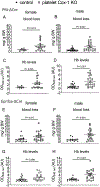
Figure 1.. Deletion of platelet Cox-1 increases blood loss in hyperlipidemic mice (Ldlr−/−) fed a HFD.
Mice after 12 weeks on a HFD were used in this experiment. Two mm of mouse tail was amputated under anesthesia and blood was collected into a glass tube containing Drabkin’s reagent for 7.5 min (female) and 15 min (male). Pf4-ΔCre - or Gp1ba-ΔCre - mediated deletion of Cox-1 in platelets significantly increased blood loss (Pf4-ΔCre, A- female, B- male; Gp1ba-ΔCre, E- female, F- male). Hb levels were significantly increased in platelet Cox-1−/− mice (Pf4-ΔCre, C- female, D- male; Gp1ba-ΔCre, G- female, H- male). Data are expressed as means ± SEMs (Mann Whitney, one-tailed, n=8-21 per group for female mice, n= 15-22 per group for male mice). Controls- Pf4-ΔCre/ Ldlr−/− or Gp1ba-ΔCre/ Ldlr−/−, platelet Cox-1 KO- Pf4-ΔCre/ Cox-1F/F/ Ldlr−/− or Gp1ba-ΔCre/ Cox-1F/F/ Ldlr−/−.
Figure 2.
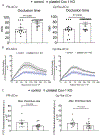
Figure 2.. Platelet Cox-1 promotes thrombogenesis in Ldlr−/− mice of both sexes.
12-14 weeks old female and male Ldlr−/− mice on a SLD were used in these experiments. The time to thrombotic carotid artery occlusion after photochemical injury was extended in female platelets Cox-1−/− hyperlipidemic mice (A- Pf4-ΔCre - left, Gp1ba-ΔCre - right). A Mann Whitney test (1-tailed) revealed a significant effect of genotype on occlusion time. Data are expressed as means ± SEMs. n=13- 15 per group. (B) Thrombogenesis in cremaster arterioles after laser-induced injury in male platelet Cox-1−/− hyperlipidemic mice. Thrombus formation was visualized in real-time with fluorescently labeled platelets as described in the Supplemental Methods. Median integrated fluorescence intensity of platelets representing thrombus formation was plotted vs time after laser-induced injury of the cremaster arteriole vessel wall. Fluorescence intensity from 10 thrombi was averaged from each mouse. Data correspond to maximal thrombus size were extracted from the fluorescence-time curves and averaged (C- Pf4-ΔCre - left, Gp1ba-ΔCre - right). A Mann Whitney test (one-tailed) revealed a significant effect of Cox-1 deletion on maximal thrombus formation. Data are expressed as means ± SEMs. n=13- 14 per group. Controls- Pf4-ΔCre/ Ldlr−/− or Gp1ba-ΔCre/ Ldlr−/−, platelet Cox-1 KO- Pf4-ΔCre/ Cox-1F/F/ Ldlr−/− or Gp1ba-ΔCre/ Cox-1F/F/ Ldlr−/−.
Figure 3.
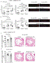
Figure 3.. Pf4-ΔCreI -mediated deletion of platelet Cox-1 promotes atherogenesis in Ldlr−/− mice while Gp1ba-ΔCre -mediated deletion restrains atherogenesis.
Aortic atherosclerotic lesion burden, represented by the percentage of lesion area stained with Sudan VI to total aortic area, was quantified by en face analysis of aortas from mice fed a HFD for 12, 24 and 36 weeks. Lesion area increased with feeding time, and Pf4-ΔCre -mediated deletion of Cox-1 increased lesion burden in mice of both sexes (A). However, Gp1ba-ΔCre -mediated Cox-1 deletion in platelets restrained lesion burden (B). For A and B, a 2-way ANOVA showed that en face aortic lesion burden was significantly affected by feeding time or deletion of Cox-1 in platelets. Bonferroni multiple comparisons test was used to test for significant differences between controls and platelets Cox-1−/− mice or feeding time. Data are expressed as means ± SEMs. *p< 0.05, n= 8-24 per group. En face aortic images of male hyperlipidemic mice on a HFD diet for 24 and 36 weeks (A- Pf4-ΔCre, B- Gp1ba-ΔCre). C- D) Aortic root lesion burden was quantified by H&E staining of cross-sections from male mice fed a HFD for 24 weeks. Area of aortic root lesions was expressed as percentage of the total area of the vessel minus the luminal area. Representative cross-sectional images are shown (right panels). Pf4-ΔCre -mediated deletion of Cox-1 increased lesion burden in male mice (C), while Gp1ba-ΔCre reduced it (D). For C and D, a Mann Whitney test (1-tailed) revealed a significant effect of Cox-1 deletion in platelets on aortic root lesion burden progression. Data are expressed as means ± SEMs. n= 8- 9 per group. Controls- Pf4-ΔCre/ Ldlr−/− or Gp1ba-ΔCre/ Ldlr−/−, platelet Cox-1 KO- Pf4-ΔCre/ Cox-1F/F/ Ldlr−/− or Gp1ba-ΔCre/ Cox-1F/F/ Ldlr−/−.
Figure 4.
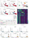
Figure 4.. Single-cell RNAseq analysis of mouse aorta revealed the presence of SEM cells in higher abundance in Pf4-ΔCre male Ldlr−/− mice fed a HFD for 24 weeks.
UMAP representation of aligned gene expression data of mouse atherosclerotic aorta from Pf4-ΔCre (A) or Gp1ba-ΔCre (B) mice fed a HFD for 24 weeks. Inflammatory SEM cells (in black rectangle) were present in a larger quantity in Pf4-ΔCre mice as compared to Gp1ba-ΔCre mice regardless of the deletion of Cox-1 in platelets. Red cells- cells that are the genotype of interest; blue cells- cells that are representative of all background transcripts. (C) UMAP showing the distribution and identify of the nine different cell clusters from mouse aorta. (D) Heatmap of the marker genes in each cluster selected as unique genes used for identification of each cluster. (E) KEGG pathway analysis of differentially expressed genes in SMCs revealed alteration in pathways associated with TCA cycle, carbon metabolism, spliceosome and protein processing in ER signaling. (E) Violin plots of the top differentially expressed genes associated with anti-inflammation showing up-regulation in Gp1ba-ΔCre -mediated deletion of Cox-1 in platelets. Top panels- Pf4-ΔCre, bottom panels- Gp1ba-ΔCre. Blue violin plots- controls, orange violin plots- platelet Cox-1 knockouts. A Mann Whitney test revealed a significant effect of Cox-1 deletion in platelets mediated by Gp1ba-ΔCre, P< 0.05 was considered significant. SEM- stem cell, endothelial cell, monocyte, SMCs- smooth muscle cells, Igha- immunoglobulin heavy chain a, Bpifa1- bacterial permeability family member A1, Scgb1a1- secretoglobin family 1A member 1, ↑ upregulation, ↓ downregulation. Controls- Pf4-ΔCre/ Ldlr−/− or ΔGp1ba-Cre/ Ldlr−/−, platelet Cox-1 KO- Pf4-ΔCre/ Cox-1F/F/ Ldlr−/− or ΔGp1ba-Cre/ Cox-1F/F/ Ldlr−/−.

References

    1. Ricciotti E, FitzGerald GA. Aspirin in the Prevention of Cardiovascular Disease and Cancer. Annu Rev Med. 2021;72:473–495. doi: 10.1146/annurev-med-051019-102940 - DOI - PubMed
    1. Patrono C, Baigent C. Role of aspirin in primary prevention of cardiovascular disease. Nat Rev Cardiol. 2019;16:675–686. doi: 10.1038/s41569-019-0225-y - DOI - PubMed
    1. Group ASC, Bowman L, Mafham M, Wallendszus K, Stevens W, Buck G, Barton J, Murphy K, Aung T, Haynes R, et al. Effects of Aspirin for Primary Prevention in Persons with Diabetes Mellitus. N Engl J Med. 2018;379:1529–1539. doi: 10.1056/NEJMoa1804988 - DOI - PubMed
    1. Hybiak J, Broniarek I, Kiryczynski G, Los LD, Rosik J, Machaj F, Slawinski H, Jankowska K, Urasinska E. Aspirin and its pleiotropic application. Eur J Pharmacol. 2020;866:172762. doi: 10.1016/j.ejphar.2019.172762 - DOI - PubMed
    1. Pietrocola F, Castoldi F, Markaki M, Lachkar S, Chen G, Enot DP, Durand S, Bossut N, Tong M, Malik SA, et al. Aspirin Recapitulates Features of Caloric Restriction. Cell Rep. 2018;22:2395–2407. doi: 10.1016/j.celrep.2018.02.024 - DOI - PMC - PubMed

Publication types

MeSH terms