. 2024 Jan;1870(1):166928.
doi: 10.1016/j.bbadis.2023.166928. Epub 2023 Oct 28.

Copper enhances aggregational toxicity of mutant huntingtin in a Drosophila model of Huntington's Disease

Affiliations

Copper enhances aggregational toxicity of mutant huntingtin in a Drosophila model of Huntington's Disease

Amanda G Lobato et al. Biochim Biophys Acta Mol Basis Dis. 2024 Jan.

Abstract

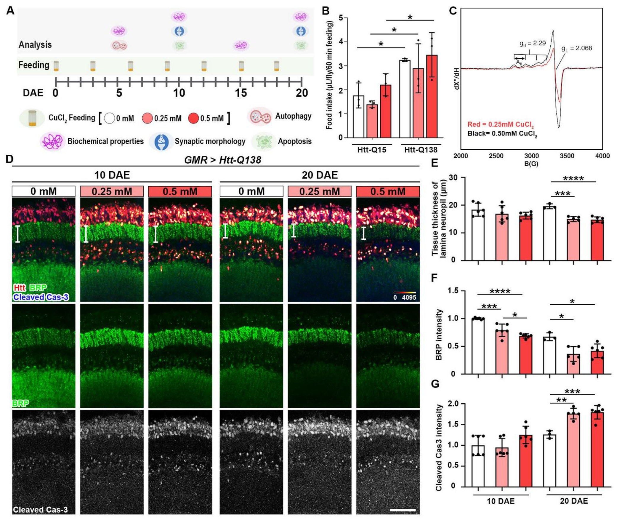
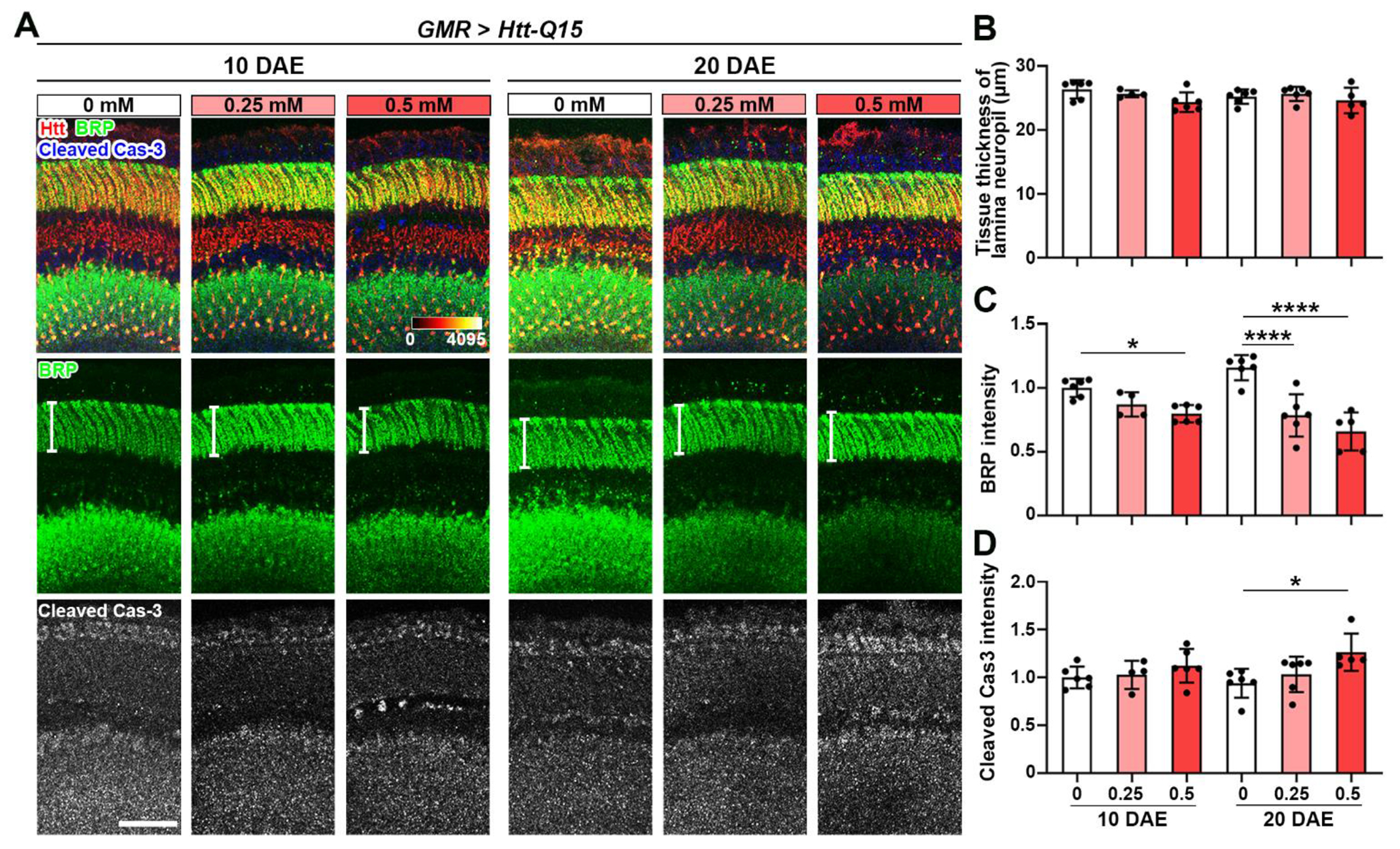
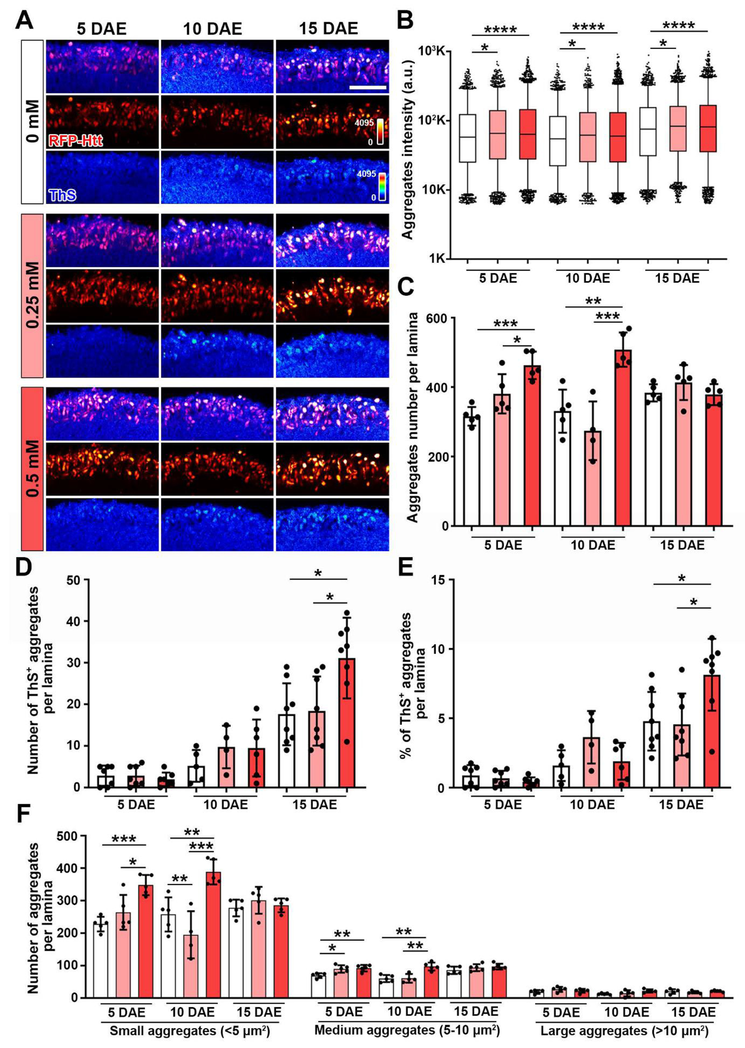
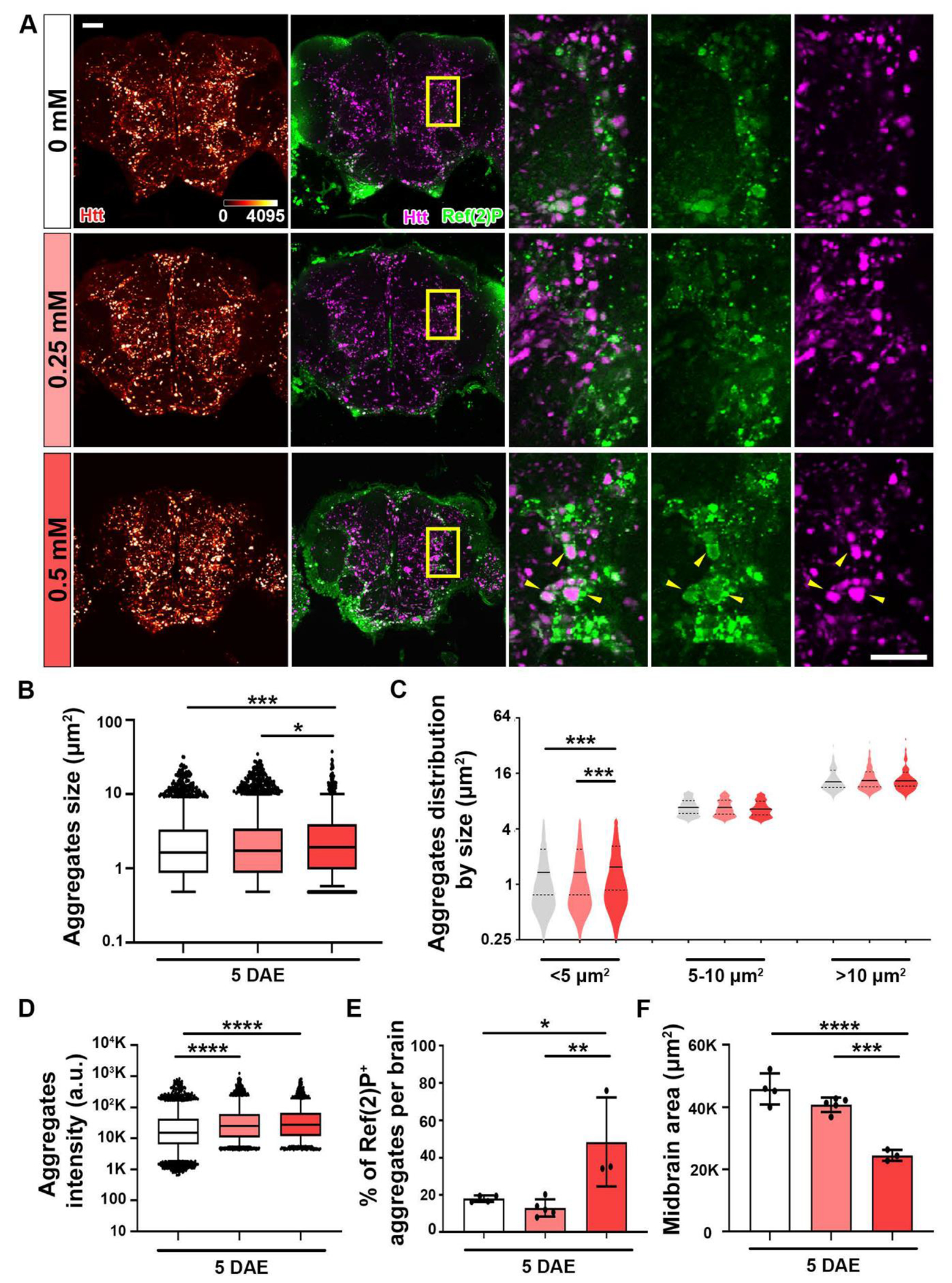
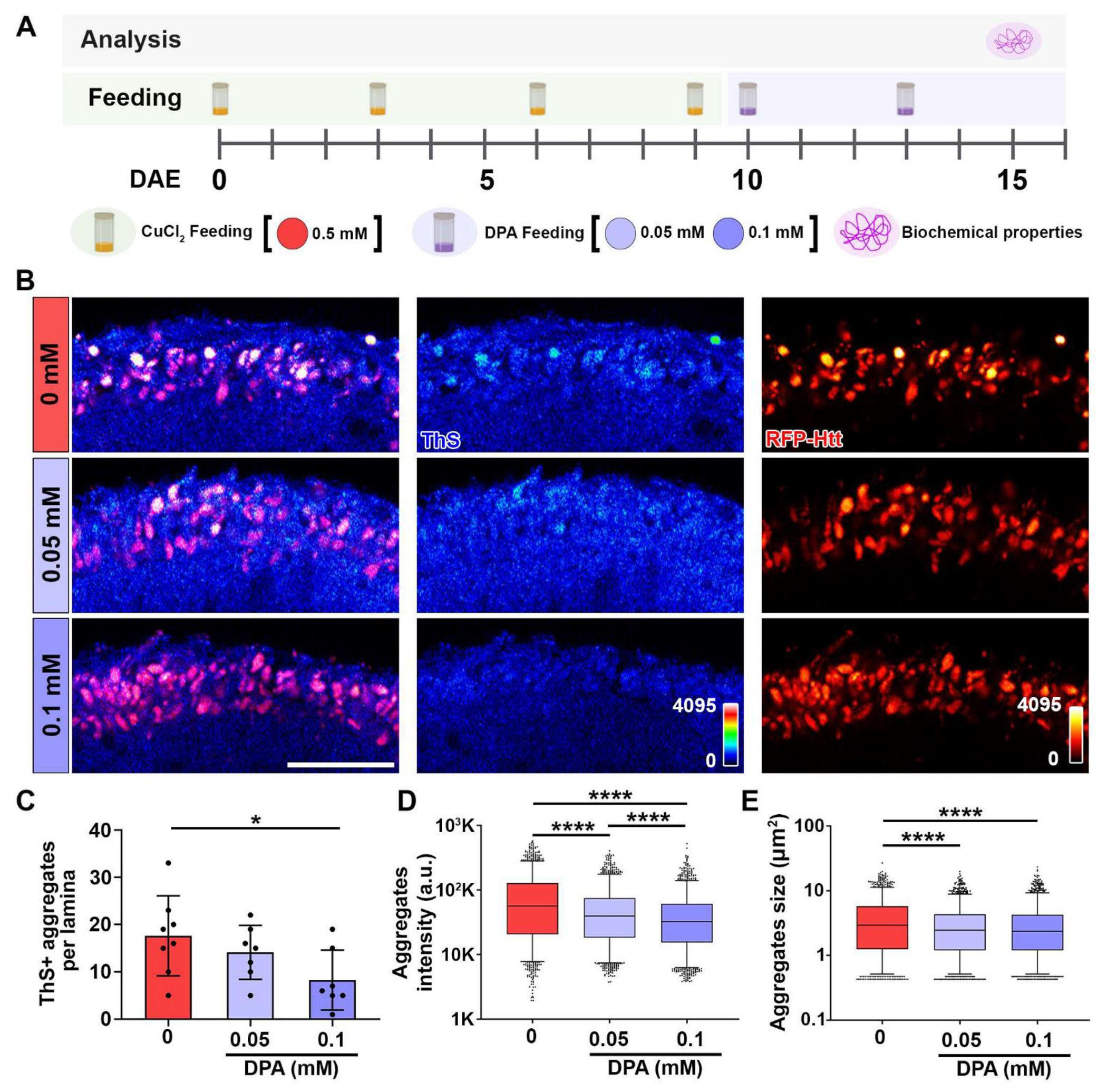
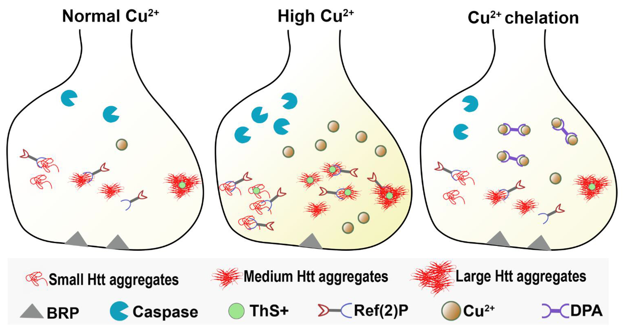
Huntington's disease (HD) is a progressive neurodegenerative disorder with clinical presentations of moderate to severe cognitive, motor, and psychiatric disturbances. HD is caused by the trinucleotide repeat expansion of CAG of the huntingtin (HTT) gene. The mutant HTT protein containing pathological polyglutamine (polyQ) extension is prone to misfolding and aggregation in the brain. It has previously been observed that copper and iron concentrations are increased in the striata of post-mortem human HD brains. Although it has been shown that the accumulation of mutant HTT protein can interact with copper, the underlying HD progressive phenotypes due to copper overload remains elusive. Here, in a Drosophila model of HD, we showed that copper induces dose-dependent aggregational toxicity and enhancement of Htt-induced neurodegeneration. Specifically, we found that copper increases mutant Htt aggregation, enhances the accumulation of Thioflavin S positive β-amyloid structures within Htt aggregates, and consequently alters autophagy in the brain. Administration of copper chelator D-penicillamine (DPA) through feeding significantly decreases β-amyloid aggregates in the HD pathological model. These findings reveal a direct role of copper in potentiating mutant Htt protein-induced aggregational toxicity, and further indicate the potential impact of environmental copper exposure in the disease onset and progression of HD.

Keywords: Aggregates; Chelation; Copper; Huntingtin; Huntington's disease.


Conflict of interest statement

Declaration of competing interest Authors declare that they have no competing interests.

Figures

Figure 1:
Figure 1:. Copper enhances Htt-induced synaptic degeneration.
(A) CuCl2 feeding paradigm. Pathological Htt-Q138 flies were driven by GMR-GAL4 and fed with different concentrations of CuCl2 until 20 DAE and assessed cellular and biochemical properties. (B) Measurement of food intake (μL/fly/60 minutes feeding) of GMR>Htt-Q15 and GMR>Htt-Q138 flies during a 1hr feeding session following 2hrs starvation. n=3 biological replicates with 4-5 flies per group. (C) X-band (9.63 GHz microwave frequency) EPR spectra for samples prepared as 0.25 mM (red) and 0.5 mM (black) CuCl2. Data were recorded at 77 K, using 6.325 mW microwave power. Markers for g-values and hyperfine splitting (A) are also shown in the figure. (D) Lamina structures at 10 DAE and 20 DAE probed for BRP (green) and cCas3 (gray). Scale bar = 30μm. (E) Quantification of tissue thickness of lamina neuropil (white bars shown on panel C). (F) Quantification of BRP intensity. (G) Quantification of cleaved caspase-3 intensity. Data are presented as mean ± SD. n=3-5, *p<0.05, **p<0.01, ***p<0.001, ****p<0.0001.
Figure 2:
Figure 2:. Copper mildly decreases active zone protein BRP in a non-pathological model.
(A) Non-pathological Htt-Q15 flies were driven by GMR-GAL4 and fed with different concentrations of CuCl2 until 10 DAE and 20 DAE. Lamina structures at 10 DAE and 20 DAE were stained for BRP (green) and cCas3 (gray). Scale bar = 30μm. (B) Quantification of tissue thickness of lamina neuropil (white bars shown on panel A). (C) Quantification of BRP intensity. (D) Quantification of cleaved caspase-3 intensity. Data are presented as mean ± SD. n=3-5, *p<0.05, **p<0.01, ***p<0.001, ****p<0.0001.
Figure 3:
Figure 3:. Copper increases ThS+ Htt aggregates.
(A) ThS staining of the lamina layer (ThS and RFP-Htt heatmaps 0-4095) at 5 DAE, 10 DAE, and 20 DAE. Scale bar: 30μm (B) Quantification of Htt aggregates intensity. Data are presented as box and whiskers plot, 5-95 percentile. (C) Quantification of Htt aggregates number per lamina. Data are presented as mean ± SD. (D) Number of ThS+ aggregates per lamina. Data are presented as mean ± SD. (E) Percentage of ThS+ aggregates per lamina. Data are presented as mean ± SD. (F) Quantification of number of aggregates per lamina separated by size: Small (<5 μm2), Medium (5-10 μm2) and Large (>10 μm2). Data are presented as mean ± SD. n=3-8, *p<0.05, **p<0.01, ***p<0.001, ****p<0.0001.
Figure 4:
Figure 4:. Copper increases Ref(2)P+ Htt aggregates.
Pathological Htt-Q138 flies driven by elav-GAL4 were fed with different CuCl2 concentrations until 5 DAE. (A) Midbrain shown with accumulation of Htt aggregates (heatmap 0-4095). Merged image of Htt aggregates (magenta) and Ref(2)P (green) staining. Yellow boxed areas are shown in higher magnification. Yellow arrows pointing at co-localization. Scale bar = 30 μm. (B) Quantification of total aggregate size. Data are presented as box and whiskers plot, 5-95 percentile. (C) Quantification of size distribution. Small aggregates are classified as <5 μm2, medium aggregates as 5-10 μm2, and large aggregates as >10 μm2. Data are presented as violin plot with quartiles. (D) Quantification of total aggregate intensity. Data are presented as box and whiskers plot, 5-95 percentile. (E) Quantification of percentage of Ref(2)P+ aggregates per lamina. Data are presented as mean ± SD. (F) Quantification of midbrain. Data are presented as mean ± SD. n=3-5, *p<0.05, **p<0.01, ***p<0.001, ****p<0.0001.
Figure 5:
Figure 5:. Chelation decreases ThS+ Htt aggregates.
(A) DPA feeding paradigm. Pathological Htt-Q138 flies driven by GMR-GAL4 were fed with 0.50 mM CuCl2 until 10 DAE, then switched to DPA feeding. Biochemical properties of aggregates were assessed at 15 DAE. (B) ThS staining of lamina layer (ThS and RFP-Htt heatmaps 0-4095) at 15 DAE. Scale bar: 30 μm. (C) Quantification of ThS+ aggregates per lamina. Data is shown as mean ± SD. (D) Quantification of aggregates intensity. Data is shown as box and whiskers plot, 5-95 percentile. (E) Quantification of aggregates size. Data is shown as box and whiskers plot, 5-95 percentile. n=6-8, *p<0.05, ****p<0.0001.
Figure 6:
Figure 6:. Copper enhances aggregational toxicity of mutant huntingtin.
Schematic model of synapse showing that under normal copper concentration, there is cytotoxicity of mutant Htt aggregates, and some co-localization of Ref(2)P with the aggregates. Under high Cu exposure, there is increased Ref(2)P clustering with Htt aggregates, increased cell death, and reduced BRP levels at the synapse. After Cu2+ chelation by DPA feeding, there is reduced Htt aggregates clustering.

References

    1. Bates G, Huntingtin aggregation and toxicity in Huntington’s disease. The Lancet, 2003. 361(9369): p. 1642–1644. - PubMed
    1. Finkbeiner S, Huntington’s Disease. Cold Spring Harb Perspect Biol, 2011. 3(6). - PMC - PubMed
    1. Labbadia J and Morimoto RI, Huntington’s disease: underlying molecular mechanisms and emerging concepts. Trends Biochem Sci, 2013. 38(8): p. 378–85. - PMC - PubMed
    1. Bachoud-Levi AC, et al., International Guidelines for the Treatment of Huntington’s Disease. Front Neurol, 2019. 10: p. 710. - PMC - PubMed
    1. Barron JC, Hurley EP, and Parsons MP, Huntingtin and the Synapse. Front Cell Neurosci, 2021. 15: p. 689332. - PMC - PubMed

Publication types

MeSH terms

Substances

LinkOut - more resources