. 2024 Apr 26;384(6694):eadf5489.
doi: 10.1126/science.adf5489. Epub 2024 Apr 26.

Ciliopathy patient variants reveal organelle-specific functions for TUBB4B in axonemal microtubules

Daniel O Dodd #  1 Sabrina Mechaussier #  2 Patricia L Yeyati  1 Fraser McPhie  1 Jacob R Anderson  3 Chen Jing Khoo  4 Amelia Shoemark  5   6 Deepesh K Gupta  7 Thomas Attard  8 Maimoona A Zariwala  9 Marie Legendre  10   11 Diana Bracht  12 Julia Wallmeier  12 Miao Gui  3 Mahmoud R Fassad  13   14 David A Parry  1 Peter A Tennant  1 Alison Meynert  1 Gabrielle Wheway  15 Lucas Fares-Taie  2 Holly A Black  16   17 Rana Mitri-Frangieh  18   19 Catherine Faucon  18 Josseline Kaplan  2 Mitali Patel  13   20 Lisa McKie  1 Roly Megaw  1   21 Christos Gatsogiannis  22 Mai A Mohamed  13   23 Stuart Aitken  1 Philippe Gautier  1 Finn R Reinholt  24 Robert A Hirst  25 Chris O'Callaghan  25 Ketil Heimdal  26 Mathieu Bottier  5 Estelle Escudier  11   18 Suzanne Crowley  27 Maria Descartes  28 Ethylin W Jabs  29   30 Priti Kenia  31 Jeanne Amiel  32   33 Giacomo Maria Bacci  34 Claudia Calogero  35 Viviana Palazzo  36 Lucia Tiberi  36 Ulrike Blümlein  37 Andrew Rogers  6 Jennifer A Wambach  7 Daniel J Wegner  7 Anne B Fulton  38 Margaret Kenna  39 Margaret Rosenfeld  40 Ingrid A Holm  41   42 Alan Quigley  43 Emma A Hall  1 Laura C Murphy  1 Diane M Cassidy  5 Alex von Kriegsheim  44 Scottish Genomes Partnership16Genomics England Research Consortium45Undiagnosed Diseases Network46Jean-François Papon  45 Laurent Pasquier  46 Marlène S Murris  47 James D Chalmers  5 Claire Hogg  6 Kenneth A Macleod  48 Don S Urquhart  48   49 Stefan Unger  48   49 Timothy J Aitman  16 Serge Amselem  10   11 Margaret W Leigh  50 Michael R Knowles  51 Heymut Omran  12 Hannah M Mitchison  13 Alan Brown  3 Joseph A Marsh  1 Julie P I Welburn  8 Shih-Chieh Ti  4 Amjad Horani  7   52 Jean-Michel Rozet  2 Isabelle Perrault  2 Pleasantine Mill  1
Affiliations

Ciliopathy patient variants reveal organelle-specific functions for TUBB4B in axonemal microtubules

Daniel O Dodd et al. Science. .

Erratum in

Abstract

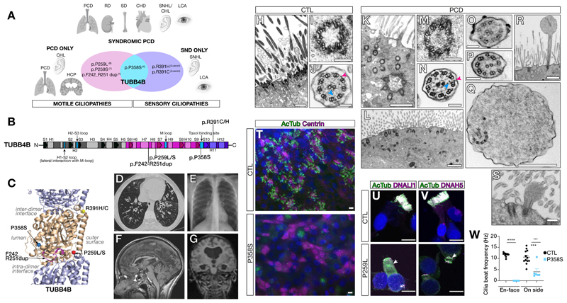
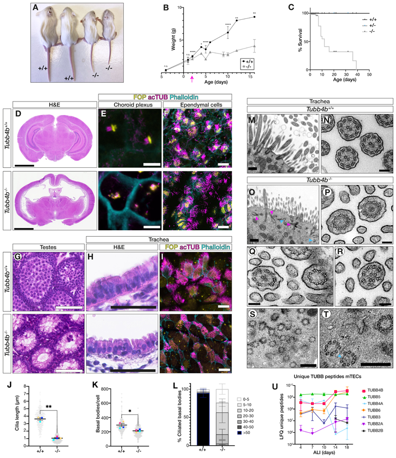
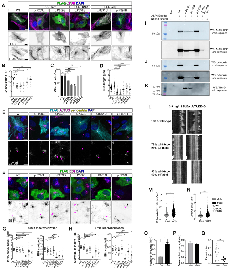
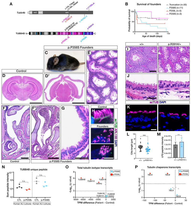
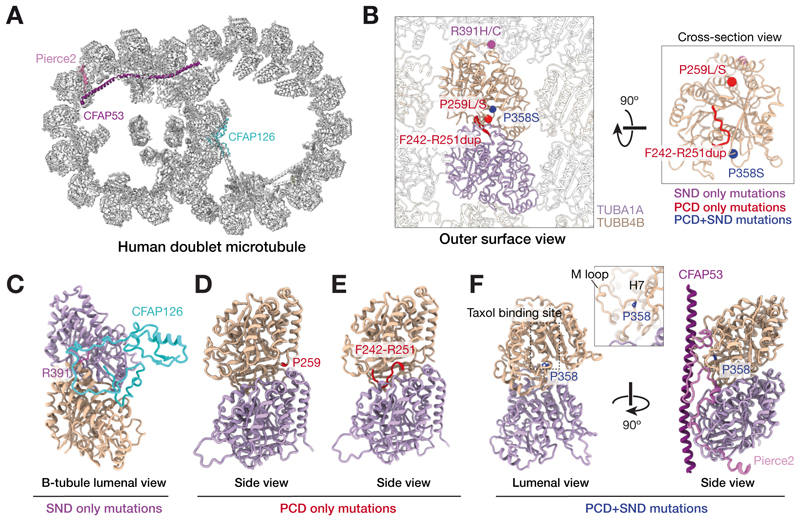
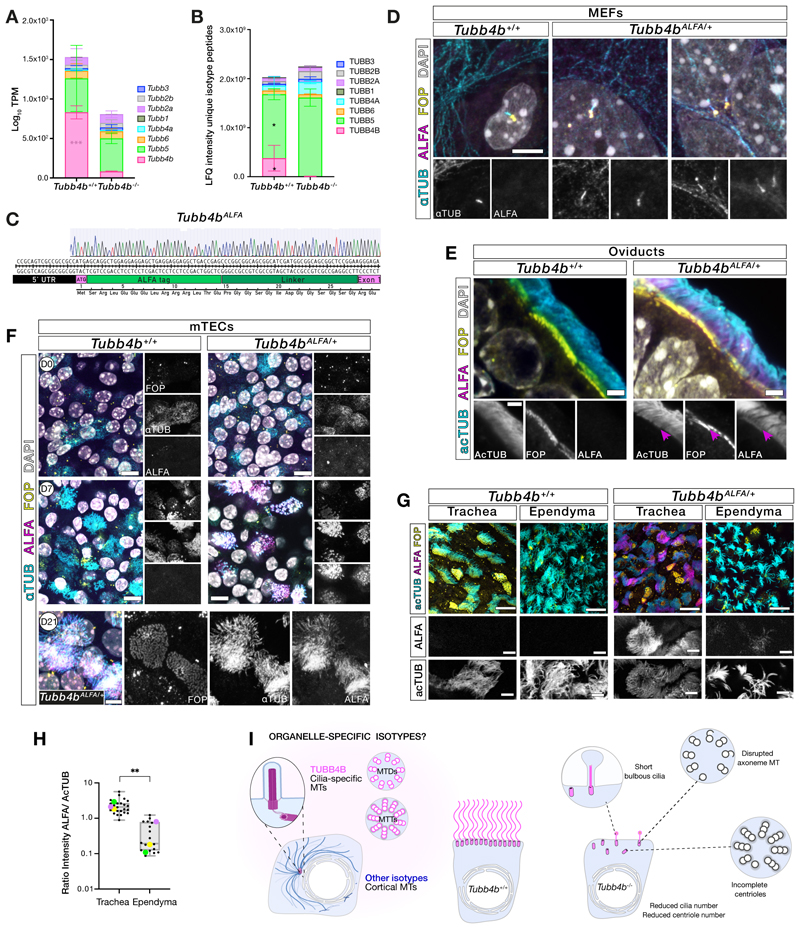
Tubulin, one of the most abundant cytoskeletal building blocks, has numerous isotypes in metazoans encoded by different conserved genes. Whether these distinct isotypes form cell type- and context-specific microtubule structures is poorly understood. Based on a cohort of 12 patients with primary ciliary dyskinesia as well as mouse mutants, we identified and characterized variants in the TUBB4B isotype that specifically perturbed centriole and cilium biogenesis. Distinct TUBB4B variants differentially affected microtubule dynamics and cilia formation in a dominant-negative manner. Structure-function studies revealed that different TUBB4B variants disrupted distinct tubulin interfaces, thereby enabling stratification of patients into three classes of ciliopathic diseases. These findings show that specific tubulin isotypes have distinct and nonredundant subcellular functions and establish a link between tubulinopathies and ciliopathies.


Conflict of interest statement

Competing interests G.W. and D.A.P. are currently employees of Illumina Inc. The other authors declare that they have no competing interests.

Figures

Figure 1
Figure 1. Distinct de novo TUBB4B variants caused PCD-only, SND-only or syndromic (PCD+SND) disease.
(A) Schematic of patient phenotypes within the ciliopathic spectrum clustered on genotypes. Abbreviations: CHL: conductive hearing loss; CHD: congenital heart disease; HCP: hydrocephaly; LCA: Leber congenital amaurosis; PCD: primary ciliary dyskinesia; RD: renal disease; SD: skeletal defects; SNHL: sensorineural hearing loss; SND: sensory-neural disorder. (B,C) Resulting changes mapped onto a protein schematic (B) or an atomic model of TUBB4B (gold) with TUBA1 (purple) (C). (D-G) Clinical features of PCD patients: (D) chest CT showing bilateral lower lobe bronchiectasis (P1); (E) X-ray showing right middle lobe atelectasis (P2); (F) midline T1 sagittal image showing irregular corpus callosum secondary to earlier hydrocephaly (shunted), no evidence of basal ganglia dysmorphology is observed, typical of tubulinopathies (P1); (G) MRI showing dilated ventricles (P8). (H-S) TEM of healthy donor (H-J) and PCD patient nasal epithelia (K-S). Patient samples showed misoriented, internally docked (K) or reduced centrioles without axonemes (L) (P3), incomplete microtubule triplets (P3) (M), and rare intact axoneme (P9) with both inner (blue arrowheads) and outer (magenta arrowheads) dynein arms (N). Missing doublets (O, P3), singlet microtubules (P, P3), disrupted axonemes (Q, P3) or rare short axonemes with bulbous tips (R, P3; S, P10) were observed. (T) Wholemount immunofluorescence of nasal epithelial cultures from unaffected parent and patient (P9). (U,V) Immunofluorescence of healthy donor or patient (P2) cells for cilia axonemes and dynein motor proteins. See Fig. S2L,M. (W) Cilia beat frequency of control and patient (P9) airway cultures. Mean ± SEM from N=3 experimental replicates. Student’s t-test: ***, p ≤ 0.001; ****, p ≤ 0.0001. Scale bars: 10 μm (T-V); 1 μm (H,K,L,R); 500 nm (Q); 125 nm (I,M), 200 nm (S) and 100 nm (J,N-P).
Figure 2
Figure 2. Tubb4b was specifically required for axoneme formation in motile ciliated tissues in vivo.
(A-C) Tubb4b-/- animals exhibited postnatal runting (A, P16), starting from P2 (B), and postnatal lethality (C). (D-F) Tubb4b-/- animals displayed hydrocephaly (D, P15). Choroid plexus multicilia were disrupted (E, P5) while ependymal cilia were largely normal (F, P5). (G-I) Tubb4b-/- animals exhibited male infertility and spermatogenesis defects (G, P22) and severe disruption of tracheal cilia by histology (H) and immunofluorescence (I). (J-L) Quantification of wholemount immunofluorescence of Tubb4b;Centrin2-eGFP neonatal (P5-P8) trachea cilia length (J), basal body number per cell (K) and percentage of ciliated basal bodies per cell (L). See Fig. S3G. N = 4 animals per genotype with (J) n >75 cilia per biological replicate and (K,L) n > 56 cells per biological replicate. (M-T) TEM from control (M,N) and Tubb4b-/- neonatal (P1-P4) mutant tracheas (O-T). Mutants showed non-docked centrioles without axonemes (magenta arrowheads) and partial centrioles (cyan arrowheads) (O). Missing microtubule doublets (P) (white arrowhead), missing central pair apparatus (R) and microtubule singlets with disrupted organization (Q). Within the mutant cytosol, partial centrioles and centrioles with microtubule doublets instead of triplets (S,T). (U) Mass spectrometry of differentiating control mTECs detected many unique β-tubulins. Scale bars: 2.5 mm (D), 5 μm (E), 20 μm (F), 100 μm (G), 50 μm (H), 10 μm (I), 500 nm (M,O), 100 nm (N,P,Q,R) and 300 nm (S,T). (B,J,K) Graphic bars: mean ± SEM derived from N>3 animals per time point. Student’s t-test ns, not significant; *, p ≤ 0.1; **, p ≤ 0.01; ***, p ≤ 0.001; ****, p ≤ 0.0001. (U) Line chart: mean ± SEM derived from N=3 experimental replicates per time point.
Figure 3
Figure 3. Disease-causing TUBB4B variants altered microtubule dynamics and ciliation.
(A) Microtubule incorporation of wild-type (WT), p.P259L, p.P259S, p.P358S, p.R391C and p.R391H TUBB4B variants overexpressed in RPE1 cells. (B) Quantification of percentage colocalization of FLAG TUBB4B variants and α-tubulin staining. (C-E) Ciliogenesis in 24 h serum-starved RPE1 cells overexpressing TUBB4B variants. Acetylated α-tubulin only channel (lower panel) with transfected cell cilia highlighted by magenta arrowhead. Quantification of (D) rates of ciliation and (E) cilia lengths. (F) Microtubule dynamics analysis of RPE1 cells overexpressing TUBB4B variants, upon repolymerization at 37 °C for 4 min. Inverted EB1 only channel (lower panel) in transfected cell highlighted by magenta arrowhead to illustrate variant effects on dynamics. (G,H) Quantification upon repolymerization at 4 (G) and 6 (H) min. See also Fig. S6. (I-K) Affinity purified (ALFA beads) lysates from stable IMCD3 cells expressing ALFA-tagged human TUBB4B variants immunoblotted against ALFA (I), α-tubulin to examine heterodimer assembly (J) and TBCD, a heterodimer assembly pathway chaperone (K). (L) TIRF microscopy time-lapse image kymographs microtubule growth dynamics from GMPCPP-stabilized wild-type seeds with varying concentrations of p.P358S containing purified tubulin TUBA1-TUBB4B heterodimers (Horizontal scale bar = 3 μm, vertical scale bar = 2.27 min). (M-Q) Data distribution and statistical analysis of microtubule-polymerization rate (M), growth length (N), nucleation frequency (O), and fraction of time for each microtubule that paused during polymerization (Q) with addition of p.P358S. Catastrophe frequency (P) is not affected. Kinetic analysis was not performed for the 50:50 as no dynamics were observed. See also Fig. S8. Scale bars: (A, E, F) 10 µm. Plots represent the mean ± SEM, N= 2 (B-D,G,H) or 3 (M-Q) experimental replicates. Ns, not significant; *, p ≤ 0.1; **, p ≤ 0.01; ***, p ≤ 0.001; ****, p ≤ 0.0001.
Figure 4
Figure 4. TUBB4B variants caused dominant disease in vivo.
(A) Schematic of patient p.P259L/S (PCD-only, red or magenta), p.P358S (syndromic PCD+LCA, blue) and p.R391H (SND-only, purple) mutations and truncating mutations (p.D249Lfs*29 and p.N256Yfs*32, null alleles (black)) on the mouse Tubb4b mRNA (top) and protein (bottom). (B-H) (B) Kaplan-Meier graph of founder mice carrying PCD-patient variants which died either spontaneously or were euthanized for health concerns. (C-H) p.P358S founders exhibited hydrocephaly (C,D’), defects in spermatogenesis (E), mucopurulent nasal plugs (F’), and loss of tracheal cilia (G,H). BL/6 age-matched controls (D,F). (I-M) Tubb4bR391H/+ animals showed no decrease in fitness or survival allowing a line to be established (see also Fig. S9). Males were infertile with defects in spermatogenesis (I). Tubb4bR391H/+ tracheal cilia histology (J), immunofluorescence, length quantified in (L). Mass spectrometry of trachea quantifying TUBB4B levels (M). (N-P) Nasal epithelial cultures from healthy controls, a PCD-only patient (red, p. P259L) or syndromic PCD+SND patient (blue, p.P358S) were used for mass spectrometry of unique TUBB4B peptides (N) or targeted RNASeq analysis of tubulin (O) or tubulin chaperone (P) transcripts. Scale bars: 2.5 mm (D), 500 μm (E), 250 μm (F), 100 μm (I), 50 μm (G), 25 μm (J) and 10 μm (H,K). (L, M) Graphic bars: mean ± SEM from N=2 biological replicates, n>20 cells/replicate (L) and N= 3 biological (M) or experimental (N) replicates. Student’s t-test: ns, not significant; ****, p ≤ 0.0001.
Figure 5
Figure 5. Structural environment of disease-causing variants of TUBB4B.
(A) Human microtubule doublet cross-section (PDB ID: 7UNG) highlighting microtubule interacting proteins (MIPs) that interact with TUBB4B residues associated with disease. Microtubule doublets are the conserved cytoskeletal element of both primary and motile cilia, consisting of complete A-tubule with 13 protofilaments and incomplete B-tubules with 10 protofilaments. (B) Orthogonal views showing disease-causing TUBB4B variants within the ciliary microtubule doublet lattice. The human tubulin isotypes (TUBA1A (purple) and TUBB4B (gold)) were determined based on the human microtubule doublet cryo-EM density map (32) and abundance in human multiciliated respiratory cells by scRNAseq (64). Variant positions are indicated with spheres colored based on their disease association. Only one TUBB4B molecule is shown in the cross-section (right), where R391 is not visible. (C) Interaction of R391 of TUBB4B with the microtubule inner protein CFAP126. (D-E) p.P259 and loop p.F242-R251 locate at the intradimer interface. (F) p.P358 locates at the taxol binding site which interacts with multiple microtubule interacting proteins (MIPs) including, for example, PIERCE2.
Figure 6
Figure 6. TUBB4B was a centriole and cilia-specific isotype.
(A) Transcriptomic data from neonatal Tubb4b tracheas of β-tubulin isotype mRNA expression profile. (B) Total proteomic quantitation of β-tubulin isotype unique LFQ peptides from neonatal Tubb4b tracheas. (C) Schematic of ALFA-tagging of endogenous Tubb4b locus to generate Tubb4bALFA mice. (D) Immunofluorescence of serum-starved primary Tubb4b+/+ and Tubb4bALFA/+ MEFs stained for centrioles (FOP, yellow), TUBB4B (ALFA, magenta) and pan-α tubulin (αTUB, cyan). (E) Immunofluorescence of P57 Tubb4b+/+ and Tubb4bALFA/+ oviduct sections stained for centrioles (FOP, yellow), TUBB4B (ALFA, magenta) and acetylated-α tubulin (AcTUB, cyan). Centriolar TUBB4B staining (magenta arrow). (F) Wholemount immunofluorescence of Tubb4b+/+ and Tubb4bALFA/+ mTEC cultures stained for centrioles (FOP, yellow), TUBB4B (ALFA, magenta) and pan-α tubulin (αTUB, cyan), at day 0 (D0), 7 (D7) or 21 (D21) post-airlift. (G,H) Comparative quantitation of endogenous TUBB4B in cilia by wholemount immunofluorescence of P8 Tubb4b+/+ (N= 2) and Tubb4bALFA/+ (N=3) matched trachea and ependyma stained for centrioles (FOP, yellow), TUBB4B (ALFA, magenta) and acetylated-α tubulin (AcTUB, cyan), acquired with identical setting. (H) Relative intensity ratio of ALFA:AcTUB in cilia with mean from each animal (color matched). This revealed significantly higher levels of TUBB4B in tracheal cilia, supporting ependymal cilia contained alternate isotypes. (I) Summary model for a cilia- and centriole-specific role for TUBB4B. Scale bars: 5 μm (D), 2 μm (E), 10 μm (F,G merged), and 5 μm (F lower row, G single channel). (A, B) Stacked graphs: mean ± SD from N=3-5 biological replicates. (H) Box and whisker plot: median ± upper and lower extremes of all samples, where colored dots represent mean of each biological replicate from N=3, n=17-23 cells. Student’s t-test: *, p ≤ 0.05; **, p ≤ 0.01; ***, p ≤ 0.005.

References

    1. Raff EC, Fackenthal JD, Hutchens JA, Hoyle HD, Turner FR. Microtubule architecture specified by a beta-tubulin isoform. Science. 1997;275:70–73. - PubMed
    1. Hoyle HD, Raff EC. Two Drosophila beta tubulin isoforms are not functionally equivalent. J Cell Biol. 1990;111:1009–1026. doi: 10.1083/jcb.111.3.1009. - DOI - PMC - PubMed
    1. Janke C, Magiera MM. The tubulin code and its role in controlling microtubule properties and functions. Nat Rev Mol Cell Biol. 2020;21:307–326. - PubMed
    1. Bahi-Buisson N, et al. The wide spectrum of tubulinopathies: what are the key features for the diagnosis? Brain. 2014;137:1676–1700. - PubMed
    1. Ti S-C, Alushin GM, Kapoor TM. Human β-Tubulin Isotypes Can Regulate Microtubule Protofilament Number and Stability. Dev Cell. 2018;47:175–190.:e5. doi: 10.1016/j.devcel.2018.08.014. - DOI - PMC - PubMed

Publication types