. 2024 May 17;384(6697):808-814.
doi: 10.1126/science.adk5518. Epub 2024 Apr 25.

Repair of CRISPR-guided RNA breaks enables site-specific RNA excision in human cells

Affiliations

Repair of CRISPR-guided RNA breaks enables site-specific RNA excision in human cells

Anna Nemudraia et al. Science. .

Abstract

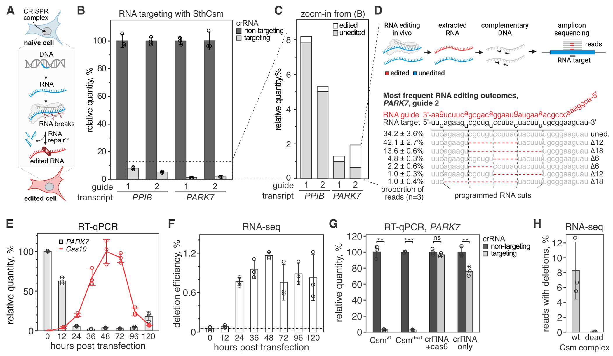
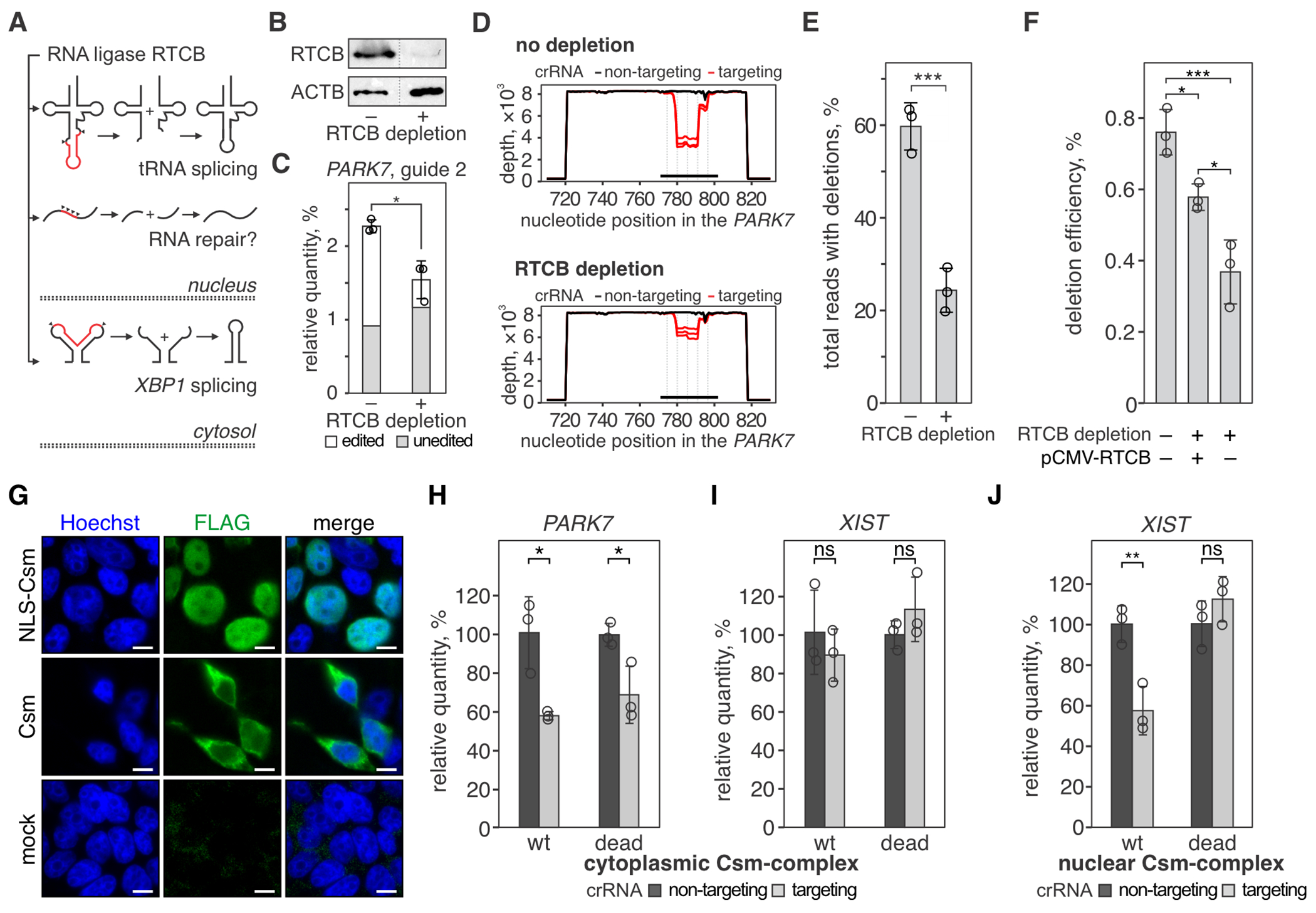
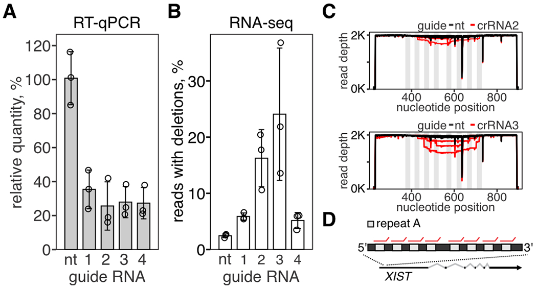
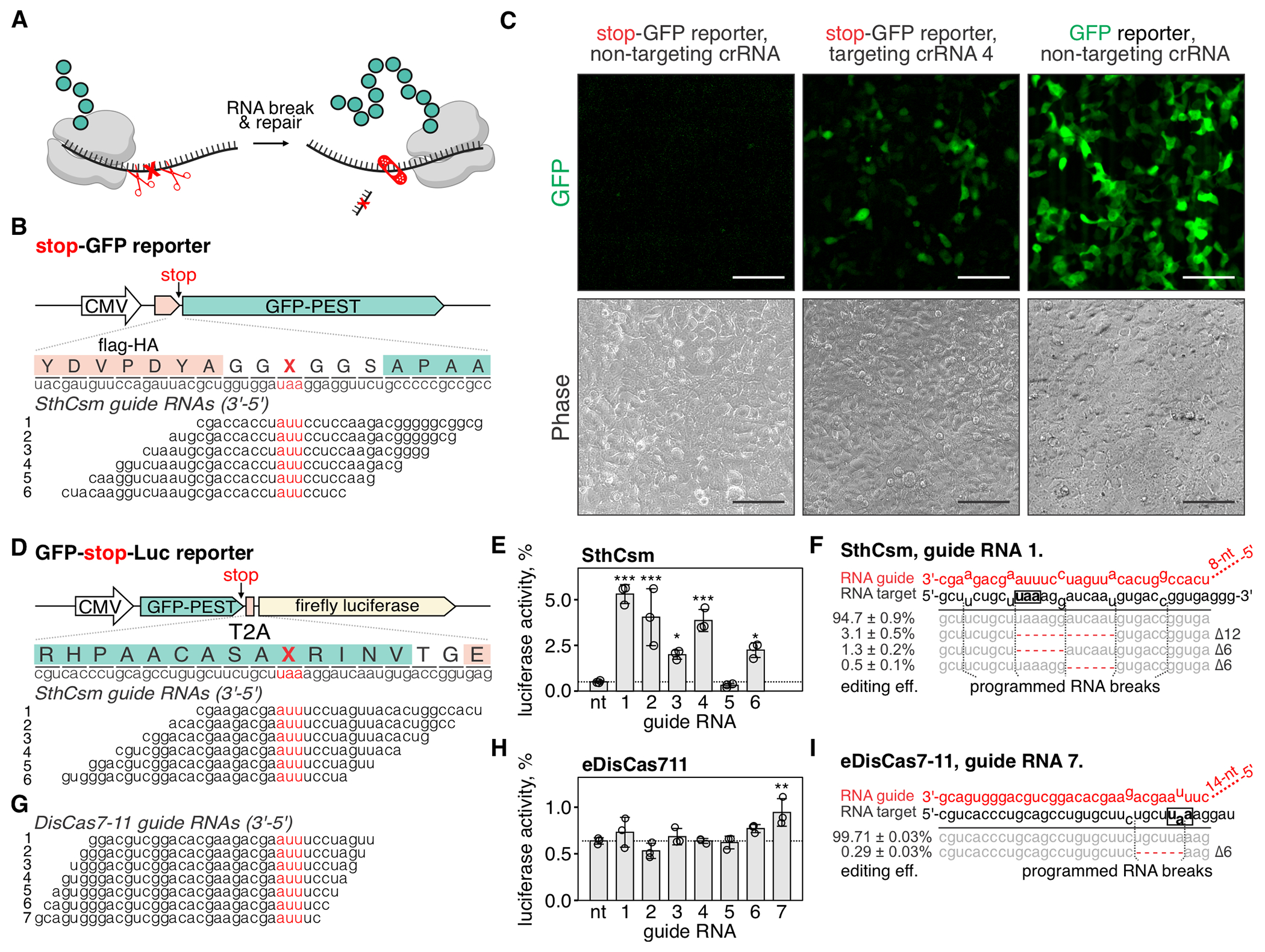
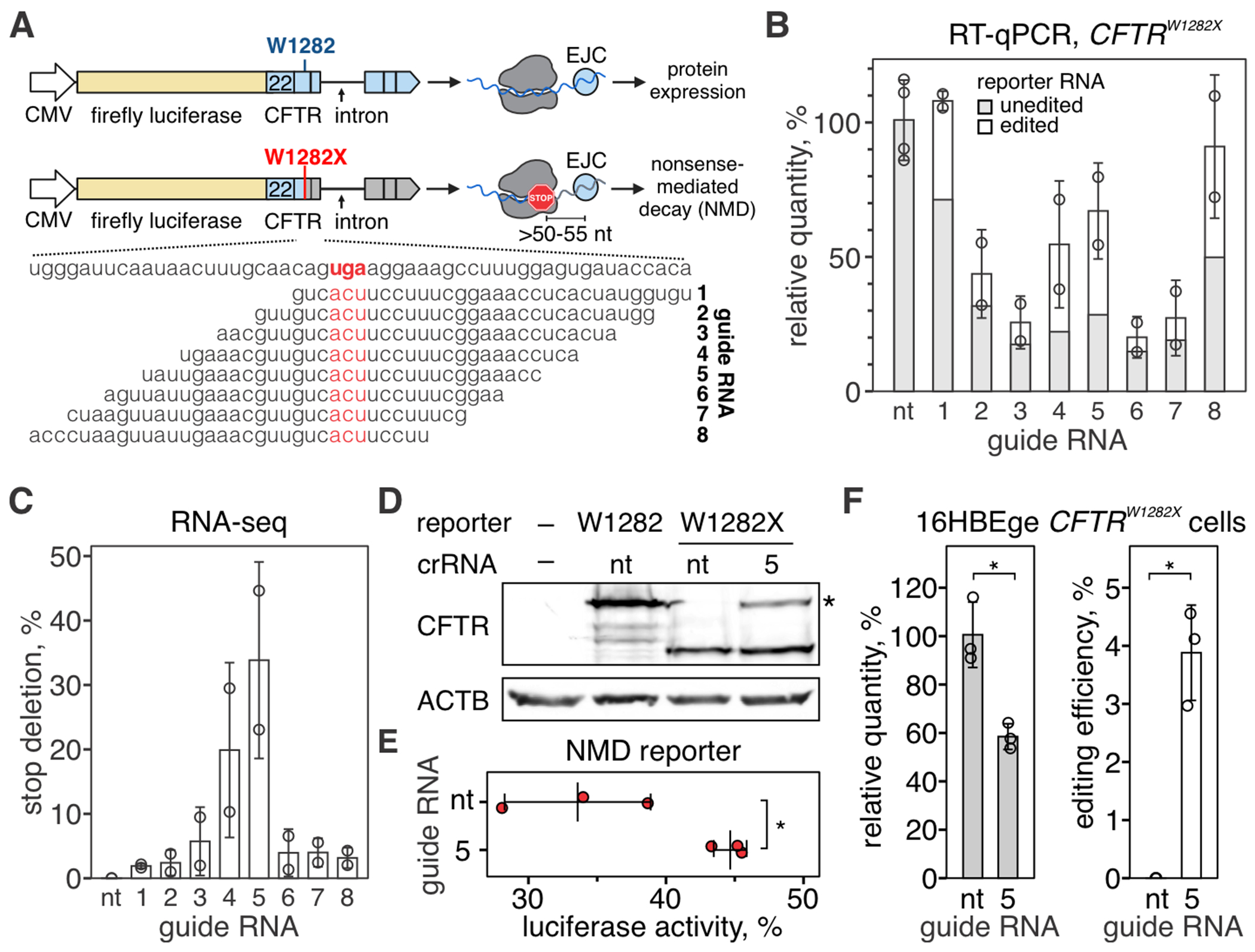
Genome editing with CRISPR RNA-guided endonucleases generates DNA breaks that are resolved by cellular DNA repair machinery. However, analogous methods to manipulate RNA remain unavailable. We show that site-specific RNA breaks generated with type-III CRISPR complexes are repaired in human cells and that this repair can be used for programmable deletions in human transcripts to restore gene function. Collectively, this work establishes a technology for precise RNA manipulation with potential therapeutic applications.


Conflict of interest statement

Competing interests: B.W. is the founder of SurGene LLC. B.W., A. Nemudryi, and A. Nemudraia are inventors of the patent applications US 63/523,592 and US 63/534,305 pertaining to use of type III CRISPR-Cas system for sequence-specific editing of RNA filed by Montana State University.

Figures

Fig. 1.
Fig. 1.. Programmable deletions of RNA with RNA-guided type III-A CRISPR complexes.
(A) Diagram of RNA editing in eukaryotic cells through sequence-specific RNA cleavage and RNA repair. (B) Human cells (293T) were transfected with plasmids encoding for NLS-tagged type III CRISPR complex of Streptococcus thermophilus (SthCsm) and RNA guides targeting PPIB or PARK7 messenger RNAs. Target transcripts were quantified with RT-qPCR, and the qPCR signal was normalized to ACTB and a non-targeting guide RNA control. Data is shown as mean ± SD of three biological replicates. (C) Zoom-in from panel B. Deep sequencing was used to quantify the proportion of signal that is derived from edited RNA. (D) Top: schematics of deep sequencing approach used to quantify RNA editing. Bottom: top five most frequent RNA editing outcomes in PARK7 transcript (guide 2). Dotted lines indicate the positions of RNA breaks by the SthCsm complex. Data is shown as mean ± SD. (E) Kinetics of PARK7 mRNA knockdown (guide 2) vs. NLS-tagged SthCsm complex expression. PARK7 qPCR signal was normalized to ACTB and 0 h time point. Cas10 expression was normalized to ACTB and maximum expression level at 48 h. Data is shown as mean ± SD of three biological replicates. (F) PARK7 qPCR products in (E) were sequenced, and deletion efficiency was calculated as [relative quantity] × [fraction of reads with deletions]. (G) PARK7 knockdown efficiencies with the “wildtype” NLS-tagged SthCsm complex (Csmwt), catalytically inactive NLS-tagged SthCsm complex (Csmdead), crRNA expressed only with Cas6 gene (no csm genes), and cRNA alone were measured using RT-qPCR. Welch’s t-test was used to compare samples expressing targeting and non-targeting crRNA. ** - p < 0.01, *** - p < 0.001, ns – non-significant. (H) RT-qPCR products in (G) were sequenced, and a fraction of reads with programmable deletions was quantified.
Fig. 2.
Fig. 2.. RTCB ligase repairs RNA cleaved by the type III CRISPR complex.
(A) Human RNA ligase RTCB joins RNA ends that are produced in tRNA splicing (top) and non-canonical XBP1 mRNA splicing (bottom) during the unfolded protein response. We hypothesized that the ligase activity of RTCB is involved in the RNA repair of CRISPR-guided RNA breaks (middle). (B) Western blot with anti-RTCB or anti-ACTB (loading control) antibodies was performed with lysates of 293T cells with (+) or without (−) RTCB depletion. See the uncropped images in fig. S3. (C) The PARK7 transcript was targeted with SthCsm (guide 2) in 293T cells with (+) or without (−) RTCB depletion. The PARK7 transcript was quantified with RT-qPCR and normalized to ACTB and non-targeting guide RNA. Data are shown as the mean ± standard deviation of three biological replicates. *P < 0.05, ***P < 0.001; one-way analysis of variance (ANOVA) with Tukey HSD post-hoc comparisons. (D) qPCR products in (C) were sequenced, and resulting reads were aligned to the reference sequence of the PARK7 transcript (NM_007262, GenBank). Graphs show sequencing depth (y-axes) at the amplified region of the transcript (x-axes). Every line shows a biological replicate (n = 3). The horizontal black bar indicates a region complementary to the guide RNA of the SthCsm complex. Vertical dotted lines mark predicted positions of RNA breaks. (E) Quantification of deletions in the target region of the PARK7 transcript. Data is shown as the mean ± standard deviation of three biological replicates. Welch’s t-test was used to compare mean values. *** p < 0.001. (F) RTCB deficient cells were transfected with a plasmid expressing RTCB (pCMV-RTCB), and the efficiency of CRISPR RNA-guided programmable deletions in PARK7 was quantified as in panels C-E. * p < 0.05; OneWelch’s t-test. (G) Immunostaining of cells expressing Flag-tagged Csm complex with (NLS-Csm) or without (Csm) NLS-tag. Scale bars are 10 μm. (H-J) Knockdown of PARK7 and XIST transcripts with cytoplasmic SthCsm (no NLS) (H, I) and knockdown of XIST with nuclear Csm (NLS-tagged) (J) was quantified with RT-qPCR and normalized to ACTB and non-targeting control. “wt” – nuclease-active SthCsm, “dead”– catalytically inactivated SthCsm (Csm3D33A mutation). *P < 0.05, ns – non-significant; Welch’s t-test. Data is shown as mean ± SD (n = 3).
Fig. 3.
Fig. 3.. Repair of concurrent RNA breaks results in large RNA excisions.
(A) Repetitive region (repeat A) in XIST transcript was targeted with SthCsm complex with four different guide RNAs. Knockdown of XIST was quantified with RT-qPCR. Data is shown as the mean of three biological replicates ± SD. B) Amplicon-seq was used to quantify programmed RNA deletions in XIST. C) Repeat A was amplified and deep-sequenced. Reads were aligned to the reference sequence (NR_001564.2, GenBank). Graphs show sequencing depth (y-axes) at the amplified region of the transcript (x-axes). Every line shows a biological replicate (n = 3). Vertical light gray rectangles indicate the position of repeats targeted by Csm complexes. D) Repeat A architecture in XIST lncRNA. Red lines show binding sites for Csm complexes.
Fig. 4.
Fig. 4.. Programmable excision of stop codons restores protein expression.
(A) Schematic representation of the proposed approach for deleting premature stop codons in human transcripts. (B) Top: schematic diagram of the stop-GFP reporter plasmid. Bottom: Six guide RNAs for SthCsm complex were designed to excise the stop codon in the gfp transcript. Underlined (red) sections of target RNA are expected to be deleted. Vertical red ticks indicate predicted sites for RNA breaks. (C) Cells were transfected with plasmids for the stop-GFP reporter and SthCsm with a non-targeting guide (left), stop-GFP reporter and SthCsm with a targeting guide (middle), or GFP reporter and SthCsm with the non-targeting guide (right). Fluorescence microscopy was used to image cells 48 h post-transfection. Scale bars – 50 μm. (D) Top: schematic diagram of the GFP-stop-Luc reporter plasmid. Bottom: Six crRNAs for SthCsm complex were designed to delete the stop codon at the 3’-end of the gfp gene. (E) Luciferase activity was measured in cell lysates 48 hours after transfection with GFP-stop-Luc and SthCsm plasmids. Luciferase activity is normalized to a control transfected with a reporter plasmid without the stop codon. Data are shown as mean ± SD of three replicates. Means were compared using one-way ANOVA, and samples with targeting guide RNAs were compared to the non-targeting control using one-tailed Dunnett’s test. *P < 0.5, **P < 0.1, ***P < 0.001. (F) Most frequent RNA editing outcomes in the sample with the most efficient rescue of luciferase activity in (B) (guide 1). Editing efficiency was quantified as mean ± SD of three biological replicates. The black box shows the stop codon that was targeted by type III CRISPR complexes. (G, H, I). The same as (D-F), but with eDisCas7-11.
Fig. 5.
Fig. 5.. Programmable excision of a non-sense mutation in the CFTR transcript restores translation.
(A) Diagram of a luciferase-based reporter for CFTRW1282X mutation (fLuc-CFTR, top) and guide RNA design (bottom). Top: Firefly luciferase was genetically linked to exons 22-27 of the CFTR cDNA, and synthetic intron sequence was inserted between exons 24 and 25. See Methods for additional details. Bottom: Eight crRNAs were tiled across the mutation (W1282X) to guide the excision of the stop codon (UGA, highlighted with red). (B) RT-qPCR was used to quantify fLuc-CFTR transcript targeted with Csm complexes. Amplicons were deep-sequenced to quantify edited vs. unedited reporter RNA. See fig. S6A for sequencing depth plots. (C) Quantification of deletions that remove stop codon (W1282X). (D) Western blot with antibodies against CFTR amino acid residues 1204-1211 with lysates from 293T cells expressing fLuc-CFTR or fLuc-CFTRW1282X) and Csm complexes with non-targeting (nt) guide RNA or CFTR-targeting guide RNA 5. See fig. S6B for uncropped images. (E) Quantification of luciferase activity in 293T cells transfected with fLuc-CFTRW1282X and Csm complexes with non-targeting guide RNA or targeting guide RNA 5. Middle bar shows mean of three biological replicates (red dots). Error bars show mean ± SD. *P < 0.05, Welch’s t-test. (F) Left: RT-qPCR was used to quantify CFTR transcript in HBE16ge CFTRW1282X cells transfected with plasmids encoding for Csm complexes with non-targeting guide RNA (nt) or CFTR-targeting guide RNA 5. Right: qPCR amplicons were deep-sequenced, and deletions removing W1282X codon were quantified. See fig. S6C for depth plot. Data is shown as the mean of three biological replicates ± SD. *P < 0.05, Welch’s t-test.

Update of

References

    1. Jinek M et al. A programmable dual-RNA-guided DNA endonuclease in adaptive bacterial immunity. Science 337, 816–821 (2012). - PMC - PubMed
    1. Gasiunas G, Barrangou R, Horvath P, Siksnys V, Cas9–crRNA ribonucleoprotein complex mediates specific DNA cleavage for adaptive immunity in bacteria. Proceedings of the National Academy of Sciences 109, (2012). - PMC - PubMed
    1. Ran FA et al. Genome engineering using the CRISPR-Cas9 system. Nature Protocols 8, 2281–2308 (2013). - PMC - PubMed
    1. van Overbeek M et al. DNA Repair Profiling Reveals Nonrandom Outcomes at Cas9-Mediated Breaks. Mol Cell 63, 633–646 (2016). - PubMed
    1. Kosicki M, Tomberg K, Bradley A, Repair of double-strand breaks induced by CRISPR–Cas9 leads to large deletions and complex rearrangements. Nature Biotechnology 36, 765–771 (2018). - PMC - PubMed