. 2024 Jul 22;59(14):1783-1793.e5.
doi: 10.1016/j.devcel.2024.04.008. Epub 2024 Apr 24.

Cryo-EM structures of membrane-bound dynamin in a post-hydrolysis state primed for membrane fission

Affiliations

Cryo-EM structures of membrane-bound dynamin in a post-hydrolysis state primed for membrane fission

John R Jimah et al. Dev Cell. .

Abstract

Dynamin assembles as a helical polymer at the neck of budding endocytic vesicles, constricting the underlying membrane as it progresses through the GTPase cycle to sever vesicles from the plasma membrane. Although atomic models of the dynamin helical polymer bound to guanosine triphosphate (GTP) analogs define earlier stages of membrane constriction, there are no atomic models of the assembled state post-GTP hydrolysis. Here, we used cryo-EM methods to determine atomic structures of the dynamin helical polymer assembled on lipid tubules, akin to necks of budding endocytic vesicles, in a guanosine diphosphate (GDP)-bound, super-constricted state. In this state, dynamin is assembled as a 2-start helix with an inner lumen of 3.4 nm, primed for spontaneous fission. Additionally, by cryo-electron tomography, we trapped dynamin helical assemblies within HeLa cells using the GTPase-defective dynamin K44A mutant and observed diverse dynamin helices, demonstrating that dynamin can accommodate a range of assembled complexes in cells that likely precede membrane fission.

Keywords: clathrin-mediated endocytosis; cryo-EM; cryo-ET; dynamin; endocytosis; membrane fission; membrane remodeling; structural biology.


Conflict of interest statement

Declaration of interests The authors declare no competing interests.

Figures
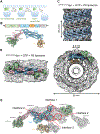
Figure 1.
Figure 1.. Cryo-EM structures of dynamin helical assemblies on lipid tubules in the super-constricted state.
(A) Sketch of membrane fission during endocytosis, as dynamin progresses through the GTPase cycle. (B) Domain organization of dynamin showing the GTPase domain (green), Bundle Signaling Element (BSE, pink), Stalk (blue), Pleckstrin homology (PH) domain (orange), and Proline Rich Domain (PRD, grey). Atomic model of a dynamin monomer generated from our super-constricted cryo-EM structure, color-coded by domain. (C) Cryo-EM map of ΔPRD dynamin-K44A incubated with GTP and organized around lipid tubules in the two-start super-constricted state (ΔPRD-K44AdynGDP). (D) Side and top views of ΔPRD-K44AdynGDP cryo-EM map. Domains of dynamin are color-coded as in (B). (E) Atomic model of ΔPRD-K44AdynGDP tetramer, with one dimer color-coded by domain and the other in grey, showing interfaces 1, 2, and 3 that drive oligomerization. See also Figures S1–S4, and Table 1.
Figure 2.
Figure 2.. GTP hydrolysis to GDP leads to super-constriction of dynamin-decorated lipid tubules.
(A) Model of GTPase domain dimer of super-constricted ΔPRD-K44AdynGDP with trans-stabilizing residues shown in boxed regions 1 and 3, and nucleotide-interacting residues in box 2. The two GTPase domains are colored green and purple, respectively and the cryo-EM density around the nucleotide is shown as a black mesh. (B) Atomic model of GDP fitted in cryo-EM map of ΔPRD-K44AdynGDP with the cryo-EM density in mesh. (C) Michaelis-Menten plot of wild type ΔPRD dynamin (black) and ΔPRD dynamin-K44A (blue) GTPase activity, in the presence of PS liposomes. Velocity measurement from three experiments shown as mean +/− SEM. (D) Overlay of the GG BSE dimer of dynamin in the GDP-bound super-constricted state (ΔPRD-K44AdynGDP); (GTPase domain in green and BSE in pink) compared to the GMPPCP-bound constricted state (ΔPRDdynPCP, grey). (E) The three-helix bundle or BSE resides in an up position in the super-constricted (GDP) state of assembled dynamin in contrast to the crystal structures of dynamin in the GDP state with the BSE in the down position (5D3Q). See also Figures S3H, Video S1.
Figure 3.
Figure 3.. The stalk and PH domains swing toward the encompassed lipid tubule to help drive super-constriction.
(A) Comparison of dynamin cryo-EM helical structures in the constricted and super-constricted states, highlighting the decrease in outer diameter from 40 nm to 36 nm and inner lumen from 7.4 nm to 3.4 nm. (B) Overlay of super-constricted ΔPRD-K44AdynGDP (domains colored as in Figure 1) and constricted ΔPRDdynPCP dimers (white) about the GTPase dimer interface. The stalk moves toward the membrane upon GTP hydrolysis; 6.3 Å for the kinked, stable monomer, (indicated by asterisk) and 9.6 Å for the flexible monomer. The variable loops of the PH domain are color-coded as shown in C. (C) Cryo-EM map (outlined in white) and model of ΔPRD-K44AdynGDP partial stalk (blue) and PH domain (orange) obtained after local refinement of ΔPRD-K44AdynGDP. PH domain variable loops 1–4 are colored red, purple, green, and blue respectively in B and C. (D) Surface map showing the electrostatic surface potential. Blue is positive and red is negative. See also Figures S2 and S3.
Figure 4.
Figure 4.. A unique amino-acid interaction observed in the super-constriction state is crucial for basal levels of endocytosis.
(A) Interactions between D406 and L402 observed in interface 3 of the GDP-bound super-constricted state, ΔPRD-K44AdynGDP. Top, ΔPRD-K44AdynGDP tetramer highlighting interface 3. Bottom, H-bond between D406 and L402 shown in model without (below) and with density in white (above). (B) Transferrin uptake assays quantifying endocytosis in cells transfected with WT, K44A, and D406A dynamin. Black bars, cells expressing dynamin plasmids (WT, K44A, D406A). Blue bars, cells not expressing dynamin plasmids. In each experiment, samples were run in triplicate (shown as dots). The error bars are standard deviation. The experiment was performed twice with comparable results. Pair Sample T-test is comparing intra-experiment triplicates, significance marked with *p=0.0007, **p=0.003. See also Figure S5C.
Figure 5.
Figure 5.. Dynamin helical polymer observed within a HeLa cell.
(A) Slice through a tomogram revealing a dynamin-decorated membrane tube with a clathrin coat at the tip. (B) Segmented tomogram with dynamin, clathrin, vesicle, and actin colored in pink, yellow, grey, and green, respectively. (C) Model of a dynamin helical polymer (rainbow-colored) fit into tomogram. Scale bar, 50 nm. (D) Measurements of outer and inner luminal diameters and the inter-rung distance of dynamin helical structures in cells. The lengths of these measurements are shown as a scatter plot and mean ±S.D. See also Videos S2–5
Figure 6.
Figure 6.. Model for dynamin organization on lipid tubules in the 1-start GTP-bound constricted state and the 2-start GDP-bound super-constricted state.
(A) Top view highlighting dynamin-mediated constriction of the underlying membrane (yellow) from an inner lumen of 7.4 nm in the ΔPRDdynPCP model to 3.4 nm in the ΔPRD-K44AdynGDP model. (B) Side view illustrating an increase in pitch upon GTP hydrolysis that allows for a second rung of dynamin to assemble. GTPase domains dimerizes (G dimer) between rungs of the helix. See also Video S6

Similar articles

Cited by

References

    1. Kozlov MM, McMahon HT, and Chernomordik LV (2010). Protein-driven membrane stresses in fusion and fission. Trends Biochem Sci 35, 699–706. 10.1016/j.tibs.2010.06.003. - DOI - PMC - PubMed
    1. Antonny B, Burd C, De Camilli P, Chen E, Daumke O, Faelber K, Ford M, Frolov VA, Frost A, Hinshaw JE, et al. (2016). Membrane fission by dynamin: what we know and what we need to know. EMBO J 35, 2270–2284. 10.15252/embj.201694613. - DOI - PMC - PubMed
    1. Ferguson SM, and De Camilli P (2012). Dynamin, a membrane-remodelling GTPase. Nat Rev Mol Cell Biol 13, 75–88. 10.1038/nrm3266. - DOI - PMC - PubMed
    1. Chen MS, Obar RA, Schroeder CC, Austin TW, Poodry CA, Wadsworth SC, and Vallee RB (1991). Multiple forms of dynamin are encoded by shibire, a Drosophila gene involved in endocytosis. Nature 351, 583–586. 10.1038/351583a0. - DOI - PubMed
    1. Hinshaw JE, and Schmid SL (1995). Dynamin self-assembles into rings suggesting a mechanism for coated vesicle budding. Nature 374, 190–192. 10.1038/374190a0. - DOI - PubMed

Publication types

LinkOut - more resources