. 2024 Apr 25;15(1):3450.
doi: 10.1038/s41467-024-47455-6.

Bat-borne H9N2 influenza virus evades MxA restriction and exhibits efficient replication and transmission in ferrets

Affiliations

Bat-borne H9N2 influenza virus evades MxA restriction and exhibits efficient replication and transmission in ferrets

Nico Joel Halwe et al. Nat Commun. .

Abstract

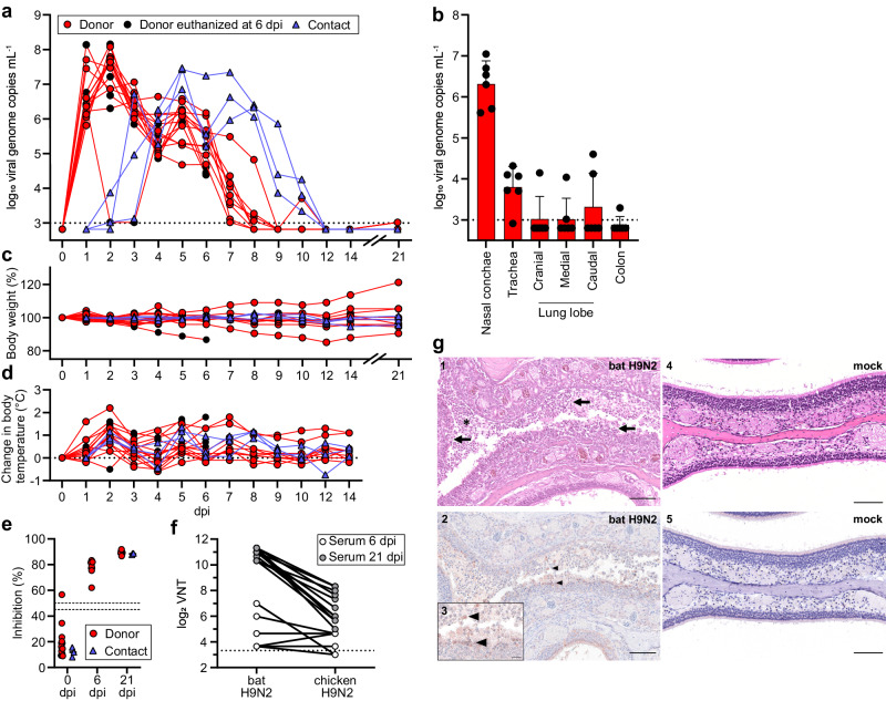
Influenza A viruses (IAVs) of subtype H9N2 have reached an endemic stage in poultry farms in the Middle East and Asia. As a result, human infections with avian H9N2 viruses have been increasingly reported. In 2017, an H9N2 virus was isolated for the first time from Egyptian fruit bats (Rousettus aegyptiacus). Phylogenetic analyses revealed that bat H9N2 is descended from a common ancestor dating back centuries ago. However, the H9 and N2 sequences appear to be genetically similar to current avian IAVs, suggesting recent reassortment events. These observations raise the question of the zoonotic potential of the mammal-adapted bat H9N2. Here, we investigate the infection and transmission potential of bat H9N2 in vitro and in vivo, the ability to overcome the antiviral activity of the human MxA protein, and the presence of N2-specific cross-reactive antibodies in human sera. We show that bat H9N2 has high replication and transmission potential in ferrets, efficiently infects human lung explant cultures, and is able to evade antiviral inhibition by MxA in transgenic B6 mice. Together with its low antigenic similarity to the N2 of seasonal human strains, bat H9N2 fulfils key criteria for pre-pandemic IAVs.


Conflict of interest statement

The Icahn School of Medicine at Mount Sinai has filed patent applications relating to influenza virus vaccines, SARS-CoV-2 serological assays and SARS-CoV-2 vaccines which list Florian Krammer as co‐inventor. Viviana Simon is also listed as co-inventor on patent applications for SARS-CoV-2 serological assays. Mount Sinai has spun out companies, Kantaro and Castlevax, to market the SARS-CoV-2 related technologies. Florian Krammer has consulted for Merck and Pfizer (before 2020), and is currently consulting for Pfizer, Seqirus, 3rd Rock Ventures, GSK and Avimex. The Krammer laboratory is also collaborating with Pfizer on animal models of SARS‐CoV‐2 and with Dynavax on universal influenza virus vaccines. All other authors declare no competing interests.

Figures

Fig. 1
Fig. 1. Ferrets are highly susceptible to bat H9N2.
a Ferrets (n = 15) were inoculated with 104.8 TCID50 of bat H9N2 IAV per animal. At 1 dpi, direct contact animals (n = 3) were co-housed. Donor ferrets euthanized at 6 dpi are indicated. Viral shedding was measured by nasal lavage. Dashed line indicates detection limit. b Organs collected from euthanized ferrets (n = 6) at 6 dpi with bat H9N2 were tested by RT-qPCR to determine viral genome copies. Dashed line indicates detection limit. Data are mean ± SD. c Changes in body weight relative to 0 dpi of bat H9N2-infected (n = 15) and contact (n = 3) ferrets were monitored throughout the course of the experiment. d Changes from the baseline body temperatures of donor and naïve contact ferrets were monitored from 0 to 14 dpi. e Ferret serum antibody titers in an IAV NP-specific ELISA at the indicated time points (0 dpi n = 18, 6 dpi n = 6, 21 dpi n = 12). Dashed lines indicates threshold between 45% and 50% inhibition. Mean antibody titers are indicated. Note that one ferret showed weak reactivity in the pre-experimental NP-ELISA, but was included in the study because of its low seropositivity, which could be due to a previously unrecognized IAV infection or an unspecific ELISA reactivity. There was no indication of H9N2 serology prior to inoculation. f Ferret neutralizing antibody titers (n = 18) against bat H9N2 and chicken H9N2. g Histopathologic findings with detection of viral antigen in the nasal mucosa of bat H9N2-infected ferrets (n = 6, panels 1–3) at 6 dpi or mock-infected ferret (n = 1, panels 4 and 5). Acute severe rhinitis with diffuse necrosis of the olfactory epithelium (arrow) and infiltrating neutrophils (asterisk) (1). Intralesional viral antigen (NP) is abundant in degenerated and desquamated epithelial cells (arrowhead) (2). The inset (3) is a higher magnification of the center of the image (2). No pathology was observed in the mock-infected ferret (4, 5). Representative images are shown. Scale bar, 100 µm (main panels), 25 µm (inset). Source data are provided as a Source Data file.
Fig. 2
Fig. 2. Bat H9N2 replicates in human lung explants and suppresses induction of MxA in MxA-transgenic mice.
a Human lung tissue explants (n = 4) were infected with human H3N2, chicken H9N2 or bat H9N2 with 1 × 106  PFU, and viral titers were determined at the indicated time points. Error bars indicate standard deviation and statistical analysis was performed using non-paired, non-parametric Kruskal-Wallis test (*p = 0.0324). Data are mean ± SD of n = 4 independent experiments. Dashed line indicates detection limit (b) At 24 hpi, human lung explants were stained for alveolar type I (AT1) (cyan) and type II (AT2) cells (yellow), CD68 indicating alveolar macrophages (green) and IAV antigens (red). Note, in chicken H9N2 and bat H9N2 infected cells, AT2 labeling was omitted for better visualization. White arrows indicate infected cells. Scale bar, 10 µm. c MDCK cells overexpressing MxA or inactive MxAT103A were infected with human-adapted pdmH1N1, avian KAN-1 (H5N1) or bat H9N2 at an MOI of 0.001, and viral titers were determined at the indicated time points. Data are mean ± SD of n = 3 independent experiments; statistical analysis was performed using two-tailed t-tests; **P = 0.01; ****P = 0.0001. Dashed line indicates detection limit. d hMxAtg/tg (n = 8) or wild-type B6 mice (n = 8) were infected with 1 × 104  PFU. Lung viral titers were determined 3 dpi. e hMxAtg/tg (n = 6) or wild-type B6 mice (n = 7) were pretreated with IFN-α 18 h prior to infection with 1 × 104 PFU. Lung viral titers were determined 3 dpi. Data are mean ± SD; statistical analysis was performed using two-tailed t-tests; ****P = 0.0001. f MxA, NP and actin protein levels in homogenized lungs from IFN-α pretreated or infected mice from (d,e) were detected by Western blot. Source data are provided as a Source Data file.

References

    1. Long JS, Mistry B, Haslam SM, Barclay WS. Host and viral determinants of influenza A virus species specificity. Nat. Rev. Microbiol. 2019;17:67–81. doi: 10.1038/s41579-018-0115-z. - DOI - PubMed
    1. Letko M, Seifert SN, Olival KJ, Plowright RK, Munster VJ. Bat-borne virus diversity, spillover and emergence. Nat. Rev. Microbiol. 2020;18:461–471. doi: 10.1038/s41579-020-0394-z. - DOI - PMC - PubMed
    1. Tong S, et al. A distinct lineage of influenza A virus from bats. Proc. Natl Acad. Sci. USA. 2012;109:4269–4274. doi: 10.1073/pnas.1116200109. - DOI - PMC - PubMed
    1. Tong S, et al. New world bats harbor diverse influenza A viruses. PLoS Pathog. 2013;9:e1003657. doi: 10.1371/journal.ppat.1003657. - DOI - PMC - PubMed
    1. Kandeil A, et al. Isolation and characterization of a distinct influenza A virus from Egyptian bats. J. Virol. 2019;93:e01059–18. doi: 10.1128/JVI.01059-18. - DOI - PMC - PubMed

Publication types

MeSH terms

Substances