. 2024 Apr 25;10(1):90.
doi: 10.1038/s41531-024-00705-2.

Ultrastructural localization of Porphyromonas gingivalis gingipains in the substantia nigra of Parkinson's disease brains

Affiliations

Ultrastructural localization of Porphyromonas gingivalis gingipains in the substantia nigra of Parkinson's disease brains

Florian Ermini et al. NPJ Parkinsons Dis. .

Abstract

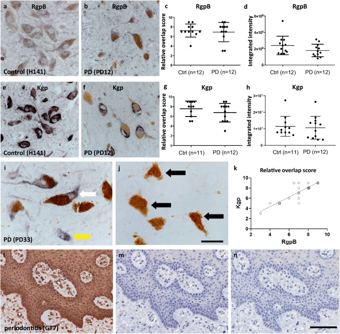
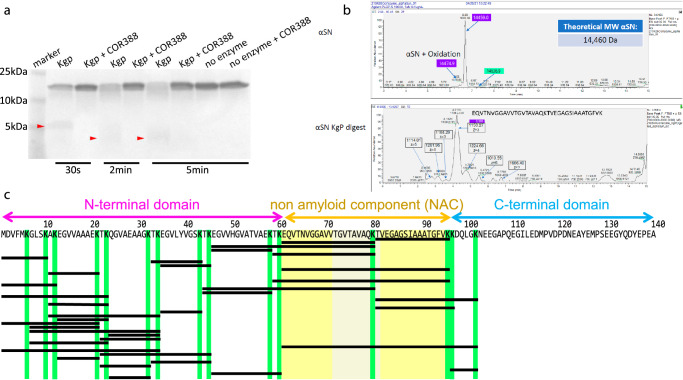
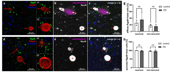
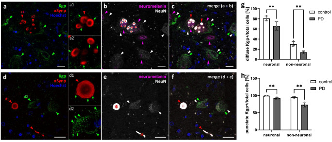
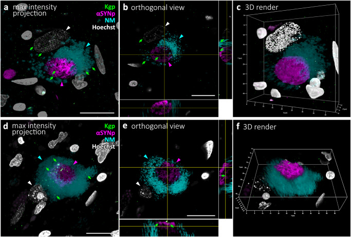
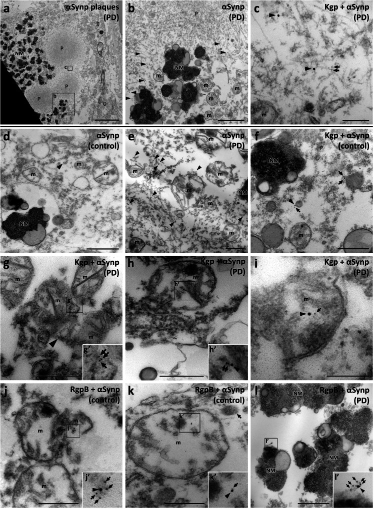
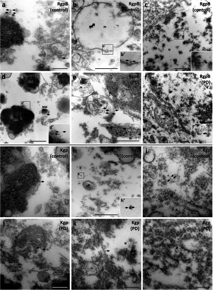
Gingipains are protease virulence factors produced by Porphyromonas gingivalis, a Gram-negative bacterium best known for its role in chronic periodontitis. Gingipains were recently identified in the middle temporal gyrus of postmortem Alzheimer's disease (AD) brains, where gingipain load correlated with AD diagnosis and tau and ubiquitin pathology. Since AD and Parkinson's disease (PD) share some overlapping pathologic features, including nigral pathology and Lewy bodies, the current study explored whether gingipains are present in the substantia nigra pars compacta of PD brains. In immunohistochemical techniques and multi-channel fluorescence studies, gingipain antigens were abundant in dopaminergic neurons in the substantia nigra of both PD and neurologically normal control brains. 3-dimensional reconstructions of Lewy body containing neurons revealed that gingipains associated with the periphery of alpha-synuclein aggregates but were occasionally observed inside aggregates. In vitro proteomic analysis demonstrated that recombinant alpha-synuclein is cleaved by lysine-gingipain, generating multiple alpha-synuclein fragments including the non-amyloid component fragments. Immunogold electron microscopy with co-labeling of gingipains and alpha-synuclein confirmed the occasional colocalization of gingipains with phosphorylated (pSER129) alpha-synuclein. In dopaminergic neurons, gingipains localized to the perinuclear cytoplasm, neuromelanin, mitochondria, and nucleus. These data suggest that gingipains localize in dopaminergic neurons in the substantia nigra and interact with alpha-synuclein.


Conflict of interest statement

F.E. and S.S.D. were employees of Cortexyme at the time the research was conducted. Dominy is an inventor on gingipain inhibitor patents, and cofounder of Lighthouse Pharmaceuticals, a company developing gingipain inhibitors as therapeutics. The other authors declare that they have no conflicts of interest.

Figures

Fig. 1
Fig. 1. IHC staining of neuromelanin-containing neurons in the SNpc of control and PD brains.
a, b CAB101 staining for RgpB in the SNpc revealed a granular cytoplasmatic stain (purple/black). Neuromelanin (brown) is clearly distinguishable from the purple black DAB-Ni reaction product in both control and PD patients. c Semiquantitative analysis of RgpB staining shows equal relative numbers of neuromelanin cells positive for RgpB in controls and PD patients. d Integrated intensity analysis of RgpB shows no difference between controls and PD (t test with Welch’s correction: p = 0.14). e, f CAB102.1 staining for Kgp in the SNpc revealed an intense cytoplasmatic stain (purple/black). Note the reduced density of neuromelanin cells in PD patients (b, f) compared to controls (a, e). g Semiquantitative analysis of Kgp staining shows equal relative numbers of neuromelanin cells positive for Kgp in controls and PD patients. h Integrated intensity analysis of Kgp shows no difference between controls and PD (t test with Welch’s correction: p = 0.58). i, j Specificity of CAB102.1 was tested by preabsorbing CAB102.1 with 10x concentration of Kgp antigen. Using non-absorbed antibody (i) both neuromelanin positive (white arrow) and neuromelanin negative (yellow arrow) cells were detected. In sections incubated with preabsorbed antibody no DAB-Ni reaction product was observed (j) and neuromelanin was observed as before (black arrows). The section shown is from the SNpc of a PD case (PD33, Table 2). Scale bar 20 µm. k The correlation of the relative overlap score of Kgp staining strongly correlated with RgpB staining (r = 0.864, p < 0.0001). l, n gingival tissue from a periodontitis patient (GT7) was used as a positive control for CAB102.1. l CAB102.1 at 1 µg/mL concentration. m CAB102.1 was substituted for recombinant rabbit IgG (1 µg/mL). n No primary antibody was used. Scale bar 100 µm. The scatter plots show the median with interquartile range (c, g) or the mean with standard deviation (d, h). The trendline in graph (k) represents a simple linear regression.
Fig. 2
Fig. 2. α-synuclein (αSyn) is a substrate for Kgp.
a Coomassie gel of recombinant αSyn exposed to 30 s, 2 min or 5 min of Kgp, with or without Kgp inhibitor COR388. Fragmented αSyn appears as a faint diffuse smear at 5 kDa and below after 30 s of Kgp digestion and appears fainter with a wider spread at 2 min and 5 min digestion (red arrowheads). b Intact mass spectrometry analysis of recombinant non-digested and Kgp-digested αSyn. In the undigested sample a single peak was observed with a predicted mass matching the theoretical mass of αSyn. In the Kgp digested sample several peaks were observed with a dominant peak matching the sequence of the non-amyloid component (NAC)-domain of αSyn. c Amino acid sequence of αSyn. The three domains (N-terminal, NAC, C-terminal) are highlighted above the sequence. Kgp protease activity was observed after lysine (K; highlighted in green) resulting in fragments defined by the position of lysine (black bars). The NAC is flanked by lysine and Kgp activity resulted in several NAC specific fragments including the highly aggregating VTGVTAVAQKTV motif (grayed yellow).
Fig. 3
Fig. 3. RgpB co-labeling using the CAB101 antibody (green) with αSynp (red), neuromelanin (magenta), and NeuN (white) in the PD SNpc.
ac RgpB shows punctate staining (green arrows) throughout the SNpc and within αSynp inclusions (red arrowheads) in neuromelanin-positive neuronal (a1) and neuromelanin and NeuN negative (a2) structures. df RgpB shows punctate staining (green arrows) within αSynp inclusions in neuromelanin negative/NeuN positive cells (d1, red arrowhead) and NeuN negative structures (d2, red arrowhead). Diffuse RgpB staining (green arrowheads) is observed outside of αSynp inclusions in neuromelanin negative/NeuN positive cells (d1, green arrowheads). αSynp staining bleeds through into the NeuN channel (b, c, e, f, red asterisk on saturated white) and was thresholded out for quantitative analysis of NeuN. Blue = Hoechst. g, h Quantification of the number of diffuse and punctate RgpB positive neuronal (NeuN positive) and non-neuronal cells relative to the total number of cells reveals no differences between neuronal or non-neuronal cells or between control or PD samples. Means with standard deviations, ns non-significant (p > 0.05, Tukey’s multiple comparison test). Scale bars are 20 µm.
Fig. 4
Fig. 4. Kgp co-labeling using the CAB102.1 antibody (green) with αSynp (red), neuromelanin (magenta), and NeuN (white) in the PD SNpc.
ac Kgp shows diffuse staining (green arrowheads) in the cytoplasm of neuromelanin-positive (magenta arrowheads) and neuromelanin-negative neuronal cells (white arrowheads). a1a2 Punctate Kgp staining (green arrows) can be observed within αSynp inclusions (red arrowheads) in neuromelanin-positive neuronal cells. df Kgp shows diffuse staining (green arrowheads) within the cytoplasm of neuromelanin-negative neuronal cells (white arrowheads). Punctate Kgp staining (green arrows) can be observed within the periphery of αSynp structures (d1) as well as within the diffuse patches of Kgp staining in the cytoplasm of neuronal SNpc cells (d2). αSynp staining bleeds through into the NeuN channel (b, c, e, f red asterisk on saturated white) and was thresholded out for quantitative analysis of NeuN. Blue = Hoechst. g, h Quantification of the number of diffuse and punctate Kgp positive neuronal (neuromelanin and NeuN positive) and non-neuronal cells relative to the total number of cells reveals significantly more punctate and diffuse Kgp in control compared to PD samples. Means with standard deviations, **p < 0.01, Tukey’s multiple comparison test. Scale bars are 20 µm.
Fig. 5
Fig. 5. 3D modeling of Kgp co-labeling using the CAB102.1 antibody (green) with αSynp (magenta), neuromelanin (cyan), and Hoechst (white) in two cells of the PD SNpc using confocal laser-scanning microscopy image stacks.
a Maximal intensity projection of an image stack depicting a neuromelanin neuron. A few Kgp puncta (green arrows) can be seen within the neuromelanin and nucleus of the cell. b Orthogonal single plane view of the neuromelanin neuron from (a). The large nucleus (white arrowhead) can be seen in the white channel. A few Kgp puncta (green arrows) can be seen on the surface of the circular αSynp inclusion (magenta) as well as within the circular αSynp aggregate (magenta arrowhead). c 3D rendering of the same cell as in a-b shown in a 23-degree rotation around the Y-axis. d Maximal intensity projection of an image stack depicting a neuromelanin neuron. e Orthogonal 2-plane view of the neuromelanin neuron from d. A few Kgp puncta (green arrows) can be seen decorating the outside of the αSynp inclusion. f 3D rendering of the same cell as in (d, e) shown in a 32-degree rotation around the X-axis.
Fig. 6
Fig. 6. Transmission electron microscopy analysis in the SNpc using double immunogold labeling with CAB101 (anti-RgpB; 6 nm gold particles, arrows) or CAB102.1 (anti-Kgp; 6 nm gold particles, arrows), and anti-αSynp (15 nm gold particles, arrowheads).
ac Kgp and αSynp in association with αSynp plaques. a An overview image of a neuromelanin-positive cell with multiple circular αSynp plaques (P). b Enlarged region from (a). αSynp immunogold labeling (black arrowheads) can be seen on the periphery of the plaques and in proximity to neuromelanin (NM) granules. c Enlarged region from (a). αSynp (black arrowheads) and Kgp (black arrows) immunogold labeling can be seen on medium electron-dense tendrils at the periphery of the plaque. d A control case with a 15 nm gold particle (black arrowhead) marking the rare presence of αSynp in the cytoplasm of a neuromelanin cell. e More frequent labeling of αSynp was observed in PD cases (black arrowheads). f Kgp (black arrows) associated with αSynp (black arrowhead) in cytoplasm near neuromelanin (NM) and mitochondria (m) (g). g Kgp (black arrows) and αSynp (black arrowhead) in proximity and within a mitochondrion (m). h, h’ Kgp (black arrows) associated with αSynp (black arrowhead) in a mitochondrion (m). i Kgp (black arrow) associated with αSynp (black arrowhead) in a mitochondrion (m). j, j’ RgpB (black arrows) associated with αSynp (black arrowhead) in a mitochondrion (m). k, k’ RgpB (black arrow) associated with αSynp (black arrowhead) in a mitochondrion (m). l, l’ Rgp (black arrows) associated with αSynp (black arrowhead) on the surface of a neuromelanin granule. NM, neuromelanin; m, mitochondria, P, αSynp plaque. Scale bars: 5 µm (a), 1 µm (b, d), 500 nm (e, f, g, h, j, k, l, m), 200 nm (c, i).
Fig. 7
Fig. 7. Immunogold labeling of RgpB and Kgp (6 nm gold particles) in control and PD neuromelanin-positive cells of the SNpc.
A similar pattern of RgpB immunogold (black arrows) was observed in samples from control (ac) and PD cases (df). Clusters of 6 nm gold particles labeling RgpB were observed in association with neuromelanin (a, d), mitochondria (b, e), and the nucleus (c, f). The pattern of Kgp immunogold (black arrow) was comparable in control (gi) and PD cases (jl). Clusters of 6 nm gold particles labeling Kgp were observed in association with neuromelanin (g), mitochondria (h, j), the nucleus (k), and rough endoplasmic reticulum (i, l). NM: neuromelanin, m: mitochondria, P: αSynp plaque, PD: Parkinson’s disease, rER; rough endoplasmic reticulum. Scale bars: 500 nm (b, c, d, e, f, h), 200 nm (a, g, i, j, k, l).

References

    1. Alam MM, et al. Alpha synuclein, the culprit in Parkinson disease, is required for normal immune function. Cell Rep. 2022;38:110090. doi: 10.1016/j.celrep.2021.110090. - DOI - PMC - PubMed
    1. DeMaagd G, Philip A. Parkinson’s disease and its management: Part 1: disease entity, risk factors, pathophysiology, clinical presentation, and diagnosis. Pharm. Ther. 2015;40:504–532. - PMC - PubMed
    1. Helmich RC, Bloem BR, Toni I. Motor imagery evokes increased somatosensory activity in Parkinson’s disease patients with tremor. Hum. Brain Mapp. 2012;33:1763–1779. doi: 10.1002/hbm.21318. - DOI - PMC - PubMed
    1. Magrinelli F, et al. Pathophysiology of motor dysfunction in Parkinson’s Disease as the rationale for drug treatment and rehabilitation. Parkinsons Dis. 2016;2016:9832839. doi: 10.1155/2016/9832839. - DOI - PMC - PubMed
    1. Spillantini MG, et al. Alpha-synuclein in Lewy bodies. Nature. 1997;388:839–840. doi: 10.1038/42166. - DOI - PubMed