. 2024 Apr 25;7(1):501.
doi: 10.1038/s42003-024-06189-3.

Absence of calcium-sensing receptor basal activity due to inter-subunit disulfide bridges

Affiliations

Absence of calcium-sensing receptor basal activity due to inter-subunit disulfide bridges

Shumin Ma et al. Commun Biol. .

Abstract

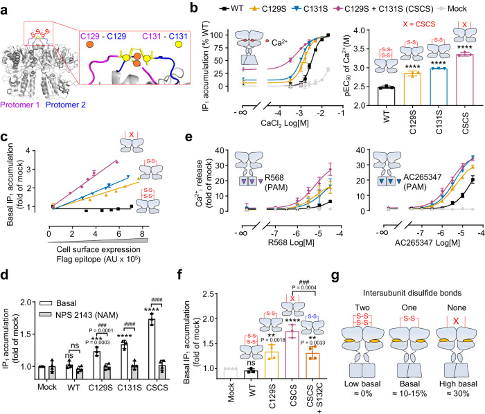
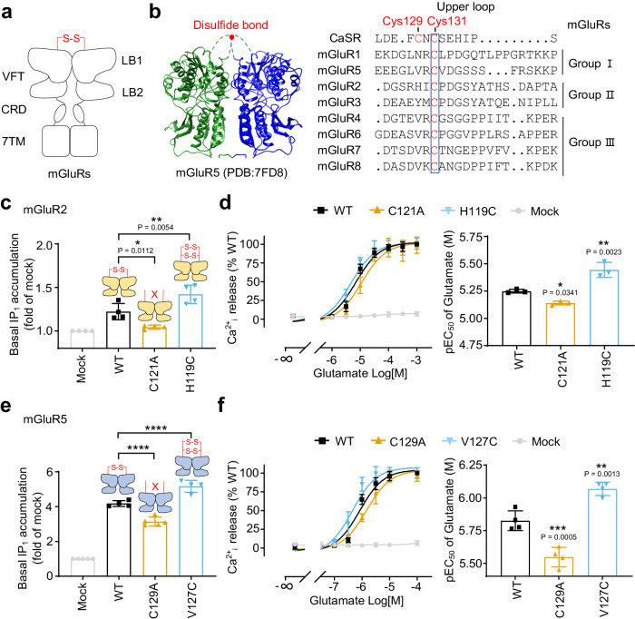
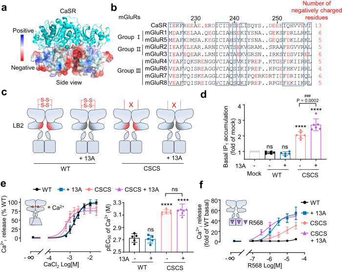
G protein-coupled receptors naturally oscillate between inactive and active states, often resulting in receptor constitutive activity with important physiological consequences. Among the class C G protein-coupled receptors that typically sense amino-acids and their derivatives, the calcium sensing receptor (CaSR) tightly controls blood calcium levels. Its constitutive activity has not yet been studied. Here, we demonstrate the importance of the inter-subunit disulfide bridges in maintaining the inactive state of CaSR, resulting in undetectable constitutive activity, unlike the other class C receptors. Deletion of these disulfide bridges results in strong constitutive activity that is abolished by mutations preventing amino acid binding. It shows that this inter-subunit disulfide link is necessary to limit the agonist effect of amino acids on CaSR. Furthermore, human genetic mutations deleting these bridges and associated with hypocalcemia result in elevated CaSR constitutive activity. These results highlight the physiological importance of fine tuning the constitutive activity of G protein-coupled receptors.


Conflict of interest statement

Philippe Rondard and Jean-Philippe Pin are involved in a collaborative team between the CNRS and Revvity (IGF, Montpellier). All other authors declare no competing interests.

Figures

Fig. 1
Fig. 1. CaSR has very low constitutive activity.
a Schematic diagram of CaSR activation mechanism. In CaSR, each subunit is composed of a VFT formed of two lobes, upper (LB1) and lower (LB2) lobe, that form Ca2+ and L-AAs binding site, a cysteine-rich domain (CRD) and a transmembrane domain (7TM) responsible for G protein activation. The two protomers are covalently linked through the two pair of inter-subunit disulfide bridges in the top of VFT. L-AAs act as pure-PAMs without direct agonist effect but could stabilizes the closed state of VFT. The active state can only be achieved by Ca2+ binding. b In both HA-tagged CaSR and mGluR5, basal inositol monophosphate (IP1) accumulation is proportional to the amount of receptors at the cell surface measured by ELISA. Data are mean ± SD from a typical experiment performed in triplicates (n = 3). c Basal IP1 accumulation for the indicated class C GPCRs. Data are mean ± SEM and normalized to the maximum response of each receptor (n = 3, 20 mM CaCl2 for CaSR, 1 mM glutamate for mGluR2 and mGluR5, 100 μM GABA for GABABR and 100 mM L-Ala with 20 mM CaCl2 for GPRC6A). d Model showing location of autosomal dominant hypocalcemic (ADH) associated mutations in the CaSR VFT active structure (PDB: 7DTV). ADH mutations are highlighted as red spheres (α carbon). e Schematic and structure (PDB: 7M3E) showing the four main VFT dimer interface in CaSR.
Fig. 2
Fig. 2. The inter-subunit disulfide bonds block the constitutive activity of CaSR.
a Close-up view of the upper loop where the two cysteine residues Cys129 (orange circle) and Cys131 (yellow circle) critical for the formation of two inter-subunit disulfide bonds (PDB: 7M3E). Of note, in this structure, only the two Cys131 forms a disulfide bridge are shown, meanwhile in another structure (PDB: 7DTV) this is the two Cys129 that forms a disulfide bridge. This variability between CaSR structures is most probably due to the high flexibility of this upper loop. b IP1 accumulation induced by CaCl2 in HEK-293 cells transfected with the CaSR WT or indicated mutants and the corresponding potencies (n = 3, pEC50 = −logEC50). c In both Flag-tagged WT receptor and indicated mutants, basal IP1 accumulation is proportional to the amount of receptors at the cell surface measured by ELISA. Data are mean ± SD from a typical experiment performed in triplicates (n = 3). d Effect of NAM NPS-2143 (10 μM, pretreated for 1.5 h) on the basal IP1 accumulation measured for the WT and indicated mutants (n = 4). e Intracellular calcium release for the WT and indicated mutants stimulated by PAM R568 (n = 5) or ago-PAM AC265347 (n = 4) in the absence of ligands. f Basal IP1 accumulation measured for the WT and indicated mutants (n = 4). g Schemes illustrating the link between the number of inter-subunit disulfide bonds and the basal activity of CaSR. Data above are mean ± SEM of at least three biologically independent experiments each performed in triplicates and normalized to mock (c, d, e, f) or the WT (b). Significance was analyzed using one-way ANOVA with Dunnett’s multiple comparisons (b, f) or two-way ANOVA with Sidak’s multiple comparisons (d) with ****P ≤ 0.0001, **P ≤ 0.01, ns for P > 0.05 versus the mock (d, f) or the WT (b), and ####P ≤ 0.0001, ###P ≤ 0.001, ns for P > 0.05 compared with indicated groups.
Fig. 3
Fig. 3. The inter-subunit disulfide bridge favors the basal activity of mGluRs.
a Schematic representation of the mGluR homodimers. b Structure of mGluR5 VFT (PBD: 7FD8) and the sequence alignment of the upper loop of the human CaSR (Cys 129 and Cys 131 are indicated) and rat mGluRs using Clustal Omega and ESPript 3. CaSR is used as reference for residue numbering and the blue box indicated the most conserved residues. c, e Basal IP1 accumulation for the WT and indicated mutants of mGluR2 (c, n = 4) of mGluR5 (e, n = 5). d, f Intracellular calcium release mediated by the indicated mutants of mGluR2 (d, n = 6) or mGluR5 (f, n = 5) upon stimulation with glutamate and the corresponding pEC50. Data above are mean ± SEM for each individual experiment and normalized to the mock (c, e) or the maximum response of WT (d, f). Significance was analyzed using one-way ANOVA with Dunnett’s multiple comparisons with ****P ≤ 0.0001, ***P ≤ 0.001, **P ≤ 0.01, and *P ≤ 0.05 versus the WT.
Fig. 4
Fig. 4. Genetic mutations at Cys129 and Cys131 favor the constitutive activity of CaSR.
a Scheme showing the reported ADH mutations at Cys129 and Cys131. b Basal IP1 accumulation measured for the WT and indicated mutants (n = 3). c, d Intracellular calcium release in WT and indicated mutants stimulated with CaCl2 and the corresponding pEC50 (n = 3–5). e, f Intracellular calcium release measured for the WT and indicated mutants stimulated by PAM R568 in the absence of ligands (n = 3–5). Data above are mean ± SEM of at least three independent experiments performed in triplicates and normalized to the mock (b) or the WT (cf). Significance was analyzed using one-way ANOVA with Dunnett’s multiple comparisons with ****P ≤ 0.0001, **P ≤ 0.01, *P ≤ 0.05, and ns for P > 0.05 versus the mock (b) or the WT (c, d).
Fig. 5
Fig. 5. L-AA binding is required for the basal activity of CSCS.
a View of the six residues involving in the L-Trp binding in the CaSR structure (PDB: 7DTW). b Basal IP1 accumulation measured for the WT and indicated mutants (n = 5). c Basal IP1 accumulation for the WT and the indicated mutants in the presence of increasing concentrations of Trp under conditions without or with three wash steps over a 3-h period (n = 9). d Cartoons illustrating the mechanism of only the homodimer formed by the CaSRC1 and CaSRC2 subunits could reach the cell surface. e Basal IP1 accumulation mediated by the indicated subunit compositions (n = 4). f Schemes illustrating how the mutations of the glutamate binding pocket (S170A) in the CaSRC1-C2 homodimer impair the basal activity of the receptor. Data above are mean ± SEM of at least three independent experiments performed in triplicates and normalized to the mock. Significance was analyzed using one-way ANOVA with Dunnett’s multiple comparisons with ****P ≤ 0.0001, ***P ≤ 0.001, **P ≤ 0.01, *P ≤ 0.05 and ns for P > 0.05 compared with indicated groups, and ####P ≤ 0.0001 versus the indicated CSCS construct containing no additional mutations.
Fig. 6
Fig. 6. Negative charges in the lower interface of the VFT limit CaSR activation but is not involved in basal activity.
a Electrostatic potential map showing charged residues in CaSR LB2 interface in the active state (PDB: 5FDK). b Sequence alignment of the LB2 interface of the human CaSR and rat mGluRs using Clustal Omega and ESPript 3. CaSR is used as reference for residue numbering and the blue boxes indicated the conserved residues. c Scheme showing constructs of where the negatively charged residues of LB2 interface were mutated into alanine (13 A) in the background of the WT and CSCS. 13 A includes mutations at residues E224, E228, E229, E231, E232, D234, D238, E241, D248, E249, E250, E251, and E257. d Basal IP1 accumulation for the CaSR the WT and indicated mutants (n = 6). e Intracellular calcium release induced by CaCl2 in the WT and indicated mutants and the corresponding pEC50 (n = 7). f Intracellular calcium release measured for the WT and indicated mutants stimulated by PAM R568 in the absence of ligands (n = 4). Data above are mean ± SEM of at least four independent experiments performed in triplicates and normalized to the mock (d) or the WT (e, f). Significance was analyzed using one-way ANOVA with Dunnett’s multiple comparisons with ****P ≤ 0.0001 and ns for P > 0.05 versus the mock (d) or the WT (e), and ###P ≤ 0.001 versus the CSCS.
Fig. 7
Fig. 7. Model of CaSR activation.
The cartoons highlight the role of inter-subunit disulfide bonds in negatively regulating CaSR activity and limiting its constitutive activity. Compared with WT, the inter-subunit disulfide bond mutants displays constitutive activity and higher potency to CaCl2.

Similar articles

Cited by

References

    1. Olofsson L, et al. Fine tuning of sub-millisecond conformational dynamics controls metabotropic glutamate receptors agonist efficacy. Nat. Commun. 2014;5:5206. doi: 10.1038/ncomms6206. - DOI - PubMed
    1. Lecat-Guillet N, et al. Concerted conformational changes control metabotropic glutamate receptor activity. Sci. Adv. 2023;9:eadf1378. doi: 10.1126/sciadv.adf1378. - DOI - PMC - PubMed
    1. Gregorio GG, et al. Single-molecule analysis of ligand efficacy in beta(2)AR-G-protein activation. Nature. 2017;547:68–73. doi: 10.1038/nature22354. - DOI - PMC - PubMed
    1. Levitz J, et al. Mechanism of assembly and cooperativity of homomeric and heteromeric metabotropic glutamate receptors. Neuron. 2016;92:143–159. doi: 10.1016/j.neuron.2016.08.036. - DOI - PMC - PubMed
    1. Berg KA, Clarke WP. Making sense of pharmacology: inverse agonism and functional selectivity. Int. J. Neuropsychopharmacol. 2018;21:962–977. doi: 10.1093/ijnp/pyy071. - DOI - PMC - PubMed

Publication types