. 2024 Apr 25;7(1):499.
doi: 10.1038/s42003-024-06192-8.

Conjugative type IV secretion systems enable bacterial antagonism that operates independently of plasmid transfer

Affiliations

Conjugative type IV secretion systems enable bacterial antagonism that operates independently of plasmid transfer

Lois Gordils-Valentin et al. Commun Biol. .

Abstract

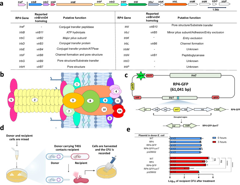
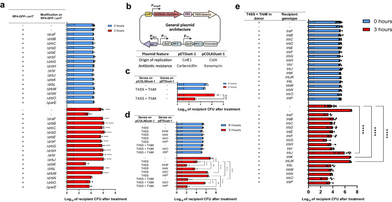
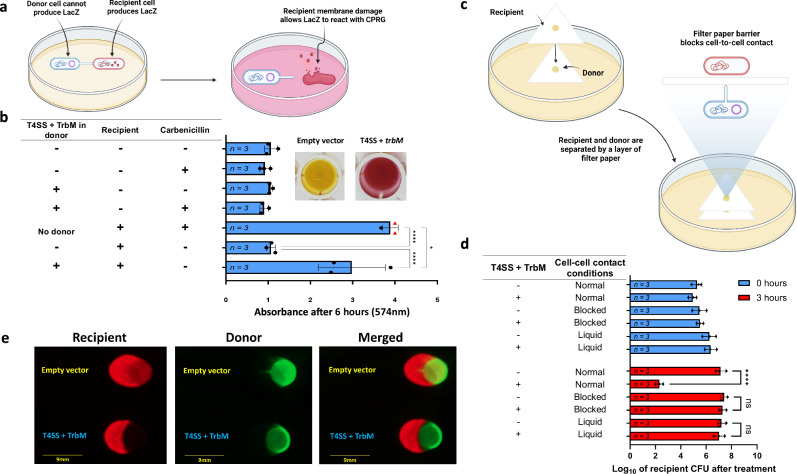
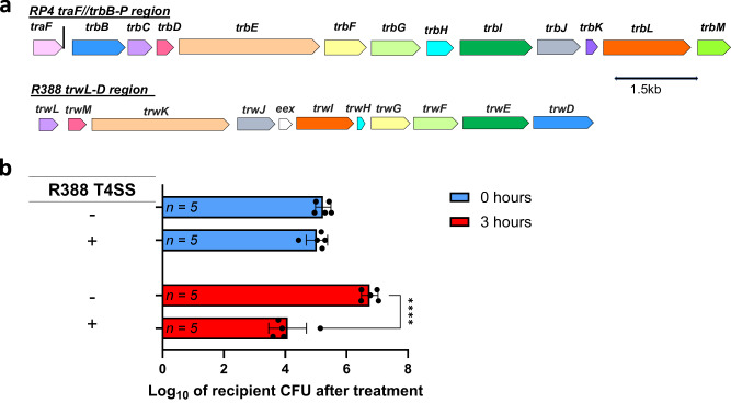
Bacterial cooperation and antagonism mediated by secretion systems are among the ways in which bacteria interact with one another. Here we report the discovery of an antagonistic property of a type IV secretion system (T4SS) sourced from a conjugative plasmid, RP4, using engineering approaches. We scrutinized the genetic determinants and suggested that this antagonistic activity is independent of molecular cargos, while we also elucidated the resistance genes. We further showed that a range of Gram-negative bacteria and a mixed bacterial population can be eliminated by this T4SS-dependent antagonism. Finally, we showed that such an antagonistic property is not limited to T4SS sourced from RP4, rather it can also be observed in a T4SS originated from another conjugative plasmid, namely R388. Our results are the first demonstration of conjugative T4SS-dependent antagonism between Gram-negative bacteria on the genetic level and provide the foundation for future mechanistic studies.


Conflict of interest statement

The authors declare no competing interest.

Figures

Fig. 1
Fig. 1. Antimicrobial potential of plasmid RP4.
a Schematic that shows the tra2 region and traF from tra1 operon of plasmid RP4. Colored blocks signify that they have been reported as part of T4SS. The table beneath summarizes these genes and their respective homologues reported in the virB/virD4 T4SS and the putative function. b Schematic of the virB/D T4SS archetype from Agrobacterium tumefaciens. The figure is modified from S. Ananiadou, D. et al. . Numbers correspond to the respective vir gene (e.g., #5 is virB5). (created with BioRender.com). c Schematic of the RP4-GFP-∆oriT plasmid alongside modifications done to the oriT and genes traXJKL inside the tra1 region using lambda red recombineering. Antibiotic resistance genes are colored red (aacIV = apramycin; tetR = tetracyline; aphA kanamycin). (created with BioRender.com). d An overview of the experimental approach (created with BioRender.com). e Recipient E. coli DA32838 CFU after 0 and 3 hours of exposure to an E. coli NEB® 10-beta donor equipped with RP4, RP4-GFP, RP4-GFP-∆oriT or pUZ8002. Plasmidless E. coli NEB® 10-beta were used as control. The initial donor CFU was approximately set to the order of 107. The raw CFU of the recipient was first log10 transformed, and the data shown is the mean of the log10 transformed data, with the sample size shown inside the corresponding bar. Error bars represent the standard deviation of the log10 transformed data. P values were obtained by doing a two-way RM ANOVA followed by Dunnett’s multiple comparisons test. **** P < 0.0001; ns, not significant.
Fig. 2
Fig. 2. Genetic determinants behind the antagonistic phenotype.
a The results of the knockout studies performed on the RP4-GFP-ΔoriT. The initial donor CFU was approximately set to the order of 107. b General plasmid architecture used for the pCOLADuet-1 and pETDuet-1-based constructs (created with BioRender.com). c The results of the T4SS reconstitution of traF/trbBCDEFGHIJKLM. d The effects of expression of trbN, trbM, trbO, and trbP on the lethality of the reconstituted T4SS. e The resistance mechanism towards the reconstituted T4SS antimicrobial was assayed using E. coli DA32838 recipients carrying either an empty pETDuet-1 vector or a T4SS-associated gene. Shown is the recipient CFU after 0 and 3 hours of treatment with E. coli NEB® 10-beta donors equipped with either the reconstituted T4SS + TrbM or an empty pCOLADuet-1 vector. The initial donor CFU for figures ce was approximately set to the order of 106. For a, ce, the raw CFU of recipients was first log10 transformed and the data shown is the mean of the log10 transformed data, with the sample size shown inside the corresponding bar. Error bars represent the standard deviation of the log10 transformed data. For a, two-way RM ANOVA were performed followed by Dunnett’s multiple comparisons test. P values shown belong to mutants that abolished the phenotype and were compared to the RP4-GFP-∆oriT control. For c, two-way RM ANOVA was performed followed by’Šídák’s multiple comparisons test. For d, two-way RM ANOVA was performed followed by Tukey’s multiple comparisons test. For e, two-way RM ANOVA was performed followed by Dunnett’s multiple comparisons test were performed. ***P < 0.001; ****P < 0.0001; ns, not significant.
Fig. 3
Fig. 3. Killing is dependent on cell-to-cell contact.
a An overview of the in situ colorimetric assay (created with BioRender.com). b The results of the in-situ assay at 574 nm. CPRG was added in the LB agar in each well. Carbenicillin was added into the well if shown as “+” in the table. The addition of carbenicillin in the corresponding wells was used as a control, as both donor and recipient E. coli strains were sensitive to carbenicillin. Red triangles in the graph indicate that the reading overflowed, so the detection limit (4.0) was used instead. Shown to the right of the graph are representative wells 16 hours after incubation in which the recipient E. coli DA32838 was exposed to an E. coli NEB® 10-beta donor containing either an empty pCOLADuet-1 vector or T4SS + TrbM on LB agar supplemented with CPRG (200 µg/mL). c Schematic of a modified mating experiment to demonstrate the antimicrobial’s dependency on cell-to-cell contact. Donor E. coli NEB® 10-beta cells are initially spotted on a piece of cellulose acetate filter paper. Afterward, a second layer is placed on top and recipient cells are spotted directly on top of where the donor was spotted (created with BioRender.com). d CFU of E. coli DA32838 recipient cells after 0 and 3 hours of exposure to donor E. coli NEB® 10-beta cells under modified treatment conditions. Cell-to-cell contact was promoted as usual in the samples denoted as “Normal”, while antimicrobial activity was assayed in conditions that blocked direct cell-to-cell contact (denoted as “Blocked” as shown in the upper panel) and in a liquid environment (denoted as “Liquid”). The donor strains either carried an empty pCOLADuet-1 vector or the reconstituted T4SS+TrbM. e CDK assay in which donor and recipient colonies were partially overlaid. The recipient strain expressed dTomato and the donor strains expressed GFP. Recipients were treated with either a donor carrying the T4SS + TrbM construct expressing GFP (labeled with “T4SS + TrbM” in the figure), or a control donor equipped with a pCOLADuet-1 vector expressing GFP (labeled with “Empty vector” in the figure). The images shown here were taken after a total of six hours of incubation (37 °C for 3 hours, followed by 30 °C for 3 hours). For the recipient strain, images were taken with an imaging system using the 515 – 545 nm excitation filter and the 568 – 617 nm emission filter. Whereas for the donor strains, images were taken using the 455–485 nm excitation filter and the 508–557 nm emission filter. Both donor and recipient strains shared the same genomic background (E. coli NEB® 10-beta). The unedited images, as well as the raw and the fluorescent images corresponding to Fig. 3e, are shown in Supplementary Fig. 11d,  12d, and  13b. For b, the raw absorbance values at 574 nm were used and the data shown is the mean, with the sample size shown inside the corresponding bar. Error bars represent the standard deviation of the absorbance values. RM one-way ANOVA was performed followed by Tukey’s multiple comparisons test. For d, the initial donor CFU was approximately set to the order of 107. The raw CFU of the recipient was first log10 transformed and the data shown is the mean of the log10 transformed data, with the sample size shown inside the corresponding bar. Error bars represent the standard deviation of the log10 transformed data. Two-way RM ANOVA was performed followed by Tukey’s multiple comparisons test. *P < 0.05; ****P < 0.0001; ns, not significant.
Fig. 4
Fig. 4. The Lethal effect is dependent on the donor-to-recipient ratio and is effective against different Gram-negative bacteria and mixed recipient populations.
a The effect of different initial donor-to-recipient ratios on the antimicrobial effect of the T4SS + TrbM donor against the E. coli DA32838 recipient. The raw CFU of recipients after the 3-hour treatment was log10 transformed and the data shown was collected from experiments performed on four different days. Each data point corresponds to a single replicate. b The efficacy of the antimicrobial against E. coli NDM-1 (BAA-2452), Enterobacter cloacae (BAA-2468), Klebsiella pneumoniae subsp. pneumoniae (derived from ATCC BAA-2524), Pseudomonas putida KT2440 (ATCC 47054) and Pseudomonas aeruginosa PAO1-LAC (ATCC 47085). c Lethal effect of the donor carrying the T4SS + TrbM against mixed recipient cell population containing E. coli DA32838 and P. putida KT2440 after 0 hours of treatment. d Lethal effect of the donor carrying the T4SS + TrbM against mixed recipient cell population containing E. coli DA32838 and P. putida KT2440 after 3 hours of treatment. e Schematic depicting the experimental setup for assays performed for figures c and d. (created with BioRender.com). For figures ad, the donor background was E. coli NEB® 10-beta. For figures bd, the initial donor CFU was approximately set to the order of 107. Whenever no recipient colonies were observed, the CFU detection limit of [(1 × 100)/5 µL]*50 µL = 10 was used to enable the subsequent log10 transformation. The raw CFU of recipients was first log10 transformed and the data shown is the mean of the log10 transformed data, with the sample size shown inside the corresponding bar. Error bars represent the standard deviation of the log10 transformed data. For b, two-way RM ANOVA was performed followed by Tukey’s multiple comparisons test. For d, ordinary one-way ANOVA followed by Šídák’s multiple comparisons test. ***P < 0.001; ****P < 0.0001; ns, not significant.
Fig. 5
Fig. 5. T4SS originated from conjugative plasmid R388 and showed antagonizing capabilities.
a Schematic depicting homologous T4SS genes between RP4 and R388 obtained from a global pairwise alignment generated by HHpred using default parameters. The amino acid sequences for the RP4 and R388 genes known to be involved in conjugation were used. Matching colors indicate homology was detected (probability value > 87%, E-value < 1.0e−8, and identity value > 15%), and white color indicates a lack of homology. For more details, refer to the supplemental material. b Antagonizing capabilities of an E. coli NEB® 10-beta donor carrying a reconstituted T4SS-containing operon sourced from R388 against an E. coli DA32838 recipient strain. The initial donor CFU was approximately set to the order of 107. The raw CFU of the recipient was first log10 transformed and the data shown is the mean of the log10 transformed data, with the sample size shown inside the corresponding bar. Error bars represent the standard deviation of the log10 transformed data. Two-way RM ANOVA was performed followed by Šídák’s multiple comparisons test. ****P < 0.0001.

Similar articles

Cited by

References

    1. Gupta, A., Gupta, R. & Singh, R. L. Microbes and Environment. in Principles and Applications of Environmental Biotechnology for a Sustainable Future 43–84 (Springer, Singapore). 10.1007/978-981-10-1866-4_3. (2017).
    1. Finlay BJ, Maberly SC, Cooper JI. Microbial diversity and ecosystem function. Oikos. 1997;80:209. doi: 10.2307/3546587. - DOI
    1. Dodds, W. K. & Whiles, M. R. Microbes and Plants. in Freshwater Ecology 211–249 (Elsevier). 10.1016/B978-0-12-813255-5.00009-0. (2020)
    1. Miransari M. Soil microbes and plant fertilization. Appl. Microbiol. Biotechnol. 2011;92:875–885. doi: 10.1007/s00253-011-3521-y. - DOI - PubMed
    1. O’Hara E, Neves ALA, Song Y, Guan LL. The role of the gut microbiome in cattle production and health: driver or passenger? Annu Rev. Anim. Biosci. 2020;8:199–220. doi: 10.1146/annurev-animal-021419-083952. - DOI - PubMed

Publication types

MeSH terms

Substances

LinkOut - more resources