. 2024 Apr 25;15(1):121.
doi: 10.1186/s13287-024-03717-0.

Synergistic effect of human uterine cervical mesenchymal stem cell secretome and paclitaxel on triple negative breast cancer

Affiliations

Synergistic effect of human uterine cervical mesenchymal stem cell secretome and paclitaxel on triple negative breast cancer

Noemi Eiro et al. Stem Cell Res Ther. .

Abstract

Background: Triple-negative breast cancer (TNBC) is the most lethal subtype of breast cancer and, despite its adverse effects, chemotherapy is the standard systemic treatment option for TNBC. Since, it is of utmost importance to consider the combination of different agents to achieve greater efficacy and curability potential, MSC secretome is a possible innovative alternative.

Methods: In the present study, we proposed to investigate the anti-tumor effect of the combination of a chemical agent (paclitaxel) with a complex biological product, secretome derived from human Uterine Cervical Stem cells (CM-hUCESC) in TNBC.

Results: The combination of paclitaxel and CM-hUCESC decreased cell proliferation and invasiveness of tumor cells and induced apoptosis in vitro (MDA-MB-231 and/or primary tumor cells). The anti-tumor effect was confirmed in a mouse tumor xenograft model showing that the combination of both products has a significant effect in reducing tumor growth. Also, pre-conditioning hUCESC with a sub-lethal dose of paclitaxel enhances the effect of its secretome and in combination with paclitaxel reduced significantly tumor growth and even allows to diminish the dose of paclitaxel in vivo. This effect is in part due to the action of extracellular vesicles (EVs) derived from CM-hUCESC and soluble factors, such as TIMP-1 and - 2.

Conclusions: In conclusion, our data demonstrate the synergistic effect of the combination of CM-hUCESC with paclitaxel on TNBC and opens an opportunity to reduce the dose of the chemotherapeutic agents, which may decrease chemotherapy-related toxicity.

Keywords: Breast cancer; Chemotherapy; Conditioned medium; Mesenchymal stem cells; Secretome; hUCESC.


Conflict of interest statement

The authors declare no conflict of interest. N.E. and F.J.V are co-inventors of a patent (“Human uterine cervical stem cell population and uses thereof”) owned by GiStem Research, of which some authors are shareholders (N.E., J.S-L, L.O.G, M.L.F-S. and F.J.V). The founding sponsors had no role in the design of this manuscript, in the collection, analyses, or interpretation of data, in the writing of the manuscript, or in the decision to publish the results.

Figures

Fig. 1
Fig. 1
(A) Relative proliferative capacity of MDA-MB-231 cells treated for 48 h with CM-hUCESC, paclitaxel (1 µM, 3 µM and 5 µM) and the combination of paclitaxel (1 µM, 3 µM and 5 µM) + CM-hUCESC. (B) Relative proliferative capacity of primary tumor cells from TNBC treated for 48 h with CM-hUCESC, paclitaxel (1 µM, 3 µM and 5 µM) and the combination of paclitaxel (1 µM, 3 µM and 5 µM) + CM-hUCESC. *p < 0.05, ***p < 0.0001
Fig. 2
Fig. 2
(A) Cell cycle analysis of MDA-MB-231 cells treated for 48 h with control (DMEM-F12 without FBS), CM-hUCESC, paclitaxel (1 µM) or the combination of paclitaxel (1 µM) + CM-hUCESC, and then subjected to flow cytometry using propidium iodide (PI). Percentage of cells (mean + standard deviation) in each phase is shown. (B) Apoptosis was determined in MDA-MB-231 cells cultured for 48 h with control (DMEM-F12 without FBS), CM-hUCESC, paclitaxel (1 µM) or the combination of paclitaxel (1 µM) + CM-hUCESC, by flow cytometry using Annexin V/PI. Annexin V+/PI- and Annexin V+/PI + indicated early and late apoptosis, respectively. (C) Invasive capacity of MDA-MB-231 cells treated for 48 h with control (DMEM-F12 without FBS), CM-hUCESC, paclitaxel (1 µM) or the combination of paclitaxel (1 µM) + CM-hUCESC in Matrigel invasion chambers. (D) Invasive capacity of MDA-MB-231 cells treated for 48 h with control (DMEM-F12 without FBS), CM-hUCESC, IgG IP and CM-hUCESC lacking TIMP-1 and − 2 (IP TIMP-1/2) in Matrigel invasion chambers. Data represent the mean ± SD. *p < 0.05; ***p < 0.0001
Fig. 3
Fig. 3
Representative images from mice treated with CM-hUCESC, paclitaxel 10 mg/kg and the combination of paclitaxel 10 mg/kg + CM-hUCESC taken at 7 and 14 days and tumor volume which was determined by measuring luminescence since day 0 until day 21
Fig. 4
Fig. 4
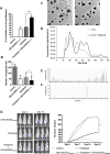
A) Relative proliferative capacity of MDA-MB-231 cells treated with control (DMEM-F12 without FBS), CM-hUCESCchemo (pre-conditioning of hUCESC with paclitaxel previously of CM production), paclitaxel (1 µM) and the combination of paclitaxel (1 µM) + CM-hUCESCchemo. B) Invasive capacity of MDA-MB-231 cells treated with control (DMEM-F12 without FBS), CM-hUCESCchemo, paclitaxel (1 µM) and the combination of paclitaxel (1 µM) + CM-hUCESCchemo. C) Transmission Electron Microscopy micrographs for control EVs (left) and paclitaxel-loaded EVs (right). (D) Size Distribution graphs by NTA for control EVs and drug loaded EVs. (E) Extracted Ion Chromatogram by HPLC-ESI-TOF at paclitaxel mass/charge for control EVs. (F) Extracted Ion Chromatogram by HPLC-ESI-TOF at paclitaxel mass/charge for drug-loaded EVs. (G) Representative images from mice treated with CM- hUCESCchemo, paclitaxel 5 mg/kg and the combination of paclitaxel 5 mg/kg + CM-hUCESC taken at 7 and 14 days and tumor volume which was determined by measuring luminescence since day 0 until day 21

References

    1. Hajdu SI. A note from history: landmarks in history of cancer, part 1. Cancer. 2011;117(5):1097–102. doi: 10.1002/cncr.25553. - DOI - PubMed
    1. DeVita VT, Jr, Chu E. A history of cancer chemotherapy. Cancer Res. 2008;68(21):8643–53. doi: 10.1158/0008-5472.CAN-07-6611. - DOI - PubMed
    1. Wani MC, Taylor HL, Wall ME, Coggon P, McPhail AT. Plant Antitumor agents. VI. The isolation and structure of taxol, a novel antileukemic and antitumor agent from Taxus brevifolia. J Am Chem Soc. 1971;93(9):2325–7. doi: 10.1021/ja00738a045. - DOI - PubMed
    1. Schirrmacher V. From chemotherapy to biological therapy: a review of novel concepts to reduce the side effects of systemic cancer treatment (review) Int J Oncol. 2019;54(2):407–19. doi: 10.3892/ijo.2018.4661. - DOI - PMC - PubMed
    1. Azim HA, Jr, de Azambuja E, Colozza M, Bines J, Piccart MJ. Long-term toxic effects of adjuvant chemotherapy in breast cancer. Ann Oncol. 2011;22(9):1939–47. doi: 10.1093/annonc/mdq683. - DOI - PubMed

Publication types

MeSH terms