. 2024 Apr 26;23(1):120.
doi: 10.1186/s12934-024-02394-1.

Methionine inducing carbohydrate esterase secretion of Trichoderma harzianum enhances the accessibility of substrate glycosidic bonds

Affiliations

Methionine inducing carbohydrate esterase secretion of Trichoderma harzianum enhances the accessibility of substrate glycosidic bonds

Yang Liu et al. Microb Cell Fact. .

Abstract

Background: The conversion of plant biomass into biochemicals is a promising way to alleviate energy shortage, which depends on efficient microbial saccharification and cellular metabolism. Trichoderma spp. have plentiful CAZymes systems that can utilize all-components of lignocellulose. Acetylation of polysaccharides causes nanostructure densification and hydrophobicity enhancement, which is an obstacle for glycoside hydrolases to hydrolyze glycosidic bonds. The improvement of deacetylation ability can effectively release the potential for polysaccharide degradation.

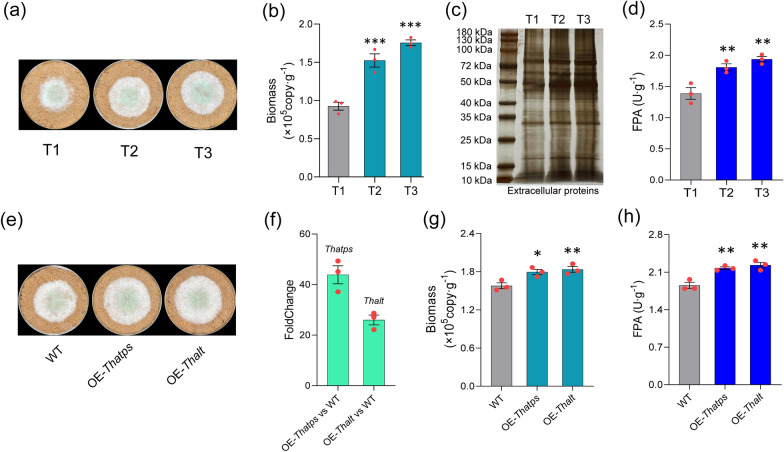
Results: Ammonium sulfate addition facilitated the deacetylation of xylan by inducing the up-regulation of multiple carbohydrate esterases (CE3/CE4/CE15/CE16) of Trichoderma harzianum. Mainly, the pathway of ammonium-sulfate's cellular assimilates inducing up-regulation of the deacetylase gene (Thce3) was revealed. The intracellular metabolite changes were revealed through metabonomic analysis. Whole genome bisulfite sequencing identified a novel differentially methylated region (DMR) that existed in the ThgsfR2 promoter, and the DMR was closely related to lignocellulolytic response. ThGsfR2 was identified as a negative regulatory factor of Thce3, and methylation in ThgsfR2 promoter released the expression of Thce3. The up-regulation of CEs facilitated the substrate deacetylation.

Conclusion: Ammonium sulfate increased the polysaccharide deacetylation capacity by inducing the up-regulation of multiple carbohydrate esterases of T. harzianum, which removed the spatial barrier of the glycosidic bond and improved hydrophilicity, and ultimately increased the accessibility of glycosidic bond to glycoside hydrolases.

Keywords: Carbohydrate esterase; Glycosidic bond accessibility; Metabolomic; Polysaccharide hydrolysis; Xylan deacetylation.


Conflict of interest statement

The authors declare that they have no competing interests.

Figures

Fig. 1
Fig. 1
Ammonium sulfate significantly improved the lignocellulose degradation of T. harzianum. a Growth of WT in rice straw medium with different ammonium sulfur addition (T1: 0%, T2: 0.5%, T3: 1.0%); b The hyphae biomass of T1, T2, and T3. Notably, the biomass of hyphae growing on straw was determined by absolute qPCR. c Extracellular proteins from each treatment (T1, T2, T3) were solubilized by using 10 mL PBS buffer, and 40 μL were subjected to SDS-PAGE and silver-stained. d The FPA of T1, T2, and T3. e The growth of WT, OE-Thatps, and OE-Thalt on MM + straw, and the strains were cultured at 28 ℃ for 4 days. f Relative to WT, and Thatps was 44-folds up-regulated in OE-Thatps, Thalt was 44-folds up-regulated in OE-Thalt. g Biomass of WT, OE-Thatps, and OE-Thalt. h FPA of WT, OE-Thatps, and OE-Thalt. Bars represented mean ± SEM, with n = 3 biological repeats; red dots resembled values from individual experiments. Student’s t-testing was conducted in (b, d, g, h). ***significant difference to WT at two-tailed P = 0.000 (b, T2), 0.000 (b, T3); **significant difference to WT at two-tailed P = 0.005 (d, T2), 0.001 (d, T3), 0.007 (g, OE-Thalt), 0.003 (h, OE-Thatps), 0.001 (h, OE-Thalt); *significant difference to WT at two-tailed P = 0.014 (g, OE-Thatps)
Fig. 2
Fig. 2
Metabolomic revealed the changes in intracellular metabolites induced by ammonium sulfate. a PCA demonstrated intra-group reproducibility and inter-group variability for T1 and T3. b The volcano plot counted and displayed log2FoldChange of the differential metabolites between T3 and T1 and log10P-value, with each point representing a differential metabolite. c Heatmap showed the significantly (P < 0.05) up/down-regulated differential metabolites identified by secondary mass spectrometry. d Match plot showed the significantly up/regulated differential metabolites (VIP-value > 1.5). e KEGG enrichment of differential metabolic pathways. f MS signal intensity of Met and AdoMet in T1 and T3. Notably these values were all significantly greater in T3 than T1. g MS signal intensity of 5mC, 3mA, and 7mG in T1 and T3. h Growth comparison of OE-ThmetH and WT on MM + straw. i Quantification of intracellular Met content and FPA. Intracellular Met content and FPA of OE-ThmetH were significantly higher than that of WT. All results were obtained from hyphae samples grown on T1 and T3, both of which contained 6 biological replicates; red dots resemble values from individual experiments. Student’s t-testing was conducted in (f, g, i), *significant difference to T1 at two-tailed P = 0.022 (g, T3: 5mC), *significant difference to WT at two-tailed P = 0.011 (i, OE-ThmetH: intracellular Met). **significant difference to WT at two-tailed P = 0.004 (i, OE-ThmetH: FPA); ***significant difference to T1 at two-tailed P = 0.000, (f, T3: Met), 0.000 (f, T3: AdoMet), 0.000 (g, T3: 3mA); ns = no statistical difference to T1 at two-tailed P = 0.454 (g, T3: 7mG)
Fig. 3
Fig. 3
WGBS revealed that ammonium-sulfate assimilates induced the upregulation of DNA methylation level. a PCA demonstrated intra-group reproducibility and inter-group variability for T1 and T3. b Profiling analysis divided the region into 20 bins, and the methylation level in each bin reflected the trend of methylation level in the genome region. c Mean methylation levels of CG, CHG, and CHH type sites in exon, intron, UTR, and intergenic gene regions for each sample, with the horizontal coordinate being the genomic element type and the vertical coordinate indicating the methylation level, and the colors (red, green, and blue) indicated the mean methylation levels of the three types of sites. d Violin plots showed the numerical distribution of CpG, CHG, and CHH methylation levels for T1 and T3, with the vertical coordinate indicating the methylation level, and the width of each violin represented the number of points that were at that methylation level. e The Venn showed the distribution of novel DMRs of T3 and the number in CpG, CHG, and CHH types. f The location of the novel DMRs of T3 in chromosome fragment and its gene function. g FoldChange of the genes that were associated with the novel DMRs in T3 relative to T1. h Growth comparison for the mutants and WT on MM + straw. i FPA of the mutants and WT grown on MM + straw at 28 °C for 4 days. Bars represent mean ± SEM, with n = 3/4 biological repeats; red dots resemble values from individual experiments. Student’s t-testing was conducted in (i). *significant difference to WT at two-tailed P = 0.017 (i, KO-Thgh92); **significant difference to WT at two-tailed P = 0.002 (i, KO-ThgsfR2); ***significant difference to WT at two-tailed P = 0.000 (i, KO-Thabe); ns = no statistical difference to WT at two-tailed
Fig. 4
Fig. 4
ChIP-seq identified the downstream genes regulated by the zinc finger transcription factor ThGsfR2. a PCA showed the difference in reads distribution between IP and Input. Notably, after data dimensionality reduction, PC1 has 100% interpretation on eigenvalue and cumulative variability. b The peak plot showed the distribution of peak reads of IP and Input. c Venn showed the distribution of peak reads over the genome functional elements. d The IGV visualization showed the location in genome of the 10 peaks with the highest enrichment of ThGsfR2, and the red column was IP, the blue column was Input, and column height indicated the signal intensity, data range was shown inside the “[]” on the left, the proximity genes for peaks were shown on the bottom. e Expression level FoldChange of the genes corresponding to peaks in OE-ThgsfR2 and KO-ThgsfR2 were compared to WT, note that we excluded the peaks located in non-gene functional regions. f Growth of OE-HP5723, OE-HP5320, OE-Thce3 and WT on MM + straw. g Biomass of OE-HP5723, OE-HP5320, OE-Thce3 and WT grown at 28 °C for 4 days. h FPA of OE-HP5723, OE-HP5320, OE-Thce3 and WT. i Motif analysis of all peaks using Homer yielded 10 ThGsfR2 binding motifs, ranked according to scoring, the last column of table showed the transcription factors predicted for motifs using JASPAR. j The conserved sequence “TCTCTCTCTC” of motif1 was presented in Thce3 promoter with 50-fold enrichment. k DNA–Protein interactions between ThGsfR2 and motif1, Thce3 promoter region R1 (−1000 to −1 bp), and R2 (−2000 to −1001 bp) were verified by Y1H assay. Bait-reporters (pAbAi::motif1, pAbAi::R1, and pAbAi::R2) could not grow in SD medium without uracil (SD-Ura) containing Aureobasidin A (AbA, 600 ng mL−1); pAbAi::motif1 + pGADT7::ThGsfR2 co-transformant and pAbAi::R2 + pGADT7::ThGsfR2 co-transformant could grow on SD-Ura-Leu containing AbA (600 ng mL−1), pAbAi::R1 + pGADT7::ThGsfR2 co-transformant could not grow on that medium. Bars represent mean ± SEM, with n = 3 biological repeats; red dots resemble values from individual experiments. Student’s t-testing was conducted in (g, h), **significant difference to WT at two-tailed P = 0.002 (g, OE-Thce3), 0.004 (h, OE-Thce3); ns = no statistical difference to WT at two-tailed P = 0.789 (g, OE-ThHP5723), 0.265 (g, OE-ThHP5320), 0.807 (h, ThHP5723), 0.077 (h, OE-ThHP5320)
Fig. 5
Fig. 5
Ammonium-sulfate induced the up-regulation of multiple CEs, which in turn improved glycosidic bond accessibility through xylan deacetylation. a HPLC results of acetate after 10-day liquid fermentation of WT and KO-ThgsfR2. The red line was WT treatments, and blue line was KO-ThgsfR2 treatments, note that the acetate mainly comes from acetyl-groups. b Peak area and concentration of acetate for WT and KO-ThgsfR2 treatments. Concentration was obtained by peak areas and standard regression equation. c Expression levels quantification of CEs genes with AS gradient (T1, T2, T3). qPCR was performed on the 7 identified CEs genes and multiple CE genes were up-regulated with the AS gradient. d HSQC spectrum of extracted xylan from WT-treated straw under T1 condition for 15 days. The signals from Xyl2Ac and Xyl3Ac could be detected. e HSQC spectrum of extracted xylan from WT-treated straw under T3 condition for 15 days, The signal peak area of Xyl2Ac was reduced and Xyl3Ac has no detectable signal. f Quantitative results of Xyl2Ac, Xyl3Ac, and XylR peak areas in the T1 and T3 NMR spectrum, which can indicate the change in acetyl content with the AS addition. The peak areas of Xyl2Ac, Xyl3Ac, and XylR were all decreased in T3 relative to T1. g T2 relaxation spectrum of T1 and T3. Low-field NMR determined the spin–spin relaxation time (T2) of straw after hyphal treated on T1 and T3 conditions for 15 days. h WSI of hyphae-treated (15 days) straw under T1 and T3 conditions, noting that the Peleg modeled WSI belonged to type II isotherm. i Degradability comparison of hyphae-treated (15 days) straw under T1 and T3 conditions. GHs (N7, A50, and XS) were used to hydrolyze hyphae-treated straw, and the affinity efficiency of GHs for polysaccharide glycosidic bonds was compared by determining the reducing sugars (40 °C and 50 °C reaction for 20 min). Note that this could compare the effects of different acetylation levels on GHs binding glycosidic bonds. Bars represent mean ± SEM, with n = 3 or 4 biological repeats; red dots resemble values from individual experiments. Student’s t-testing was conducted in (b, c, i), *significant difference to T1 at two-tailed P = 0.002 (i, T3: A50-50 ℃). ***significant difference to WT at two-tailed P = 0.000 (b, KO-ThgsfR2: peak area), 0.000 (b, KO-ThgsfR2: acetate). ***significant difference to T1 at two-tailed P = 0.000 (i, T3: XS-50 ℃). ns = no statistical difference to T1 at two-tailed P = 0.335 (i, T3: N7-40 ℃), 0.495 (i, T3: A50-40 ℃), 0.113 (i, T3: XS-40 ℃), 0.267 (i, T3: N7-50 ℃)
Fig. 6
Fig. 6
Schematic diagram of ammonium-sulfate induced up-regulation of multiple CEs and thereby increased glycosidic bond accessibility. Normally, the transcription factor ThGsfR2 inhibited ThCE3 expression by competitive binding of the functional region of Thce3 promoter. After the ammonium-sulfate addition, AS would be transported into the cell by SULTR and AMT, and ammonium ions were converted to NH3 by deprotonation, while SO42− was reduced into S2− by ATPS. Pyruvate produced by glycolysis bound NH3 to produce alanine (Ala). Ala would be converted to O-acetyl homoserine (OAHS) by multi-enzyme catalysis. Subsequently, OAHS would be converted to homocysteine (HCY) catalyzed by the O-acetyl homoserine sulfhydrylase, and further catalyzed by 5-methyltetrahydrofolate-homocysteine methyltransferase (metH) to produce the terminal assimilates methionine (Met). Met was converted to AdoMet by ATP activation and induced methylation of the ThgsfR2 promoter, leading to transcriptional repression of ThgsfR2. This allowed Thce3 to be released from the repression of ThGsfR2, exhibiting a significant up-regulation of transcriptional level. In addition, multiple CEs were induced to be up-regulated by unknown inducers generated by AS assimilation. Up-regulation of CEs enhanced polysaccharide deacetylation, which in turn increased hydrophilicity and removed the spatial barrier of glycosidic bonds. 1. Acetylation of xylose residues prevented glycoside hydrolases from glycosidic bonds. 2. CEs catalyzed deacetylation of the xylose acetyl group. 3. Deacetylation removed the spatial barrier of glycosidic bonds and increased the accessibility of the glycosidic bond to glycoside hydrolases

Similar articles

Cited by

References

    1. Rubin EM. Genomics of cellulosic biofuels. Nature. 2008;454:841–845. doi: 10.1038/nature07190. - DOI - PubMed
    1. Xia YW, Wang JF, Guo CX, Xu HH, Wang W, Yang MZ, Shen QR, Zhang RF, Miao YZ. Exploring the multi-level regulation of lignocellulases in the filamentous fungus Trichoderma guizhouense NJAU4742 from an omics perspective. Microb Cell Fact. 2022;21:144. doi: 10.1186/s12934-022-01869-3. - DOI - PMC - PubMed
    1. Garron ML, Henrissat B. The continuing expansion of CAZymes and their families. Curr Opin Chem Biol. 2019;53:82–87. doi: 10.1016/j.cbpa.2019.08.004. - DOI - PubMed
    1. Woo SL, Hermosa R, Lorito M, Monte E. Trichoderma: a multipurpose, plant-beneficial microorganism for eco-sustainable agriculture. Nat Rev Microbiol. 2023;21:312–326. doi: 10.1038/s41579-022-00819-5. - DOI - PubMed
    1. Sun X, Zhang X, Huang H, Wang Y, Tu T, Bai Y, Wang Y, Zhang J, Luo H, Yao B. Engineering the cbh1 promoter of Trichoderma reesei for enhanced protein production by replacing the binding sites of a transcription repressor ACE1 to those of the activators. J Agric Food Chem. 2020;68:1337–1346. doi: 10.1021/acs.jafc.9b05452. - DOI - PubMed

Supplementary concepts

LinkOut - more resources