

Fetal Gestational Age Estimation Using Ultrasonic Transverse Cerebellar Diameter in a Sub-Saharan African Population
- PMID: 38665343
- PMCID: PMC11040476
- DOI: 10.4103/jmu.jmu_116_22
Fetal Gestational Age Estimation Using Ultrasonic Transverse Cerebellar Diameter in a Sub-Saharan African Population
Abstract
Background: To explore the relationship between fetal Transverse Cerebellar Diameter (TCD) and menstrual gestational age (GA) and to generate normative references (nomogram) of the fetal TCD in some pregnant women in Southwest Nigeria.
Methods: Four hundred pregnant women with a singleton fetus between 14 and 38 weeks GA were enrolled. The TCD and other biometric parameters (biparietal diameter, head circumference, abdominal circumference, and femur length) as well as the cerebellar appearance were analyzed and correlated with the GA.
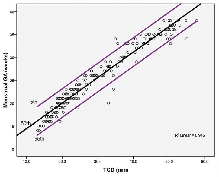
Results: The mean TCD increased from 13.3 ± 0.3 mm at 14 weeks to 52.3 ± 3.3 mm at 38 weeks of pregnancy. A strong positive correlation was observed between TCD and GA, which was best represented by a linear regression equation: Predicted GA = 0.557 × TCD + 8.840. The regression analysis indicated a statistically significant strong positive relationship between TCD and GA (r = 0.972 and P < 0.001). The cerebellar appearance based on shape and echogenicity was graded into Grade I: 230 fetuses (57.5%); Grade II: 74 fetuses (18.5%) and Grade III: 96 fetuses (24.0%). Median GA and TCD were 21 weeks and 21.2 mm for Grade I; 29 weeks and 35.5 mm for Grade II; and 35 weeks and 48.1 mm for Grade III, respectively.
Conclusion: The TCD increased in a linear fashion with advancing GA in the evaluated fetuses. The TCD is, therefore, a good marker for GA estimation. There is a gradual ultrasonographic change in fetal cerebellar appearance with advancing gestation.
Keywords: Fetal biometry; gestational age; pregnancy; transverse cerebellar diameter; ultrasonography.
Copyright: © 2023 Journal of Medical Ultrasound.
Conflict of interest statement
There are no conflicts of interest.
Figures
Similar articles
-
Relationship between ultrasound estimated fetal gestational age and cerebellar appearance in healthy pregnant Nigerian women.Ann Afr Med. 2015 Jul-Sep;14(3):132-6. doi: 10.4103/1596-3519.149908. Ann Afr Med. 2015. PMID: 26021393
-
Accuracy of Transverse Cerebellar Diameter in Estimating Gestational Age in the Second and Third Trimester: A Prospective Study in Saudi Arabia.Diagnostics (Basel). 2025 Apr 29;15(9):1130. doi: 10.3390/diagnostics15091130. Diagnostics (Basel). 2025. PMID: 40361948 Free PMC article.
-
Estimation of gestational age using trans-cerebellar diameter: a sonographic study of a cohort of healthy pregnant women of Igbo ethnic origin in a suburb of Lagos, southwest Nigeria.J Ultrasound. 2021 Mar;24(1):41-47. doi: 10.1007/s40477-020-00448-9. Epub 2020 Mar 19. J Ultrasound. 2021. PMID: 32193743 Free PMC article.
-
Transverse cerebellar diameter: a reliable predictor of gestational age.Afr Health Sci. 2020 Dec;20(4):1927-1932. doi: 10.4314/ahs.v20i4.51. Afr Health Sci. 2020. PMID: 34394259 Free PMC article.
-
Determination of the Accuracy of Transcerebellar Diameter in Estimating Gestational Age in the Second and Third Trimesters of Pregnancy.Cureus. 2024 Jun 27;16(6):e63292. doi: 10.7759/cureus.63292. eCollection 2024 Jun. Cureus. 2024. PMID: 39070496 Free PMC article.
References
-
- Skinner C, Mount CA. Treasure Island, Florida, USA: StatPearls Publishing; 2022. [Last accessed on 2022 Oct 26]. Sonography Assessment of Gestational Age. Available from:https://www.ncbi.nlm.nih.gov/books/NBK570610/ - PubMed
-
- Oboro VO, Bello TO, Oyeniran AO. First trimester sonographic dating formula for the Nigerian obstetric population. West Afr J Radiol. 2012;19:1–4.
-
- Chawanpaiboon S, Kanokpongsakdi S, Vantanasiri C. Predicting fetal intrauterine growth retardation by using reference centile charts. Siriraj Med J. 2017;60:316–9.
LinkOut - more resources
Full Text Sources