. 2023;6(1):288.
doi: 10.1038/s42005-023-01412-9. Epub 2023 Oct 11.

Femtosecond multimodal imaging with a laser-driven X-ray source

Affiliations

Femtosecond multimodal imaging with a laser-driven X-ray source

Adam Doherty et al. Commun Phys. 2023.

Abstract

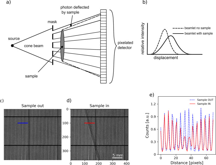
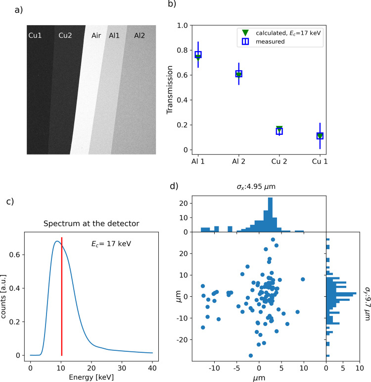
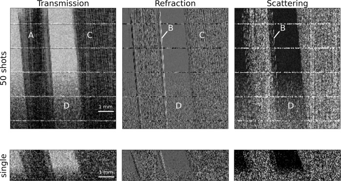
Laser-plasma accelerators are compact linear accelerators based on the interaction of high-power lasers with plasma to form accelerating structures up to 1000 times smaller than standard radiofrequency cavities, and they come with an embedded X-ray source, namely betatron source, with unique properties: small source size and femtosecond pulse duration. A still unexplored possibility to exploit the betatron source comes from combining it with imaging methods able to encode multiple information like transmission and phase into a single-shot acquisition approach. In this work, we combine edge illumination-beam tracking (EI-BT) with a betatron X-ray source and present the demonstration of multimodal imaging (transmission, refraction, and scattering) with a compact light source down to the femtosecond timescale. The advantage of EI-BT is that it allows multimodal X-ray imaging technique, granting access to transmission, refraction and scattering signals from standard low-coherence laboratory X-ray sources in a single shot.

Keywords: Applied physics; Plasma physics.


Conflict of interest statement

Competing interestsThe authors declare no competing interests.

Figures

Fig. 1
Fig. 1. Edge illumination principle.
a An absorbing mask splits the X-ray beam into beamlets which are imaged with a pixelated detector. b the presence of the sample between the mask and the detector changes the beamlets’ shape. c, d Cropped detail (2.7 × 3.1 mm) of transmission images of the mask used in this experiment without (c) and with (d) sample (detail of a leaf margin). e line profiles from c and d of the beamlets with (solid red line) and without sample (dashed blue line). See Supplementary Note 1 Fig. S1 for the full-size raw images.
Fig. 2
Fig. 2. Schematic of the experiment at ALLS.
The laser is focused onto a gas jet inside the interaction chamber. The accelerated electrons are swept away by magnets after the gas jet. The laser is stopped by an appropriate beam-block, while the X-ray propagates outside the chamber. The mask is placed after the chamber exit window, the pixelated X-ray detector just downstream. The sample is mounted between the mask and the detector.
Fig. 3
Fig. 3. Source characterisation.
a Transmission image of the X-ray beam through a set of metal filters as recorded by the detector: transmission through 136 and 68 μm of copper (Cu1 and Cu2) and through 50 and 100 μm of aluminium (Al1 and Al2) are shown. b The transmission is compared to that of a synchrotron-like spectrum with 17 keV critical energy. The error bars indicate the standard deviation. c Calculated X-ray spectrum for a critical energy of 17 keV as seen at the detector: the laser beam-stop, exit window and air before the detector absorbs the low energy part of the spectrum. The average energy of 10.3 keV is marked with the vertical red line. d Source position fluctuation obtained from 100 consecutive shots. See Supplementary Note 3 for further information.
Fig. 4
Fig. 4. Retrieved multimodal images.
Transmission, refraction, and scattering were retrieved for a leaf (ac) and two electrochemical samples (pristine, df, and after electrochemical deposition, gi). The horizontal lines present in all images are artefacts due to horizontal bridges that interrupt the vertical grid every 1.5 mm to reinforce the mechanical strength of the free-standing mask. Avoiding a substrate minimises photon loss by absorption.
Fig. 5
Fig. 5. From multi to single-shot imaging.
A sample made of carbon fibres (A) wrapped in Kapton tape (B), bamboo wood (C) on the top of a foam layer (D) was imaged, acquired without dithering, and retrieved by decreasing the number of cumulated images from 50 to single shot. The field of view is 7.3 (H) x7.8 (V) mm. The vertical binning is 2 and 8, respectively, for the 50 and single-shot images.

References

    1. Wilkins SW, Gureyev TE, Gao D, Pogany A, Stevenson AW. Phase-contrast imaging using polychromatic hard X-rays. Nature. 1996;384:335–338. doi: 10.1038/384335a0. - DOI
    1. Pfeiffer F, Weitkamp T, Bunk O, David C. Phase retrieval and differential phase-contrast imaging with low-brilliance X-ray sources. Nat. Phys. 2006;2:258–261. doi: 10.1038/nphys265. - DOI
    1. Morgan, K. S., Paganin, D. M. & Siu, K. K. W. X-ray phase imaging with a paper analyzer. Appl. Phys. Lett.100, 124102 (2012).
    1. Zanette I, et al. Speckle-based X-ray phase-contrast and dark-field imaging with a laboratory source. Phys. Rev. Lett. 2014;112:253903. doi: 10.1103/PhysRevLett.112.253903. - DOI - PubMed
    1. Olivo, A. & Speller, R. A coded-aperture technique allowing X-ray phase contrast imaging with conventional sources. Appl. Phys. Lett.91, 074106 (2007).