. 2024 Apr 5;4(4):1315-1322.
doi: 10.1021/jacsau.4c00213. eCollection 2024 Apr 22.

Gauging Iron-Sulfur Cubane Reactivity from Covalency: Trends with Oxidation State

Affiliations

Gauging Iron-Sulfur Cubane Reactivity from Covalency: Trends with Oxidation State

Liam Grunwald et al. JACS Au. .

Abstract

We investigated room-temperature metal and ligand K-edge X-ray absorption (XAS) spectra of a complete redox series of cubane-type iron-sulfur clusters. The Fe K-edge position provides a qualitative but convenient alternative to the traditional spectroscopic descriptors used to identify oxidation states in these systems, which we demonstrate by providing a calibration curve based on two analytic methods. Furthermore, high energy resolution fluorescence detected XAS (HERFD-XAS) at the S K-edge was used to measure Fe-S bond covalencies and record their variation with the average valence of the Fe atoms. While the Fe-S(thiolate) covalency evolves linearly, gaining 11 ± 0.4% per bond and hole, the Fe-S(μ3) covalency evolves asystematically, reflecting changes in the magnetic exchange mechanism. A strong discontinuity manifested for superoxidation to the all-ferric state, distinguishing its electronic structure and its potential (bio)chemical role from those of its redox congeners. We highlight the functional implications of these trends for the reactivity of iron-sulfur cubanes.


Conflict of interest statement

The authors declare no competing financial interest.

Figures

Scheme 1
Scheme 1. Molecular Structure of the [Fe4S4(CysS)4]n– Cofactor Model, Kn[Fe4S4(DmpS)4] (Where n = 0-4)
R refers to 2,6-dimesitylphenyl (Dmp).
Figure 1
Figure 1
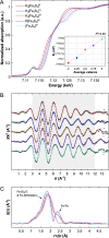
(A) Normalized Fe K-edge XAS spectra of the Kn[Fe4S4(DmpS)4] (n = 0–4) powdered samples measured at room temperature. The inset shows the correlation between the average Fe oxidation state (average valence) and the energy of the inflection point (E0); a linear fit of these data is presented as a dotted gray line, indicating an increase in E0 of 0.57 ± 0.03 eV per 1-electron oxidation. (B) Unfiltered EXAFS spectra of the Kn[Fe4S4(DmpS)4] (n = 0–4; gray dots) in k-space, and the corresponding fits (colored lines). The range over which the fit was carried out is shaded in gray. (C) FT-EXAFS spectra (k2-weighted) of Kn[Fe4S4(DmpS)4] (n = 0–4).
Figure 2
Figure 2
Normalized S K-edge HERFD-XAS spectra of Kn[Fe4S4(DmpS)4] (n = 0–4) recorded on powdered samples at room temperature in vacuo.
Figure 3
Figure 3
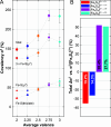
(A) Variation of covalency, α2, and its individual contributions with the average valence of the Fe-atom. While the total α2 is shown as colored diamonds ([Fe4S4]4+: cyan, [Fe4S4]3+: magenta, [Fe4S4]2+: yellow, [Fe4S4]1+: blue, [Fe4S4]0: red), contributions arising from sulfide- and thiolate-bonding are presented as dot and triangle shaped markers, respectively. Dotted gray lines are intended to guide the eye. Approximate error bars are shown in gray color; for details on the error estimation, refer to the Supporting Information. (B) Net change in the covalency upon redox transformations of the [Fe4S4]2+ cubane per Fe-atom: superreduction to [Fe4S4]0 (red), ferredoxin (Fd) type reduction to [Fe4S4]1+ (blue), high-potential iron–sulfur protein (HiPIP) type oxidation to [Fe4S4]3+ (magenta) and superoxidation to [Fe4S4]4+ (cyan). The covalency change relative to the resting oxidation state ([Fe4S4]2+ in percent (%) is indicated on the individual bars: white font for covalency decrease, black font for covalency increase.
Figure 4
Figure 4
H-bonds (dashed green; values in Å; given as distance from the H-bond donor to the acceptor atom) detected in the second sphere of the FeS cofactor in the crystal structure of thermochromatium tepidum HiPIP (PDB code 5WQR(49)) in the reduced ([Fe4S4]2+) oxidation state. H-bonds were calculated according to the criteria described by Mills and Dean, relaxing the angle and distance tolerances by 20° and 0.4 Å, respectively. This figure was generated using the ChimeraX program suite.,

References

    1. Beinert H.; Holm R. H.; Münck E. Iron-Sulfur Clusters: Nature’s Modular, Multipurpose Structures. Science 1997, 277 (5326), 653–659. 10.1126/science.277.5326.653. - DOI - PubMed
    1. Raanan H.; Pike D. H.; Moore E. K.; Falkowski P. G.; Nanda V. Modular origins of biological electron transfer chains. Proc. Natl. Acad. Sci. U. S. A. 2018, 115 (6), 1280–1285. 10.1073/pnas.1714225115. - DOI - PMC - PubMed
    1. Sharma S.; Sivalingam K.; Neese F.; Chan G. K.-L. Low-energy spectrum of iron–sulfur clusters directly from many-particle quantum mechanics. Nat. Chem. 2014, 6 (10), 927–933. 10.1038/nchem.2041. - DOI - PubMed
    1. Teo R. D.; Rousseau B. J. G.; Smithwick E. R.; Di Felice R.; Beratan D. N.; Migliore A. Charge Transfer between [4Fe4S] Proteins and DNA Is Unidirectional: Implications for Biomolecular Signaling. Chem. 2019, 5 (1), 122–137. 10.1016/j.chempr.2018.09.026. - DOI - PMC - PubMed
    1. Kubas A. Characterization of charge transfer excited states in [2Fe–2S] iron–sulfur clusters using conventional configuration interaction techniques. Theor. Chem. Acc. 2020, 139 (7), 120.10.1007/s00214-020-02635-7. - DOI

LinkOut - more resources