. 2024 May 13;45(18):1662-1680.
doi: 10.1093/eurheartj/ehae205.

Myocardial reperfusion injury exacerbation due to ALDH2 deficiency is mediated by neutrophil extracellular traps and prevented by leukotriene C4 inhibition

Affiliations

Myocardial reperfusion injury exacerbation due to ALDH2 deficiency is mediated by neutrophil extracellular traps and prevented by leukotriene C4 inhibition

Kun Yang et al. Eur Heart J. .

Abstract

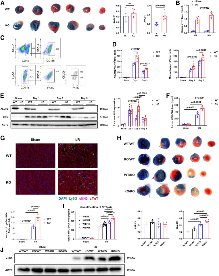
Background and aims: The Glu504Lys polymorphism in the aldehyde dehydrogenase 2 (ALDH2) gene is closely associated with myocardial ischaemia/reperfusion injury (I/RI). The effects of ALDH2 on neutrophil extracellular trap (NET) formation (i.e. NETosis) during I/RI remain unknown. This study aimed to investigate the role of ALDH2 in NETosis in the pathogenesis of myocardial I/RI.

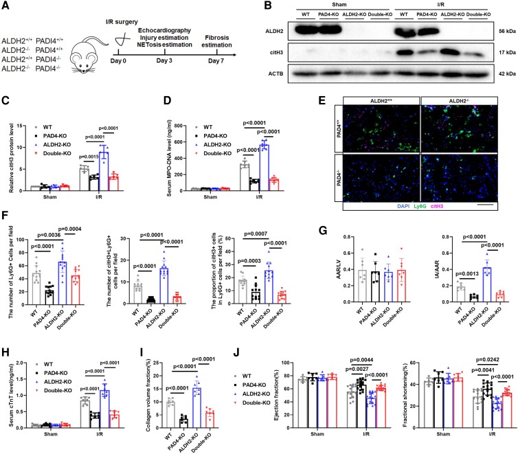
Methods: The mouse model of myocardial I/RI was constructed on wild-type, ALDH2 knockout, peptidylarginine deiminase 4 (Pad4) knockout, and ALDH2/PAD4 double knockout mice. Overall, 308 ST-elevation myocardial infarction patients after primary percutaneous coronary intervention were enrolled in the study.

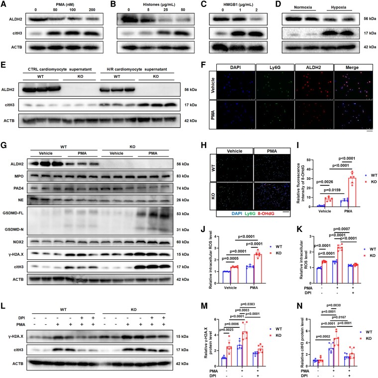
Results: Enhanced NETosis was observed in human neutrophils carrying the ALDH2 genetic mutation and ischaemic myocardium of ALDH2 knockout mice compared with controls. PAD4 knockout or treatment with NETosis-targeting drugs (GSK484, DNase1) substantially attenuated the extent of myocardial damage, particularly in ALDH2 knockout. Mechanistically, ALDH2 deficiency increased damage-associated molecular pattern release and susceptibility to NET-induced damage during myocardial I/RI. ALDH2 deficiency induced NOX2-dependent NETosis via upregulating the endoplasmic reticulum stress/microsomal glutathione S-transferase 2/leukotriene C4 (LTC4) pathway. The Food and Drug Administration-approved LTC4 receptor antagonist pranlukast ameliorated I/RI by inhibiting NETosis in both wild-type and ALDH2 knockout mice. Serum myeloperoxidase-DNA complex and LTC4 levels exhibited the predictive effect on adverse left ventricular remodelling at 6 months after primary percutaneous coronary intervention in ST-elevation myocardial infarction patients.

Conclusions: ALDH2 deficiency exacerbates myocardial I/RI by promoting NETosis via the endoplasmic reticulum stress/microsomal glutathione S-transferase 2/LTC4/NOX2 pathway. This study hints at the role of NETosis in the pathogenesis of myocardial I/RI, and pranlukast might be a potential therapeutic option for attenuating I/RI, particularly in individuals with the ALDH2 mutation.

Keywords: Aldehyde dehydrogenase 2; Leukotriene C4 receptor antagonist; Microsomal glutathione S-transferase 2; Myocardial ischaemia/reperfusion injury; NETosis.


Figures

Structured Graphical Abstract
The ALDH2 deletion increases myocardial DAMP release during myocardial I/RI. Undergoing the stimulation of DAMPs, ALDH2 deletion induces NOX2-dependent NET formation by upregulating the ER stress/Mgst2/LTC4 pathway, resulting in aggravated myocardial I/RI. ALDH2, aldehyde dehydrogenase 2; DAMP, damage-associated molecular pattern; I/RI, ischaemia/reperfusion injury; NOX2, NADPH oxidase 2; NET, neutrophil extracellular trap; ER, endoplasmic reticulum; Mgst2, microsomal glutathione S-transferase 2; LTC4, leukotriene C4; ROS, reactive oxygen species.
Figure 1
Figure 1
The human ALDH2 rs671 (Glu504Lys) polymorphism predicts NETosis vulnerability. (A) Human peripheral neutrophils were isolated from healthy volunteers. ALDH2 rs671 genotypes (GG, GA, and AA, n = 5 of each genotype) were treated by PMA. (B) The MPO–DNA complex levels were assessed in the human neutrophils treated by PMA or vehicle for 2 h (n = 5 of each group, two-way ANOVA using Tukey’s multiple comparisons test). Immunofluorescence staining of the neutrophils treated by PMA for 2 h with staining for CD66b and nuclei. (C) Quantification (n = 5 of each group, one-way ANOVA using Tukey’s multiple comparisons test) and (D) representative immunofluorescence images of the human neutrophils treated by PMA for 2 h with staining for CD66b (green) and nuclei (blue). Scale bar, 20 μm. (E) Quantification (n = 5 of each group, one-way ANOVA using Tukey’s test) and representative bands of citH3 protein measured by Western blot assay. The data were presented as the means ± SD of five independent biological experiments. ALDH2, aldehyde dehydrogenase 2; NETosis, neutrophil extracellular trap (NET) formation; PMA, phorbol 12-myristate 13-acetate; citH3, citrullinated histone H3
Figure 2
Figure 2
Increased NETosis during myocardial I/RI in ALDH2-KO mice. (AG) Myocardial I/R surgery (45 min of ischaemia followed by reperfusion) was performed in wild-type mice and ALDH2-KO (KO) mice. (A) Representative images and quantification of cross-sections stained with TTC and Evans blue to determine the extent of I/RI at 3 days post-I/R (n = 5 per group, unpaired two-tailed Student's t-test). (B) The serum cTnT levels were measured by ELISA assay (n = 6 of each group, two-way ANOVA using Tukey’s multiple comparisons test). (C) Gating strategy for flow cytometric analysis of leucocyte population isolated from the hearts of WT and KO subjected to sham operation or I/R surgery at 1 and 3 days post-I/R. (D) Flow cytometry–based quantification of neutrophils and macrophages (n = 6 of each group, two-way ANOVA using Tukey’s multiple comparisons test). (E) Representative bands and quantification of citH3 protein from the hearts of WT and KO subjected to sham operation or I/R surgery at 1, 3, and 5 days post-I/R. (sham n = 3 of each group; I/R Days 1, 3, and 5, n = 6 of each group, two-way ANOVA using Sidak’s multiple comparisons test). (F) The serum MPO–DNA complex levels were assessed in WT and KO mice 3 days post-I/R (n = 6 of each group, two-way ANOVA using Tukey’s multiple comparisons test). (G) Representative immunofluorescence images and quantification of WT and KO of hearts at 3 days post-I/R with staining for Ly6G, cTnT, citH3, and nuclei (n = 4 of each group, two-way ANOVA using Tukey’s multiple comparisons test). Scale bar, 50 μm. (HJ) Myocardial I/R surgery (45 min of ischaemia followed by reperfusion) was performed in WT/WT, KO/WT, WT/KO, and KO/KO mice (sham group: per n = 4; I/R group: per n = 10). (H) Representative images and quantification of cross-sections stained with TTC and Evans blue to determine the extent of I/RI at 3 days (n = 4 of each group, one-way ANOVA using Tukey’s multiple comparisons test). (I) The serum MPO–DNA complex levels were measured by ELISA assay (sham n = 4 of each group; I/R n = 7 of each group, two-way ANOVA using Sidak’s multiple comparisons test). (J) Quantification of citH3 protein measured by Western blot assay. The data were presented as the means ± SD of at least four independent biological experiments. ALDH2, aldehyde dehydrogenase 2; NETosis, neutrophil extracellular trap (NET) formation; I/RI, ischaemia/reperfusion injury; TTC, triphenyl tetrazolium chloride; cTnT, cardiac troponin T; WT, wild type; KO, knockout; citH3, citrullinated histone H3
Figure 3
Figure 3
NETosis plays a core role in the ALDH2 deficiency-mediated exacerbation of myocardial I/RI. Myocardial I/R surgery (45 min of ischaemia followed by reperfusion) was performed in wild-type (WT) mice and global PAD4 knockout mice (PAD4-KO) and global ALDH2/PAD4 double knockout mice (double-KO). (A) Experimental strategy of myocardial I/R model. (B) Representative bands and (C) quantification of citH3 protein from WT, PAD4-KO, ALDH2-KO, and double-KO of sham hearts and hearts 3 days post-I/R (n = 5 of each group, two-way ANOVA using Tukey’s multiple comparisons test). (D) The serum MPO–DNA complex levels were assessed in WT, PAD4-KO, ALDH2-KO, and double-KO mice at 3 days post-I/R (n = 7 of each group, two-way ANOVA using Tukey’s multiple comparisons test). (E) Representative immunofluorescence images and (F) quantification of Ly6G+ cell and citH3+Ly6G+ cell in WT, PAD4-KO, ALDH2-KO, and double-KO of hearts at 3 days post-I/R with staining for Ly6G, cTnT, citH3, and nuclei (n = 12 of each group, one-way ANOVA using Tukey’s multiple comparisons test). Scale bar, 100 μm. (G) Quantification of cross-sections stained with TTC and Evans blue (n = 7 of each group, one-way ANOVA using Tukey’s multiple comparisons test). (H) The serum cTnT levels were measured by ELISA assay (n = 7 of each group, two-way ANOVA using Tukey’s multiple comparisons test). (I) The degree of fibrosis was assessed by Masson's trichrome staining (n = 7 of each group, one-way ANOVA using Tukey’s multiple comparisons test). (J) Echocardiography was performed to assess EF and FS of the left ventricle in WT, PAD4-KO, ALDH2-KO, and double-KO groups subjected to sham operation or I/R surgery at 3 days post-I/R (n = 7 of each sham group and n = 15 of each I/R group, two-way ANOVA using Sidak’s multiple comparisons test). The data were presented as the means ± SD of at least five independent biological experiments. NETosis, neutrophil extracellular trap (NET) formation; ALDH2, aldehyde dehydrogenase 2; I/RI, ischaemia/reperfusion injury; PAD4, peptidylarginine deiminase 4; Ly6G, lymphocyte antigen 6 complex locus G6D; citH3, citrullinated histone H3; cTnT, cardiac troponin T; TTC, triphenyl tetrazolium chloride; EF, ejection fraction; FS, fractional shortening
Figure 4
Figure 4
Myocardial I/RI leads to the increased release of DAMPs in ALDH2-KO mice. (A) Propidium iodide (PI) was added to detect the loss of plasma membrane integrity, and fluorescent images were obtained by confocal microscopy. Quantification of PI+cTnT+ cells elevated died cardiomyocytes (n = 4 of each group, two-way ANOVA using Tukey’s multiple comparisons test). Scale bar, 100μm. The cellular supernatant (B) HMGB1 and (C) nucleosomes were measured by ELISA assay (n = 4 of each group, two-way ANOVA using Tukey’s multiple comparisons test). (D) Representative bands of necroptosis protein p-RIP3 and p-MLKL in WT and KO cardiomyocytes treated by control or H/R. These cell supernatants were used to treat neutrophils. (E) Representative immunofluorescence images and quantification of Ly6G+citH3+ double-positive cells with staining for Ly6G, citH3, and nuclei (n = 4 of each group, two-way ANOVA using Tukey’s multiple comparisons test). Scale bar, 50 μm. (F) Representative bands of citH3 protein. (G) Quantification of HMGB1, p-RIP3, and p-MLKL proteins measured by Western blot assay. The serum (H) nucleosome and (I) HMGB1 levels were measured by ELISA assay (n = 4 of each group, two-way ANOVA using Tukey’s multiple comparisons test). The data were presented as the means ± SD of four independent biological experiments. ALDH2, aldehyde dehydrogenase 2; I/RI, ischaemia/reperfusion injury; DAMPs, damage-associated molecular patterns; cTnT, cardiac troponin T; HMGB1, high mobility group box 1; p-RIP3, phosphorylated receptor-interacting protein 3; p-MLKL, phosphorylated mixed lineage kinase domain-like pseudokinase; H/R, hypoxia/reoxygenation; Ly6G, lymphocyte antigen 6 complex locus G6D; citH3, citrullinated histone H3
Figure 5
Figure 5
ALDH2 deficiency promotes NOX2-dependent NETosis in neutrophils. (A–D) ALDH2 and citH3 protein levels were obtained from WT neutrophils stimulated with PMA (0, 50, 100, and 200 nM), histones (0, 5, 25, and 50 μg/mL), HMGB1 (0, 1, and 2 μg/mL), and hypoxia for 4 h. (E) The WT cardiomyocytes were treated by control or H/R, and the supernatant was used to treat WT and KO neutrophils. Representative bands of citH3 protein. (F) Representative immunofluorescence images of neutrophils with staining with Ly6G, ALDH2, and nuclei. Scale bar, 50 μm. WT and KO neutrophils were treated by PMA (100 nM). (G) NETosis relevant proteins MPO, NE, NOX2, PAD4, GSDMD-FL/-N, γ-H2A.X, and citH3 were assessed in neutrophils. (H) Representative immunofluorescence images and (I) quantification of 8-OHdG in neutrophils with staining for Ly6G, 8-OHdG, and nuclei (n = 6 of each group, two-way ANOVA using Tukey’s multiple comparisons test). Scale bar, 50 μm. (J) Fluorescence intensity of ROS in neutrophils. WT and KO neutrophils were treated by DPI (10 μM) and PMA (100 nM; n = 6 of each group, two-way ANOVA using Tukey’s multiple comparisons test). (K) Fluorescence intensity of ROS in neutrophils (n = 6 of each group, two-way ANOVA using Tukey’s multiple comparisons test). (L) Representative bands and (M and N) quantification of citH3 and γ-H2A.X proteins (n = 6 of each group, two-way ANOVA using Tukey’s multiple comparisons test). The data were presented as the means ± SD of at least six independent biological experiments. ALDH2, aldehyde dehydrogenase 2; NETosis, neutrophil extracellular trap (NET) formation; NOX2, NADPH oxidases 2; citH3, citrullinated histone H3; PMA, phorbol 12-myristate 13-acetate; HMGB1, high mobility group box 1; H/R, hypoxia/reoxygenation; 8-OHdG, 8-hydroxy-2′-deoxyguanosine; Ly6G, lymphocyte antigen 6 complex locus G6D; MPO, myeloperoxidase; NE, neutrophil elastase; PAD4, peptidylarginine deiminase 4; GSDMD, gasdermin D; γ-H2A.X, gamma-histone 2A family member X; DPI, diphenyleneiodonium chloride; ROS, reactive oxygen species
Figure 6
Figure 6
ALDH2 deficiency-induced NOX2-dependent NETosis is mediated by the ER stress/ microsomal glutathione S-transferase 2 (Mgst2)/LTC4 pathway. Proteomic analysis of WT and KO neutrophils stimulated by PMA for 2 h. (A) The heatmap showing the most significant enrichment proteins. (B) The volcano plot showing the most significant enrichment proteins. (C) ER stress/Mgst2/LTC4/NOX2 pathway is activated in ALDH2-deficienct neutrophils. (D) Representative bands of proteins Mgst2, NOX2, γ-H2A.X, and citH3 in WT and KO neutrophils treated by vehicle or PMA. (E) Representative immunofluorescence images and (F) quantification of LTC4 in neutrophils with staining for Ly6G, LTC4, and nuclei (n = 5 of each group, two-way ANOVA using Tukey’s multiple comparisons test). Scale bar, 50 μm. WT and KO neutrophils were treated by pranlukast (10 μM) and PMA (100 nM). (G) Representative bands and (H–J) quantification of proteins NOX2, γ-H2A.X, and citH3 (n = 4 of each group, two-way ANOVA using Tukey’s multiple comparisons test). The data were presented as the means ± SD of at least four independent biological experiments. ALDH2, aldehyde dehydrogenase 2; NOX2, NADPH oxidases 2; NETosis, neutrophil extracellular trap (NET) formation; ER, endoplasmic reticulum; Mgst2, microsomal glutathione S-transferase 2; LTC4, leukotriene C4; PMA, phorbol 12-myristate 13-acetate; γ-H2A.X, gamma-histone 2A family member X; citH3, citrullinated histone H3; Ly6G, lymphocyte antigen 6 complex locus G6D
Figure 7
Figure 7
LTC4 receptor antagonist pranlukast ameliorates myocardial I/RI in ALDH2-KO mice via NETosis inhibition. Myocardial I/R surgery (45 min of ischaemia followed by reperfusion) was performed in wild-type (WT) mice and ALDH2-KO mice; sham operation mice as control. The mice were given vehicle or pranlukast treatment for 3 days after surgery. (A) Experimental strategy of myocardial I/R model. (B) Representative bands of proteins ALDH2 and citH3 in the infarct area of hearts from I/R mice at 3 days post-I/R. (C) The serum MPO–DNA complex levels and (D) the serum LTC4 levels were assessed by ELISA assay (n = 6 of each group, two-way ANOVA using Tukey’s multiple comparisons test). (E) Representative immunofluorescence images and (F) quantification of Ly6G+cells and Ly6G+citH3+ double-positive cells with staining for Ly6G, citH3, and nuclei (n = 12 of each group, two-way ANOVA using Tukey’s multiple comparisons test). Scale bar, 50 μm. (G) Serum cTnT levels were assessed by ELISA assay (n = 6 of each group, two-way ANOVA using Tukey’s multiple comparisons test). (H) Representative images and quantification of cross-sections stained with TTC and Evans blue to determine the extent of I/RI at 3 days post-I/R (n = 6 of each group, two-way ANOVA using Tukey’s multiple comparisons test). (I) The degree of fibrosis was assessed by Masson's trichrome staining (n = 6 of each group, two-way ANOVA using Tukey’s multiple comparisons test). (J) Echocardiography was performed to assess EF and FS of the left ventricle in WT + vehicle, WT + pranlukast, KO + vehicle, and KO + pranlukast groups subjected to sham operation or I/R surgery at 3 days post-I/R (n = 7 of each sham group and n = 15 of each I/R group, two-way ANOVA using Sidak’s multiple comparisons test). The data were presented as the means ± SD of at least six independent biological experiments. LTC4, leukotriene C4; ALDH2, aldehyde dehydrogenase 2; NETosis, neutrophil extracellular trap (NET) formation; I/RI, ischaemia/reperfusion injury; citH3, citrullinated histone H3; MPO, myeloperoxidase; Ly6G, lymphocyte antigen 6 complex locus G6D; cTnT, cardiac troponin T; TTC, triphenyl tetrazolium chloride; EF, ejection fraction; FS, fractional shortening
Figure 8
Figure 8
High MPO–DNA complex and LTC4 levels predict adverse LVR in STEMI patients undergoing pPCI. (A) Flowchart of study participants. (B) The serum MPO–DNA complex levels and (C) the serum LTC4 levels were assessed by ELISA assay. (D) Receiver operating characteristic curve analysis of MPO–DNA and LTC4 for predicting LVR prevalence. (E) Receiver operating characteristic curve analysis of myocardial infarction risk model and its modifications for predicting LVR prevalence. The data were presented as the means ± SD. LVR is defined as a relative increase in LVEDVi ≥20% within 6 months post pPCI. MPO, myeloperoxidase; LTC4, leukotriene C4; STEMI, ST-elevation myocardial infarction; pPCI, primary percutaneous coronary intervention; LVR, left ventricular remodelling; LVEDVi, left ventricular end-diastolic volume index; CMR, cardiac magnetic resonance; IS, infarct size; LVLS, left ventricular longitudinal systolic strain

References

    1. Tsao CW, Aday AW, Almarzooq ZI, Alonso A, Beaton AZ, Bittencourt MS, et al. Heart disease and stroke statistics—2022 update: a report from the American Heart Association. Circulation 2022;145:e153–639. 10.1161/CIR.0000000000001052 - DOI - PubMed
    1. Cung TT, Morel O, Cayla G, Rioufol G, Garcia-Dorado D, Angoulvant D, et al. Cyclosporine before PCI in patients with acute myocardial infarction. N Engl J Med 2015;373:1021–31. 10.1056/NEJMoa1505489 - DOI - PubMed
    1. Hausenloy DJ, Yellon DM. Myocardial ischemia-reperfusion injury: a neglected therapeutic target. J Clin Invest 2013;123:92–100. 10.1172/JCI62874 - DOI - PMC - PubMed
    1. Yellon DM, Hausenloy DJ. Myocardial reperfusion injury. N Engl J Med 2007;357:1121–35. 10.1056/NEJMra071667 - DOI - PubMed
    1. Liao Y-H, Xia N, Zhou S-F, Tang T-T, Yan X-X, Lv B-J, et al. Interleukin-17A contributes to myocardial ischemia/reperfusion injury by regulating cardiomyocyte apoptosis and neutrophil infiltration. J Am Coll Cardiol 2012;59:420–9. 10.1016/j.jacc.2011.10.863 - DOI - PMC - PubMed

Publication types

MeSH terms

Substances