. 2024 Mar 26;13(4):215.
doi: 10.3390/biology13040215.

Increased Vulnerability to Ferroptosis in FUS-ALS

Affiliations

Increased Vulnerability to Ferroptosis in FUS-ALS

Muhammad Ismail et al. Biology (Basel). .

Abstract

Ferroptosis, a regulated form of cell death characterized by iron-dependent lipid peroxide accumulation, plays a pivotal role in various pathological conditions, including neurodegenerative diseases. While reasonable evidence for ferroptosis exists, e.g., in Parkinson's disease or Alzheimer's disease, there are only a few reports on amyotrophic lateral sclerosis (ALS), a fast progressive and incurable neurodegenerative disease characterized by progressive motor neuron degeneration. Interestingly, initial studies have suggested that ferroptosis might be significantly involved in ALS. Key features of ferroptosis include oxidative stress, glutathione depletion, and alterations in mitochondrial morphology and function, mediated by proteins such as GPX4, xCT, ACSL4 FSP1, Nrf2, and TfR1. Induction of ferroptosis involves small molecule compounds like erastin and RSL3, which disrupt system Xc- and GPX4 activity, respectively, resulting in lipid peroxidation and cellular demise. Mutations in fused in sarcoma (FUS) are associated with familial ALS. Pathophysiological hallmarks of FUS-ALS involve mitochondrial dysfunction and oxidative damage, implicating ferroptosis as a putative cell-death pathway in motor neuron demise. However, a mechanistic understanding of ferroptosis in ALS, particularly FUS-ALS, remains limited. Here, we investigated the vulnerability to ferroptosis in FUS-ALS cell models, revealing mitochondrial disturbances and increased susceptibility to ferroptosis in cells harboring ALS-causing FUS mutations. This was accompanied by an altered expression of ferroptosis-associated proteins, particularly by a reduction in xCT expression, leading to cellular imbalance in the redox system and increased lipid peroxidation. Iron chelation with deferoxamine, as well as inhibition of the mitochondrial calcium uniporter (MCU), significantly alleviated ferroptotic cell death and lipid peroxidation. These findings suggest a link between ferroptosis and FUS-ALS, offering potential new therapeutic targets.

Keywords: amyotrophic lateral sclerosis; cell death; mitochondria; oxidative damage.


Conflict of interest statement

The authors declare that they have no competing interests related to the direct applications of this research.

Figures

Figure 1
Figure 1
Mitochondrial depolarization in FUS-mutated cells. (A) HeLa cells were stained with TMRE and MitoTracker deep red for live-cell imaging. Scale bar = 10 µm (B) The mean TMRE signal intensity was quantified using MitoTracker deep red as counterstain for detection of mitochondria. Data are from 6 independent biological replicates (n = 6). See also Supplementary Figure S1. Data indicated as mean ± s.d. * p < 0.05. (two-way ANOVA).
Figure 2
Figure 2
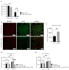
Increased production of reactive oxygen species within the mitochondria of FUS HeLa-mutated cells. Scale bar = 10 µm (A) HeLa cells were stained with MitoSOX Red (MSR) and MitoTracker deep red for live-cell imaging. (B) The mean MSR signal intensity was quantified using MitoTracker deep red as counterstain for detection of mitochondrial ROS. Data shown are from 3 independent biological replicates (n = 3). Data indicated as mean ± s.d. ** p < 0.01. (two-way ANOVA), from three independent experiments.
Figure 3
Figure 3
Inhibition of GPX4 and xCT enhances the vulnerability to ferroptosis, particularly in FUS-mutated cells. (A,B) Microscope images of both HeLa-FUS-WT-eGFP and HeLa-FUS-P525L-eGFP of untreated and treated with different concentration of RSL3 and erastin with or without Lip-1. Scale bar = 100 μm. (C,D) Dose-dependent toxicity of oxidative cell death-inducing agents (RSL3 and erastin). Both cell lines were treated with increasing concentrations of GPX4 inhibitor RSL3 and system xCT inhibitor erastin with or without Lip-1. Data shown represent the mean ± s.d. * p < 0.05, ** p < 0.01, **** p < 0.0001. (two-way ANOVA), of n = 3 wells of a 96-well plate, from three independent experiments. Cell viability was measured after 24 h of treatment.
Figure 4
Figure 4
ALS causing FUS mutation leads to misregulation of the protein expression of established ferroptotic factors. (A) Immunoblot analysis of both HeLa-FUS-WT and HeLa-FUS-P525L cell lines of FUS, GPX4, XCT, FSP1, and ACSL4 using specific antibodies. See also Supplementary Figure S2. (B) Quantification of the expression of the key ferroptotic proteins normalized to the reference gene B-Actin. Data shown from three independent experiments represent the mean ± s.d. ns = non-significant * p < 0.05, ** p < 0.01, *** p < 0.001; (two-way ANOVA), of n = 3. (C,D) Both cell lines were treated with increasing concentrations of erastin with or without the ferroptosis inhibitor DFO (100 µM). Cell viability was analyzed using Presto Blue method 24 h after the treatment. Data shown represent the mean ± s.d. ** p < 0.01; (two-way ANOVA), of n = 3 wells of a 96-well plate, from three independent experiments.
Figure 5
Figure 5
Cellular redox defense system is impaired by FUS mutations, which can be mitigated by mitochondrial calcium uniporter inhibition. (A) Luciferase-based assay used to analyze total glutathione (GSH) and oxidized glutathione (GSSG) levels in both cell lines. Data shown represent the mean ± s.d. ns > 0.9999, * p < 0.05; (two-way ANOVA), normalized with vehicle (untreated control) of n = 3 wells of a 96-well plate, from three independent experiments. (B) Live-cell fluorescence imaging of lipid peroxides in HeLa-FUS-WT and HeLa-FUS-P525L. Scale bars = 100 µm. (C) Lipid peroxides florescence intensity relative to baseline without any treatment in HeLa-FUS-WT compared to HeLa-FUS-P525L. (D,E) Lipid peroxides fluorescence intensity after 20 µM erastin treatment for 24 h with and without Ru265 (50 µM) and Ru265 (100 µM) in both HeLa-FUS-WT and HeLa-FUS-P525L. Control untreated (no erastin and no Ru265). Data are from a minimum of 3 stage positions. For statistical analysis, each stage position counted as one data entry. Data are mean ± s.d., * p < 0.05, ** p < 0.01, **** p < 0.0001; (two-way ANOVA), of n = 3 wells of a 96-well plate, from three independent experiments.
Figure 6
Figure 6
Human-induced pluripotent stem cell-derived neural progenitor cell (NPC)-generated motor neurons (MN) show higher susceptibility to ferroptosis. (A) Brightfield images of both FUS- WT and FUS-P525L smNPCs-generated MN with and without treatment of RSL3. Scale bar = 100 pixels. (B) is the magnification area of A. (C,D) Dose-dependent toxicity of oxidative cell death. Both cell lines were treated with increasing concentrations of RSL3 and erastin. Data shown represent the mean ± s.d. * p < 0.05, ** p < 0.01, **** p < 0.0001; (two-way ANOVA), of n = 3 wells of a 96-well plate, from three independent experiments. Cell viability was measured after 48 h.

References

    1. Dixon S.J., Lemberg K.M., Lamprecht M.R., Skouta R., Zaitsev E.M., Gleason C.E., Patel D.N., Bauer A.J., Cantley A.M., Yang W.S., et al. Ferroptosis: An iron-dependent form of nonapoptotic cell death. Cell. 2012;149:1060–1072. doi: 10.1016/j.cell.2012.03.042. - DOI - PMC - PubMed
    1. Friedmann Angeli J.P., Schneider M., Proneth B., Tyurina Y.Y., Tyurin V.A., Hammond V.J., Herbach N., Aichler M., Walch A., Eggenhofer E., et al. Inactivation of the ferroptosis regulator Gpx4 triggers acute renal failure in mice. Nat. Cell Biol. 2014;16:1180–1191. doi: 10.1038/ncb3064. - DOI - PMC - PubMed
    1. Stockwell B.R., Friedmann Angeli J.P., Bayir H., Bush A.I., Conrad M., Dixon S.J., Fulda S., Gascon S., Hatzios S.K., Kagan V.E., et al. Ferroptosis: A Regulated Cell Death Nexus Linking Metabolism, Redox Biology, and Disease. Cell. 2017;171:273–285. doi: 10.1016/j.cell.2017.09.021. - DOI - PMC - PubMed
    1. Forcina G.C., Dixon S.J. GPX4 at the Crossroads of Lipid Homeostasis and Ferroptosis. Proteomics. 2019;19:e1800311. doi: 10.1002/pmic.201800311. - DOI - PubMed
    1. Shi Z., Zhang L., Zheng J., Sun H., Shao C. Ferroptosis: Biochemistry and Biology in Cancers. Front. Oncol. 2021;11:579286. doi: 10.3389/fonc.2021.579286. - DOI - PMC - PubMed