. 2024 Apr 10;13(4):347.
doi: 10.3390/antibiotics13040347.

Anticandidal Activity of a Siderophore from Marine Endophyte Pseudomonas aeruginosa Mgrv7

Affiliations

Anticandidal Activity of a Siderophore from Marine Endophyte Pseudomonas aeruginosa Mgrv7

Essam Kotb et al. Antibiotics (Basel). .

Abstract

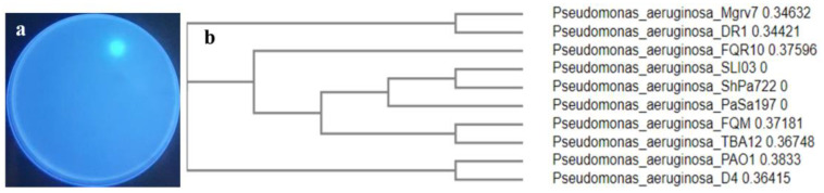
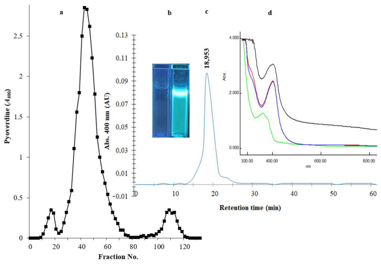
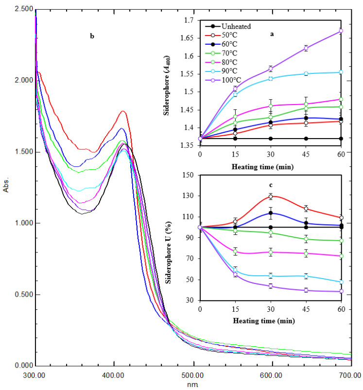
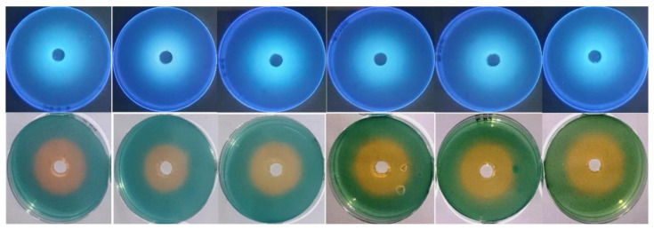
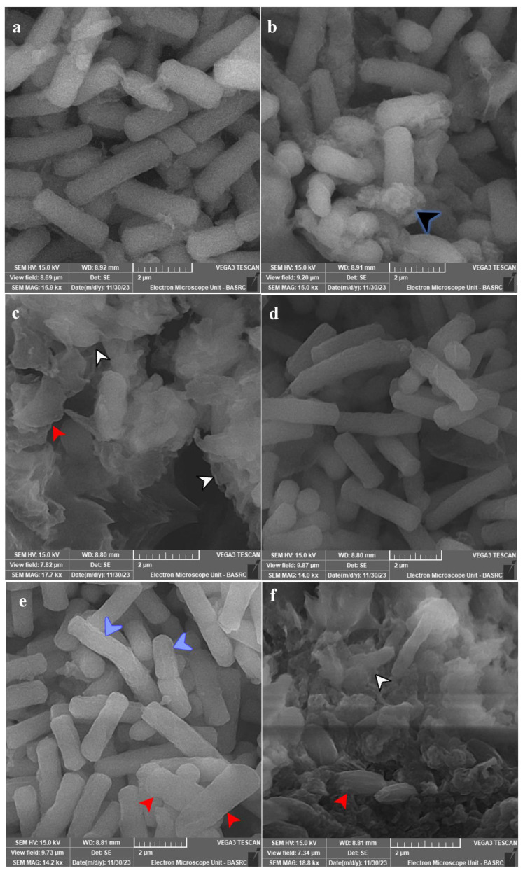
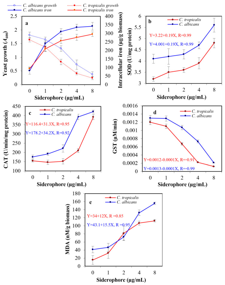
An endophytic symbiont P. aeruginosa-producing anticandidal siderophore was recovered from mangrove leaves for the first time. Production was optimal in a succinate medium supplemented with 0.4% citric acid and 15 µM iron at pH 7 and 35 °C after 60 h of fermentation. UV spectra of the acidic preparation after purification with Amberlite XAD-4 resin gave a peak at 400 nm, while the neutralized form gave a peak at 360 nm. A prominent peak with RP-HPLC was obtained at RT 18.95 min, confirming its homogeneity. It was pH stable at 5.0-9.5 and thermally stable at elevated temperatures, which encourages the possibility of its application in extreme environments. The minimum inhibitory concentration (MIC) and minimum fungicidal concentration (MFC) against Candida spp. Were in the range of 128 µg/mL and lower. It enhanced the intracellular iron accumulation with 3.2-4.2-fold (as judged by atomic absorption spectrometry) with a subsequent increase in the intracellular antioxidative enzymes SOD and CAT. Furthermore, the malondialdehyde (MDA) concentration due to cellular lipid peroxidation increased to 3.8-fold and 7.3-fold in C. albicans and C. tropicalis, respectively. The scanning electron microscope (SEM) confirmed cellular damage in the form of roughness, malformation, and production of defensive exopolysaccharides and/or proteins after exposure to siderophore. In conclusion, this anticandidal siderophore may be a promising biocontrol, nonpolluting agent against waterborne pathogens and pathogens of the skin. It indirectly kills Candida spp. by ferroptosis and mediation of hyperaccumulation of iron rather than directly attacking the cell targets, which triggers the activation of antioxidative enzymes.

Keywords: Candida; Pseudomonas aeruginosa; antimicrobial; biocontrol; mangrove; siderophore.


Conflict of interest statement

The authors declare that the research was conducted in the absence of any commercial or financial relationships that could be construed as a potential conflict of interest.

Figures

Figure 1
Figure 1
Showing the selected siderophore-producing isolate Mgrv7. Panel (a) represents its fluorescence under UV illumination (wavelength 365 nm) when grown on CAS agar of pH 7.0 for 48 h at 25 °C. Panel (b) represents the phylogenetic tree based on partial 16SrDNA gene sequence (Accession number PP024541).
Figure 2
Figure 2
The optimized parameters influencing siderophore productivity; medium type (a), organic acid supplementation (b), iron concentration (c), growth phase (d), medium pH (e), and incubation temperature (f). The preliminary production of siderophore in liquid medium was performed by inoculation of two milliliters of 108 colony forming units/mL Luria-Bertani (LB) broth in 25 mL of Kings B of pH 6.0 in a 100 mL volume Erlenmeyer flask. Incubation was conducted at 25 °C for 48 h at 120 rpm. The fermented broth was centrifuged at 5000 rpm for 20 min. Siderophores were estimated in cell-free supernatants; direct quantitative measurements were performed at wavelength 400 nm, with each unit quantified as the amount of siderophore producing an increase in absorbance of 0.1. Error bars represent the means ± standard deviations (n = 3). One-way analysis of variance (ANOVA) test indicated that the differences between means were significant since the p-values were lower than 0.5.
Figure 3
Figure 3
Panel (a) represents the chromatogram of PVD purification through an Amberlite XAD-4 column (2.5 × 20 cm2). The elution of immobilized PVD was performed with 50% (v/v) methanol at a speed of 5 mL/min. Panel (b) represents two cuvettes under a UV lamp of wavelength 365 nm; the left cuvette contains the elution solvent only, and the right cuvette contains the purified PVD. Panel (c) HPLC-visible chromatograms of the purified siderophore analyzed by reverse-phase HPLC using absorbance determination at wavelength 400 nm. Panel (d) represents the UV spectra of the 0.2 µm filtrate (black line), the chloroform extraction layer (red line), the acidified form of PVD at pH 3.0 (blue line), and the neutralized form of PVD at pH 7.0 (green line).
Figure 4
Figure 4
Time course of thermal treatment of tested siderophore at different temperatures for 60 min (a). Heating changed the characteristic color and fluorescence of siderophore gradually with maximum change at 100 °C after 60 min of exposure. UV spectra of heated preparations after 1 h of exposure (b). Heating changed the absorption peak from 414 nm to 410–412 nm. In addition, the optical density increased in comparison with the non-heated preparation. The reactivity of treated preparations at A630 followed a regression pattern at 70–100 °C and a stimulative effect at 50–60 °C (c). Error bars represent means ± standard deviations (n = 3). Some lines had no error bars because the value was 100%. Two-way ANOVA for panel a revealed that both temperature and time had effects on siderophore in form of A400, as the p-values were 0.0002 and 0.000003, respectively. This agrees with the values of correlation coefficient between the time and A400 under different temperatures, which were between 0.88 and 0.97, meaning that it was a very strong positive correlation. The correlation between temperature and A400 under different times was between 0.89 and 0.92, meaning that it was a very strong positive correlation. In panel (c), two-way ANOVA revealed that temperature had an effect on siderophore in the form of A630 as the p-value was 0.0000009, while there was insufficient evidence to conclude that A630 depends on the time since the p-value was 0.06 > 0.05. These ensure a correlation between the temperature and A630 under different time values where the correlation coefficient was between −0.66 and −0.76, which means that there was a strong negative correlation. The relation between time and A630 seemed to be unstable since for lower temperatures, the correlation between time and A630 was a weak positive correlation (between 0.2 and 0.4), while for higher temperatures, it was a very strong negative correlation (between −0.80 and −0.99).
Figure 5
Figure 5
pH stability of siderophore at pH values 5.0, 6.0, 7.0, 8.0, 9.0, and 9.5 from left to right direction. Siderophore preparations were adjusted to these pH values, and CAS agars were prepared at the same range. Each well was supplied with 0.5 mL of siderophore preparation. Incubation was conducted at 35 °C for 24 h. The appearance of orange halos on CAS agars and their fluorescence under UV were taken as indicative of their remaining activity and stability at the tested pH values.
Figure 6
Figure 6
SEM micrographs for tested species of Candida. Panel (a) represents untreated C. tropicalis ATCC 13803. Panels (b,c) show the treated cells with 10 µg PVD/mL after 12 and 24 h. Panel (d) represents the untreated cells of C. albicans ATCC 14053. Panels (e,f) show the treated cells with 10 µg PVD/mL after 12 and 24 h. Growth was allowed in Yeast Extract Peptone-Dextrose (YPD) broth for 24 h at 37 °C and 120 rpm shaking speed. C. tropicalis showed production of exopolysaccharides and/or proteins (black arrowhead) after 12 h of exposure, which were massive with cellular lysis (white arrowhead) and loss of shape (red arrowhead) after 24 h of exposure. C. albicans showed more surface roughness (blue arrowhead) after 12 h of exposure, which was more obvious after 24 h of exposure with loss of shape (red arrowhead) and cellular lysis (white arrowhead).
Figure 7
Figure 7
Effect of the subinhibitory concentrations of siderophore on the growth and intracellular iron accumulation in C. albicans and C. tropicalis (a). In addition, the ROS accumulation was indicated by direct assessment of the activity of intracellular antioxidative enzymes, superoxide dismutase (SOD, (b)), catalase (CAT, (c)), glutathione S-transferase (GST, (d)), and lipid peroxidation by release of malondialdehyde (MDA) from the cellular organelles and contents (e). The statistical study for panel a proved that increased iron internalization had a negative effect on the growth of C. albicans and C. tropicalis, as the correlation coefficients were −0.81 and −0.92, respectively. The effect upon C. tropicalis was higher, as the increase in siderophore concentration led to a greater increase in iron internalization since the concentration of siderophore had a positive correlation with iron accumulation (0.73 and 0.78, respectively). The relation between SOD, CAT, GST, MDA, and the growth of both Candida were studied in the form of linear regression (equations are represented in figures).

Similar articles

Cited by

References

    1. Brown A.J.P., Haynes K., Gow N.A.R., Quinn J. Stress responses in Candida. In: Calderone R.A., Clancy C.J., editors. Candida and Candidiasis. 2nd ed. ASM Press; Washington, DC, USA: 2012. pp. 225–242. - DOI
    1. Zuza-Alves D.L., Silva-Rocha W.P., Chaves G.M. An update on Candida tropicalis based on basic and clinical approaches. Front. Microbiol. 2017;8:1927. doi: 10.3389/fmicb.2017.01927. - DOI - PMC - PubMed
    1. Chen Y., Yang W., Zou G., Chen S., Pang J., She Z. Bioactive polyketides from the mangrove endophytic fungi Phoma sp. SYSU-SK-7. Fitoterapia. 2019;139:104369. doi: 10.1016/j.fitote.2019.104369. - DOI - PubMed
    1. Wang S., He G., Chen M., Zuo T., Xu W., Liu X. The role of antioxidant enzymes in the ovaries. Oxidative Med. Cell. Longev. 2017;2017:36–51. doi: 10.1155/2017/4371714. - DOI - PMC - PubMed
    1. Wu S.H., He J., Li X.N., Huang R., Song F., Chen Y.W., Miao C.P. Guaiane sesquiterpenes and isopimarane diterpenes from an endophytic fungus Xylaria sp. Phytochemistry. 2014;105:197–204. doi: 10.1016/j.phytochem.2014.04.016. - DOI - PubMed

LinkOut - more resources