. 2024 Apr 9;13(8):656.
doi: 10.3390/cells13080656.

Protecting Orthopaedic Implants from Infection: Antimicrobial Peptide Mel4 Is Non-Toxic to Bone Cells and Reduces Bacterial Colonisation When Bound to Plasma Ion-Implanted 3D-Printed PAEK Polymers

Affiliations

Protecting Orthopaedic Implants from Infection: Antimicrobial Peptide Mel4 Is Non-Toxic to Bone Cells and Reduces Bacterial Colonisation When Bound to Plasma Ion-Implanted 3D-Printed PAEK Polymers

Hedi Verena Kruse et al. Cells. .

Abstract

Even with the best infection control protocols in place, the risk of a hospital-acquired infection of the surface of an implanted device remains significant. A bacterial biofilm can form and has the potential to escape the host immune system and develop resistance to conventional antibiotics, ultimately causing the implant to fail, seriously impacting patient well-being. Here, we demonstrate a 4 log reduction in the infection rate by the common pathogen S. aureus of 3D-printed polyaryl ether ketone (PAEK) polymeric surfaces by covalently binding the antimicrobial peptide Mel4 to the surface using plasma immersion ion implantation (PIII) treatment. The surfaces with added texture created by 3D-printed processes such as fused deposition-modelled polyether ether ketone (PEEK) and selective laser-sintered polyether ketone (PEK) can be equally well protected as conventionally manufactured materials. Unbound Mel4 in solution at relevant concentrations is non-cytotoxic to osteoblastic cell line Saos-2. Mel4 in combination with PIII aids Saos-2 cells to attach to the surface, increasing the adhesion by 88% compared to untreated materials without Mel4. A reduction in mineralisation on the Mel4-containing surfaces relative to surfaces without peptide was found, attributed to the acellular portion of mineral deposition.

Keywords: 3D printing; antimicrobial peptide; biofilm; filament deposition modelling; infection prevention; orthopaedic implant; plasma immersion ion implantation; polyether ether ketone; polyether ketone; selective laser sintering.


Conflict of interest statement

The authors declare no conflicts of interest.

Figures

Figure 1
Figure 1
Water contact angles of untreated (UT) and plasma immersion ion implantation (PIII)-treated PEEK sheet, FDM-printed PEEK, and SLS-printed PEK surfaces, arranged in order of increasing roughness. Shown are mean values and standard deviation (n ≤ 6). The measurement for PIII-treated SLS PEK is 0°. Above the graph are photographic images of a 4 µL (PEEK sheet) or a 1 µL (FDM PEEK and SLS PEK) water droplet on the respective surfaces. **** p ≤ 0.0001.
Figure 2
Figure 2
Effectiveness of Mel4 bound to PAEK specimens against S. aureus using inactive peptide Mel1 as control. (A) The microbial counts in logarithms of the colony-forming units (CFUs)/mL on PEEK sheet, FDM PEEK, and SLS PEK surfaces with respect to untreated (UT) and plasma immersion ion implantation (PIII)-treated surfaces with either no peptide, Mel1 or Mel4 immobilised to them. Shown are the mean values and standard deviation of 3 replicates. (B) All available data from graph A combined, equally weighted, normalised to UT and graphed irrespective of the substrate material. Mel4 shows a 1.8 log reduction on the UT surface and a 3.7 log reduction on PIII-treated surfaces. Mel1 does not significantly increase or decrease bacterial adhesion on UT or PIII-treated surfaces. Some significance bars are omitted for readability. ** p ≤ 0.01; **** p ≤ 0.0001 (C) Mode of S. aureus adhesion to PAEK specimens. Note: SEM micrographs are not quantitatively interpretable. Scale bar = 10 µm.
Figure 3
Figure 3
Viability of Saos-2 cells cultured for 24 and 72 h with free Mel4 in solution as a function of the concentration. Each point represents the mean value and the standard deviation of 3 replicates. The lines correspond to linear regression curves. Immob.: Concentration used to immobilise Mel4 to PAEK specimens. * p ≤ 0.05; *** p ≤ 0.001.
Figure 4
Figure 4
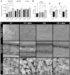
Saos-2 cell adhesion to PAEK surfaces. (A) Cell adhesion to PEEK sheet, FDM PEEK, and SLS PEK surfaces either untreated (UT) or plasma immersion ion implantation (PIII)-treated with either no peptide, or Mel4 immobilised to them. The mean value and standard deviation of 2–3 replicates are shown. (B) Effect of PIII and Mel4 on cell adhesion, irrespective of substrate material, obtained by combining data from A, equally weighted and normalised to UT. (C) Effect of presence of Mel4 obtained by combining data from A, irrespective of substrate material and treatment status, equally weighted and normalised to no peptide. (D) Overall effect of PIII treatment obtained by combining data from (A), irrespective of substrate material and presence of peptide, equally weighted and normalised to UT. * p ≤ 0.05; ** p ≤ 0.01; *** p ≤ 0.001. (E) SEM micrographs of Soas-2 cells adhered to PAEK specimens after 48 h of incubation. Scale bar = 30 µm.
Figure 5
Figure 5
Initial cell proliferation of Saos-2 cells on PAEK surfaces. Results are shown for PEEK sheet, FDM PEEK, and SLS PEK surfaces either untreated (UT) or plasma immersion ion implantation (PIII)-treated with either no peptide or Mel4 immobilised to them. Each point represents the mean value and the standard deviation of 3 replicates. There was no significant reduction in the growth at each time point.
Figure 6
Figure 6
Saos-2 cell mineralisation on PAEK surfaces. (A) Mineralisation per million Saos-2 cells assessed by PrestoBlue and Alizarin Red S assays. Results are shown for PEEK sheet, FDM PEEK, and SLS PEK surfaces either untreated (UT) or plasma immersion ion implantation (PIII)-treated with either no peptide or Mel4 immobilised to them. The mean value and standard deviation of 2–3 replicates are shown. (B) Effect of PIII and Mel4 on mineralisation, irrespective of substrate material, obtained by combining data from A, equally weighted and normalised to UT. (C) Effect of presence of Mel4 obtained by combining data from A, irrespective of substrate material and treatment status, equally weighted and normalised to no peptide. (D) Overall effect of PIII treatment obtained by combining data from (A), irrespective of substrate material and presence of peptide, equally weighted and normalised to UT.* p ≤ 0.05; ** p ≤ 0.01; *** p ≤ 0.001. **** p < 0.0001. (E) Light microscopic images of Alizarin Red S staining. 40× magnification. Scale bar = 0.5 mm.
Figure 7
Figure 7
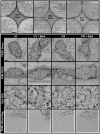
Morphology of Saos-2 cells after 35 days in culture on PAEK specimens and after performance of PrestoBlue and Alizarin Red S assays. SEM images reveal cellular and acellular contributions to mineralisation. Top panel: Overview SEM micrographs of Saos-2 growth on PAEK specimens. Saos-2 cells formed clusters, typical for late-stage differentiation. On rougher FDM PEEK and PEK specimens, the clusters occupy valleys created by the 3D process within the surface. Scale bar = 1 mm. Lower panels: Interaction of cells with the respective surface. PIII-treated samples show better coverage of individual cells on the surface independent of the presence of Mel4. Bottom panel: Magnified areas from PEEK sheet micrographs (white dotted boxes). Close-up images reveal surface features on specimen not containing Mel4. Scale bar = 30 µm. UT: Untreated, PIII: Plasma Immersion Ion Implantation.
Figure 8
Figure 8
Illustrating the different modes of attachment of Mel4 to untreated (UT) and PIII-treated surfaces (using the example of a PEEK surface). A partial negative charge (δ) on the untreated surface adsorbs Mel4 by attracting its positive charge (+), orienting it along the surface. Meanwhile, PIII-generated radicals buried in the subsurface diffuse to the surface and bind the peptide from solution. The positively charged nitrogen ions, implanted during PIII treatment, give the surface overall a more positive charge that may help to erect the peptide into an orthogonal orientation, more favourable for antimicrobial action.

References

    1. Ribeiro M., Monteiro F.J., Ferraz M.P. Infection of orthopedic implants with emphasis on bacterial adhesion process and techniques used in studying bacterial-material interactions. Biomatter. 2012;2:176–194. doi: 10.4161/biom.22905. - DOI - PMC - PubMed
    1. Shukla A., Fleming K.E., Chuang H.F., Chau T.M., Loose C.R., Stephanopoulos G.N., Hammond P.T. Controlling the release of peptide antimicrobial agents from surfaces. Biomaterials. 2010;31:2348–2357. doi: 10.1016/j.biomaterials.2009.11.082. - DOI - PubMed
    1. Seo M.D., Won H.S., Kim J.H., Mishig-Ochir T., Lee B.J. Antimicrobial peptides for therapeutic applications: A review. Molecules. 2012;17:12276–12286. doi: 10.3390/molecules171012276. - DOI - PMC - PubMed
    1. Browne K., Chakraborty S., Chen R., Willcox M.D.P., Black D.S., Walsh W.R., Kumar N. A new era of antibiotics: The clinical potential of antimicrobial peptides. Int. J. Mol. Sci. 2020;21:7047. doi: 10.3390/ijms21197047. - DOI - PMC - PubMed
    1. Tummanapalli S.S., Willcox M.D. Antimicrobial resistance of ocular microbes and the role of antimicrobial peptides. Clin. Exp. Optom. 2021;104:295–307. doi: 10.1111/cxo.13125. - DOI - PubMed

Publication types

MeSH terms

LinkOut - more resources