Review
. 2024 Apr 12;13(8):670.
doi: 10.3390/cells13080670.

Exosomes in Vascular/Neurological Disorders and the Road Ahead

Affiliations
Review

Exosomes in Vascular/Neurological Disorders and the Road Ahead

Faisal A Alzahrani et al. Cells. .

Abstract

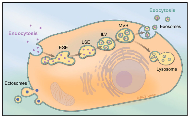
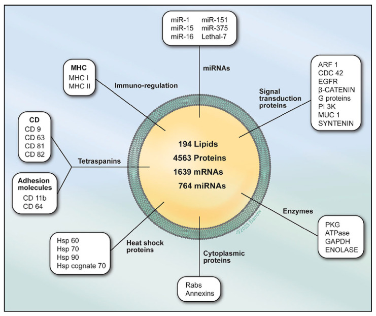
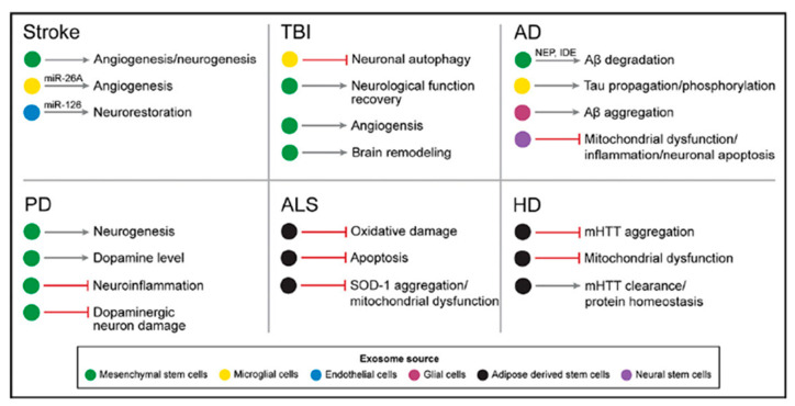
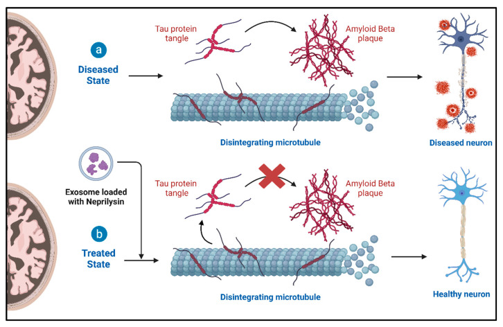
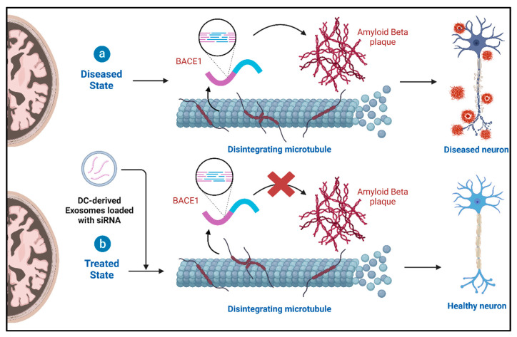
Neurodegenerative diseases, such as Alzheimer's disease (AD), Parkinson's disease (PD), amyotrophic lateral sclerosis (ALS), Huntington's disease (HD), stroke, and aneurysms, are characterized by the abnormal accumulation and aggregation of disease-causing proteins in the brain and spinal cord. Recent research suggests that proteins linked to these conditions can be secreted and transferred among cells using exosomes. The transmission of abnormal protein buildup and the gradual degeneration in the brains of impacted individuals might be supported by these exosomes. Furthermore, it has been reported that neuroprotective functions can also be attributed to exosomes in neurodegenerative diseases. The potential neuroprotective functions may play a role in preventing the formation of aggregates and abnormal accumulation of proteins associated with the disease. The present review summarizes the roles of exosomes in neurodegenerative diseases as well as elucidating their therapeutic potential in AD, PD, ALS, HD, stroke, and aneurysms. By elucidating these two aspects of exosomes, valuable insights into potential therapeutic targets for treating neurodegenerative diseases may be provided.

Keywords: Alzheimer’s; exosomes; neurological diseases; stroke; vascular diseases.


Conflict of interest statement

The authors declare no conflicts of interest.

Figures

Figure 1
Figure 1
Exosomes form through the cellular uptake of extracellular components via endocytosis and plasma membrane invagination. This process initiates the creation of early-sorting endosomes (ESEs) that may bud independently or fuse with preformed ESEs from the ER, TGN, or mitochondria. Fusion with the ER and TGN allows endocytic cargo access to ESEs, progressing to the formation of late-sorting endosomes (LSEs). LSEs undergo invagination, generating intraluminal vesicles (ILVs) that include diverse constituents from various sources. ILVs of different sizes and content result based on invagination volume. LSEs evolve into multivesicular bodies (MVBs), representing future exosomes. MVBs can fuse with autophagosomes for lysosomal degradation or directly fuse with lysosomes. Alternatively, MVBs can travel to the plasma membrane via the cell’s cytoskeleton, docking with MVB-docking proteins on the luminal side. This leads to exocytosis, releasing exosomes with a lipid bilayer orientation akin to the plasma membrane, contributing to the extracellular vesicle pool. Figure is used with permission from the Barrow Neurological Institute, Phoenix, Arizona, USA.
Figure 2
Figure 2
Exosomes serve as a cell-to-cell transit system within the human body, exhibiting diverse and multifunctional roles. These extracellular vesicles, produced by all cells, encapsulate nucleic acids [12,13], proteins [11,14], lipids [15,16], and metabolites [17,18]. Functioning as crucial mediators of intercellular communication, both in close proximity and over long distances, exosomes play pivotal roles in influencing various facets of cell biology. Their impact extends across health and disease, highlighting their significance in orchestrating cellular interactions and contributing to the regulation of cellular processes. Figure is used with permission from the Barrow Neurological Institute, Phoenix, AZ, USA.
Figure 3
Figure 3
Role of exosomes in various neurodegenerative diseases. Figure is used with permission from the Barrow Neurological Institute, Phoenix, AZ, USA.
Figure 4
Figure 4
Treatment of AD with neprilysin-loaded exosomes.
Figure 5
Figure 5
Treatment of AD with siRNA-loaded exosomes.
Figure 6
Figure 6
Direct pathways of nose-to-brain exosome delivery.
Figure 7
Figure 7
Enhancement of neurovascular remodeling after stroke and TBI by MSC-derived exosomes.

References

    1. Kalluri R. The biology and function of exosomes in cancer. J. Clin. Investig. 2016;126:1208–1215. doi: 10.1172/JCI81135. - DOI - PMC - PubMed
    1. Van Niel G., d’Angelo G., Raposo G. Shedding light on the cell biology of extracellular vesicles. Nat. Rev. Mol. Cell Biol. 2018;19:213–228. doi: 10.1038/nrm.2017.125. - DOI - PubMed
    1. Mathieu M., Martin-Jaular L., Lavieu G., Théry C. Specificities of secretion and uptake of exosomes and other extracellular vesicles for cell-to-cell communication. Nat. Cell Biol. 2019;21:9–17. doi: 10.1038/s41556-018-0250-9. - DOI - PubMed
    1. Kahlert C., Kalluri R. Exosomes in tumor microenvironment influence cancer progression and metastasis. J. Mol. Med. 2013;91:431–437. doi: 10.1007/s00109-013-1020-6. - DOI - PMC - PubMed
    1. Bebelman M.P., Smit M.J., Pegtel D.M., Baglio S.R. Biogenesis and function of extracellular vesicles in cancer. Pharmacol. Ther. 2018;188:1–11. doi: 10.1016/j.pharmthera.2018.02.013. - DOI - PubMed

MeSH terms

LinkOut - more resources