Review
. 2024 Apr 18;14(8):838.
doi: 10.3390/diagnostics14080838.

Multi-Modality Imaging in Vasculitis

Affiliations
Review

Multi-Modality Imaging in Vasculitis

Mohamed N Allam et al. Diagnostics (Basel). .

Abstract

Systemic vasculitides are a rare and complex group of diseases that can affect multiple organ systems. Clinically, presentation may be vague and non-specific and as such, diagnosis and subsequent management are challenging. These entities are typically classified by the size of vessel involved, including large-vessel vasculitis (giant cell arteritis, Takayasu's arteritis, and clinically isolated aortitis), medium-vessel vasculitis (including polyarteritis nodosa and Kawasaki disease), and small-vessel vasculitis (granulomatosis with polyangiitis and eosinophilic granulomatosis with polyangiitis). There are also other systemic vasculitides that do not fit in to these categories, such as Behcet's disease, Cogan syndrome, and IgG4-related disease. Advances in medical imaging modalities have revolutionized the approach to diagnosis of these diseases. Specifically, color Doppler ultrasound, computed tomography and angiography, magnetic resonance imaging, positron emission tomography, or invasive catheterization as indicated have become fundamental in the work up of any patient with suspected systemic or localized vasculitis. This review presents the key diagnostic imaging modalities and their clinical utility in the evaluation of systemic vasculitis.

Keywords: Behçet’s disease; Cogan syndrome; Doppler ultrasound; Kawasaki disease; Takayasu’s arteritis; angiography; computed tomography; eosinophilic granulomatosis with polyangiitis; giant cell arteritis; granulomatosis with polyangiitis; immunoglobulin G—related disease; magnetic resonance imaging; polyarteritis nodosa; positron emission tomography; vasculitis.


Conflict of interest statement

The authors declare no conflict of interest.

Figures
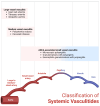
Figure 1
Figure 1
Classification of systemic vasculitides. Created with Biorender.com.
Figure 2
Figure 2
A 75-year-old man with known giant cell arteritis. Axial double inversion recovery MR images (A,B) demonstrating concentric mural thickening of the thoracic aorta (arrows). Axial T1-weighted images after intravenous contrast administration (C,D) demonstrating mild enhancement to the aortic wall more pronounced in the arch and descending aorta (arrowheads), consistent with active vasculitis. Images of chest CTA (E,F) showing concentric mural thickening of the ascending aorta, aortic arch and descending aorta (arrows).
Figure 3
Figure 3
A 60-year-old woman with giant cell arteritis. Coronal images of attenuation corrected F-18 FDG PET (A), CT without contrast (B) and fused PET/CT (C) demonstrating moderate intensity linear increased tracer activity in the wall of the ascending aorta and proximal arch (arrows), most consistent with active vasculitis.
Figure 4
Figure 4
A 78-year-old man with polymyalgia rheumatica and active vasculitis. Maximum intensity projection images of FDG PET (A) demonstrating abnormal radiotracer accumulation in multiple joints (shoulders, elbows, hands, hips, knees and feet) in keeping with polyarticular inflammatory arthropathy (asterisks). Axial images of fused PET CT at the level of the descending aorta (B) and abdominal aorta (C) showing near concentric tracer avid mural thickening to the aorta (arrows), most consistent with active vasculitis.
Figure 5
Figure 5
A 28-year-old man with active Takayasu’s disease. Axial (A), coronal (B) and sagittal (C) images of chest CTA demonstrating concentric mural thickening involving the ascending aorta, arch, descending aorta and proximal right brachiocephalic artery (arrows) and small reactive pericardial effusion (arrowhead).
Figure 6
Figure 6
A 32-year-old woman with history of Takayasu’s disease. Coronal maximum intensity projection image of neck CTA (A) showing complete occlusion to the left common carotid artery (arrow). Sagittal images of CT without contrast (B) and F-18 FDG PET/CT (C) demonstrating distal descending aorta stent (arrows) with no pathological abdominal wall radiotracer accumulation consistent with sequela of chronic arteritis with no evidence of acute inflammation.
Figure 7
Figure 7
A 54-year-old woman with history of Takayasu’s disease. Invasive catheterization with injection of contrast in the aorta at the origin of the left (bovine) common carotid artery; stenoses at the origin of the left internal and external carotid arteries are demonstrated (arrow), and the collateral vessels that they give rise to. The right common carotid and right subclavian arteries are completely occluded. The left vertebral and left subclavian arteries are occluded.
Figure 8
Figure 8
An 80-year-old woman with polyarteritis nodosa presenting with gastrointestinal bleeding. Coronal maximum intensity projection images of abdomen CTA at three different levels demonstrating pseudoaneurysms of the celiac and superior mesenteric artery branches secondary to arteritis. (A) Pseudoaneurysm of the gastroduodenal artery and jejunal branch of the superior mesenteric artery (arrow) and contrast extravasation into the proximal small bowel loop consistent with active GI bleed (arrowhead). (B) Pseudoaneurysm of the left gastric artery (arrow). (C) Pseudoaneurysm of the distal aspect of the splenic artery (arrow).
Figure 9
Figure 9
A 35-year-old woman with Kawasaki’s disease. Double oblique MPR images of coronary CTA (A,B) showing diffuse aneurysmal dilation to the LAD (arrow), large caliber first diagonal branch (arrowhead) and RCA (asterisk).
Figure 10
Figure 10
A 48-year-old man with granulomatosis with polyangiitis. CT chest without contrast (A) demonstrating a 1.5 cm solid non-calcified pulmonary nodule in the basal left lower lobe (arrow). CT of the facial bones without contrast (B) showing diffuse mucosal thickening with near complete opacification of the maxillary sinuses with erosion of the medial wall of the maxillary sinuses and nasal septum (asterisks).
Figure 11
Figure 11
A 50-year-old woman with known granulomatosis with polyangiitis. Axial images of F-18 FDG PET/CT at time of presentation (AC) demonstrating a tracer avid mass like consolidative opacity in the basal segments of the lower lobe of the right lung (arrows). Axial images of subsequent F-18 FDG PET/CT approximately 6 months after initiation of immunosuppressive therapy (DF) demonstrating known right lung consolidation is smaller and significantly less tracer avid (arrows).
Figure 12
Figure 12
A 75-year-old woman with Behcet’s disease. Axial images of attenuation corrected F-18 FDG PET (A), CT without contrast (B) and fused PET/CT (C) demonstrating concentric tracer avid mural thickening to the ascending aorta involving the root (arrow) as well as the main pulmonary artery (arrowhead), consistent with active vasculitis.
Figure 13
Figure 13
A 53-year-old woman with Behcet’s myopericarditis. Short axis (A) and two-chamber (B) inversion recovery images of cardiac MR approximately 10 min after intravenous administration of gadolinium demonstrating mid myocardium and epicardium abnormal hyperintense signal consistent with delayed myocardial enhancement in the inferior and inferolateral segments of the left ventricle extending from base to the apex (arrows). In addition, there is delayed enhancement of the pericardium overlying same segments (arrowhead).
Figure 14
Figure 14
65-year-old woman with IgG4-related aortitis. Axial images of F-18 FDG attenuation corrected PET (A), CT without contrast (B) and fused PET/CT (C) demonstrating tracer avid concentric mural thickening involving the abdominal aorta (arrows) and underlying calcified atherosclerosis. No evidence of retroperitoneal fibrosis.

References

    1. Jennette J.C., Falk R.J., Bacon P.A., Basu N., Cid M.C., Ferrario F., Flores-Suarez L.F., Gross W.L., Guillevin L., Hagen E.C., et al. 2012 Revised International Chapel Hill Consensus Conference Nomenclature of Vasculitides. Arthritis Rheum. 2013;65:1–11. doi: 10.1002/art.37715. - DOI - PubMed
    1. Watts R.A., Robson J. Introduction, epidemiology and classification of vasculitis. Best Pract. Res. Clin. Rheumatol. 2018;32:3–20. doi: 10.1016/j.berh.2018.10.003. - DOI - PubMed
    1. Cid M.C., Campo E., Ercilla G., Palacin A., Vilaseca J., Villalta J., Ingelmo M. Immunohistochemical analysis of lymphoid and macrophage cell subsets and their immunologic activation markers in temporal arteritis: Influence of Corticosteroid Treatment. Arthritis Rheumatol. 1989;32:884–893. doi: 10.1002/j.2326-5205.1989.tb00020.x. - DOI - PubMed
    1. Maz M., Chung S.A., Abril A., Langford C.A., Gorelik M., Guyatt G., Archer A.M., Conn D.L., Full K.A., Grayson P.C., et al. 2021 American College of Rheumatology/Vasculitis Foundation Guideline for the Management of Giant Cell Arteritis and Takayasu Arteritis. Arthritis Rheumatol. 2021;73:1349–1365. doi: 10.1002/art.41774. - DOI - PubMed
    1. Sugihara T., Hasegawa H., Uchida H.A., Yoshifuji H., Watanabe Y., Amiya E., Maejima Y., Konishi M., Murakawa Y., Ogawa N., et al. Associated factors of poor treatment outcomes in patients with giant cell arteritis: Clinical implication of large vessel lesions. Arthritis Res. Ther. 2020;22:72. doi: 10.1186/s13075-020-02171-6. - DOI - PMC - PubMed

LinkOut - more resources