. 2024 Mar 28;10(4):233.
doi: 10.3390/gels10040233.

In Vivo Evaluation of Wound Healing Efficacy of Gel-Based Dressings Loaded with Pycnogenol™ and Ceratothoa oestroides Extracts

Affiliations

In Vivo Evaluation of Wound Healing Efficacy of Gel-Based Dressings Loaded with Pycnogenol™ and Ceratothoa oestroides Extracts

Andreas Vitsos et al. Gels. .

Abstract

Ceratothoa oestroides and French maritime pine bark (Pycnogenol™) extracts are considered promising therapeutic agents in wound healing. This study explores the healing efficacy of composite dressings containing these extracts, aiming to enhance their stability and effectiveness, utilizing a low-temperature vacuum method for producing Sodium Alginate-Maltodextrin gel dressings. Surgical wounds were inflicted on SKH-hr2 hairless mice. Dressings were loaded with Pycnogenol™ and/or C. oestroides extracts and assessed for their efficacy. Wound healing was primarily evaluated by clinical and histopathological evaluation and secondarily by Antera 3D camera and biophysical measurements. Dressings were stable and did not compromise the therapeutic properties of C. oestroides extract. All interventions were compared to the C. oestroides ointment as a reference product. Most of the wounds treated with the reference formulation and the C. oestrodes dressing had already closed by the 15th day, with histological scores of 7 and 6.5, respectively. In contrast, wounds treated with Pycnogenol™, either alone or in combination with C. oestroides, did not close by the end of the experiment (16th day), with histological scores reaching 15 in both cases. Furthermore, treatment with 5% Pycnogenol™ dressing appeared to induce skin thickening and increase body temperature. The study underscores the wound healing potential of C. oestroides extracts and highlights the need for further research to optimize Pycnogenol™ dosing in topical applications.

Keywords: Ceratothoa oestroides; Pinus pinaster; Pycnogenol™; composite wound dressings; gel dressings; wound healing.


Conflict of interest statement

The authors declare that there are no conflicts of interest.

Figures

Figure 1
Figure 1
Photographic Capture of Gel Dressings. On the right, a dressing devoid of any active ingredients is highlighted, while on the left, a dressing containing both C. oestroides and Pycnogenol™ is depicted.
Figure 2
Figure 2
Weight variation of different dressings. Weight measurements are presented in grams (g). The combined dressings (in black) exhibited a higher mean and median weight compared to all other dressings, followed by the Pycnogenol™ dressings (in brown). The dressings containing C. oestroides (magenta) and the control dressings (red) demonstrated approximately equal weights.
Figure 3
Figure 3
SEM images of the different dressings.
Figure 4
Figure 4
Measurements of the experimental animals’ temperature at the beginning and end of the experiment were maintained at the same levels, apart from Pycnogenol™, which showed a slight increase. * Statistical significance between starting and ending measurements was observed for Pycnogenol™ groups (p < 0.05).
Figure 5
Figure 5
Measurements of the transepidermal water loss (TEWL) before wound infliction and at the end of the study. * Statistical significance between starting and ending measurements was observed in all groups (p < 0.05).
Figure 6
Figure 6
Measurements of the hydration before wound infliction and at the end of the study. No statistically significant differences were observed between the different groups or starting and ending points (p > 0.1).
Figure 7
Figure 7
Skin thickness measurements at the start and conclusion of the experiment. * Statistical significance (p = 0.0011) reported between starting and ending measurements regarding Pycnogenol™ dressing treatment.
Figure 8
Figure 8
Wound surface area measurements throughout the experiment. Significant differences were observed as follows: by the 13th day, between the reference ointment treatment and the control dressing and the Pycnogenol™ dressings (p < 0.01); by the 15th day, the reference ointment treatment, compared to C. oestroides dressing (p < 0.02). Pycnogenol™ dressings, showed a statistically significant (worsening) compared to the control; between the reference ointment and C. oestroides dressing from the 6th day until the final day (p < 0.001).
Figure 9
Figure 9
Wound volume measurements throughout the experiment. Significant differences were observed as follows: C. oestroides dressing and reference ointment showed significant differences compared to the other interventions (p < 0.001); Pycnogenol™ dressing showed significant worsening compared to the control and the C. oestroides treatments (p < 0.001).
Figure 10
Figure 10
Wound depth measurements throughout the experiment. Significant differences were observed as follows: C. oestroides dressing and reference ointment showed a significant difference compared to the other interventions (p < 0.001); Pycnogenol™ dressing showed significant worsening compared to the control and the C. oestroides treatments (p < 0.001).
Figure 11
Figure 11
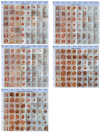
Photodocumentation throughout the experiment (days 1, 3, 6, 8, 10, 13, 15, and 16) display an evaluation of wound healing in the five treatment groups: control dressing; reference ointment; C. oestroides dressing; Pycnogenol™ dressing; C. oestroides + Pycnogenol dressing.
Figure 12
Figure 12
Skin sections at the end of the experimental procedure, stained with hematoxylin–eosin (100×). White arrows depict non-inflamed dermis; black arrows depict moderate to intense inflammatory elements; ellipses depict incomplete healing and parakeratosis.
Figure 13
Figure 13
Custom-built apparatus for dressings production. On the right, there is a heated and thermostatically controlled vacuum chamber; in the middle, the vapors’ concentrating trap; and on the left, a vacuum pump.

References

    1. Almadani Y.H., Vorstenbosch J., Davison P.G., Murphy A.M. Wound Healing: A Comprehensive Review. Semin. Plast. Surg. 2021;35:141–144. doi: 10.1055/s-0041-1731791. - DOI - PMC - PubMed
    1. Kolimi P., Narala S., Nyavanandi D., Youssef A.A.A., Dudhipala N. Innovative Treatment Strategies to Accelerate Wound Healing: Trajectory and Recent Advancements. Cells. 2022;11:2439. doi: 10.3390/cells11152439. - DOI - PMC - PubMed
    1. Senni K., Pereira J., Gueniche F., Delbarre-Ladrat C., Sinquin C., Ratiskol J., Godeau G., Fischer A.-M., Helley D., Colliec-Jouault S. Marine polysaccharides: A source of bioactive molecules for cell therapy and tissue engineering. Mar. Drugs. 2011;9:1664–1681. doi: 10.3390/md9091664. - DOI - PMC - PubMed
    1. Ramalingam S., Chandrasekar M.J.N., Nanjan M.J. Plant-based Natural Products for Wound Healing: A Critical Review. Curr. Drug. Res. Rev. 2022;14:37–60. doi: 10.2174/2589977513666211005095613. - DOI - PubMed
    1. Fan B., Dun S.H., Gu J.Q., Guo Y., Ikuyama S. Pycnogenol Attenuates the Release of Proinflammatory Cytokines and Expression of Perilipin 2 in Lipopolysaccharide-Stimulated Microglia in Part via Inhibition of NF-κB and AP-1 Activation. PLoS ONE. 2015;10:e0137837. doi: 10.1371/journal.pone.0137837. - DOI - PMC - PubMed

LinkOut - more resources