. 2024 Mar 26;22(4):147.
doi: 10.3390/md22040147.

Attenuation of Nicotine Effects on A549 Lung Cancer Cells by Synthetic α7 nAChR Antagonists APS7-2 and APS8-2

Affiliations

Attenuation of Nicotine Effects on A549 Lung Cancer Cells by Synthetic α7 nAChR Antagonists APS7-2 and APS8-2

Ahmad Joukhan et al. Mar Drugs. .

Abstract

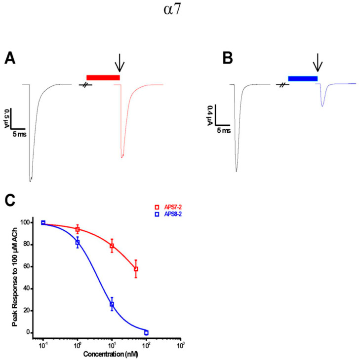
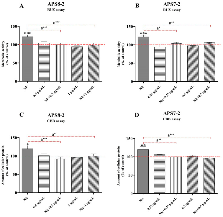
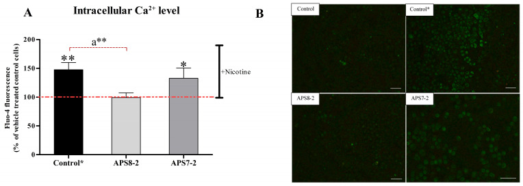
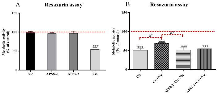
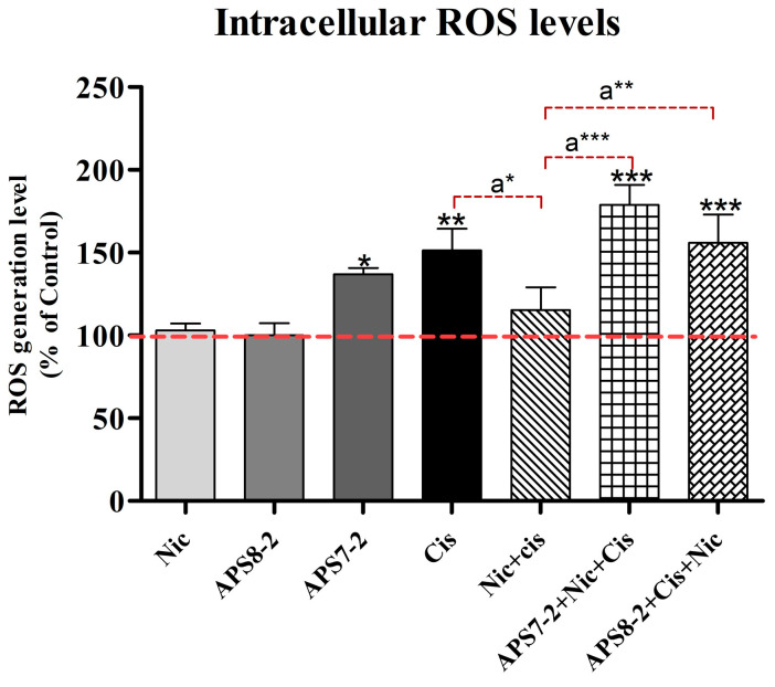
Nicotine binds to nicotinic acetylcholine receptors (nAChRs) that are overexpressed in different cancer cells, promoting tumor growth and resistance to chemotherapy. In this study, we aimed to investigate the potential of APS7-2 and APS8-2, synthetic analogs of a marine sponge toxin, to inhibit nicotine-mediated effects on A549 human lung cancer cells. Our electrophysiological measurements confirmed that APS7-2 and APS8-2 act as α7 nAChR antagonists. APS8-2 showed no cytotoxicity in A549 cells, while APS7-2 showed concentration-dependent cytotoxicity in A549 cells. The different cytotoxic responses of APS7-2 and APS8-2 emphasize the importance of the chemical structure in determining their cytotoxicity on cancer cells. Nicotine-mediated effects include increased cell viability and proliferation, elevated intracellular calcium levels, and reduced cisplatin-induced cytotoxicity and reactive oxygen species production (ROS) in A549 cells. These effects of nicotine were effectively attenuated by APS8-2, whereas APS7-2 was less effective. Our results suggest that APS8-2 is a promising new therapeutic agent in the chemotherapy of lung cancer.

Keywords: APS7-2; APS8-2; marine toxin; nAChR antagonist; nicotinic acetylcholine receptor.


Conflict of interest statement

The authors declare no conflicts of interest.

Figures

Figure 1
Figure 1
Electrophysiological characterization of APS7-2 and APS8-2 on human heterologous α7 nAChRs expressed in X. laevis oocytes. Agonist-evoked responses of human α7 nAChRs using 100 μM acetylcholine (black arrow), first applied alone and then co-applied with (A) 10 nMAPS7-2 (red bar) or (B) 10 nM APS8-2 (blue bar). (C) The data are normalized to the peak amplitude of current recorded without APS (100%) and presented as mean ± SEM (n = 5 oocytes).
Figure 2
Figure 2
Cytotoxicity of APS8-2 and APS7-2 on A549 human adenocarcinoma lung cancer cells after 24 h exposure. Cytotoxicity was measured using RUZ, NRU, and CBB assays. (A) The cytotoxicity of APS8-2 was measured in the range of 0.5 µg/mL to 100 µg/mL, while (B) for APS7-2 it was in the range of 0.25 µg/mL to 10 µg/mL. (C) Microscopic images (20×) of A549 cells, including control, 0.5 µg/mL APS8-2, and 0.5 µg/mL APS7-2; scale bar represents 100 μm. Measurements were normalized to the untreated control (dashed line) as the mean percentage (±SD). Data were statistically analyzed by ANOVA with Bonferroni multiple comparisons post-test. The asterisks indicate a significant difference compared to the untreated control: *** corresponds to p < 0.001.
Figure 3
Figure 3
Evaluation of the effects of nicotine (Nic), APS8-2, APS7-2, and the combination of APS8-2 or APS7-2 with nicotine on the viability and proliferation of A549 cells. A549 cell viability and proliferation were assessed using (A,B) RUZ assay for cell viability and (C,D) CBB assay for cell proliferation. A549 cells were pretreated with 1 µM nicotine for 24 h, then treated with 0.25 µg/mL APS7-2, 0.5 µg/mL APS7-2, 0.5 µg/mL APS8-2, or 1 µg/mL APS8-2 for another 24 h. Measurements were normalized to the untreated control (dashed line) as the mean percentage (±SD). The data were statistically analyzed by ANOVA with Bonferroni multiple comparisons post-test. Compared with the untreated control (* equals p < 0.05; ** equals p < 0.01; *** equals p < 0.001); compared with nicotine-treated cells (a* equals p < 0.05; a** equals p < 0.01; a*** equals p < 0.001).
Figure 4
Figure 4
Intracellular Ca2+ level in A549 cells after 100 s of adding nicotine. (A) Cells were pretreated with APS compounds (1 µg/mL APS7-2 and 1 µg/mL APS8-2) for 24 h in a serum-deprived medium. In the control, cells were not pretreated with APS compounds, and fluorescence was measured after adding 1 mM nicotine. (B) Fluo-4 fluorescence intensity was measured in A549 cells. Control was measured after 100 s without adding nicotine. Control* (after adding nicotine) and A549 cells pretreated with APS7-2 and APS8-2 were measured after 100 s of adding nicotine. The scale bar represents 100 μm. The measurements were normalized to untreated cells (control) before adding nicotine, and they are presented as the mean percentage (±SD). Each treatment was conducted two times independently with three replications, and fluorescence data were taken from at least 30 individual cells per repetition. The data were analyzed using ANOVA with multiple comparisons and the Bonferroni post-test, and statistical significance was determined using p-values. A comparison was made between the treated cells with nicotine and the untreated control, with * indicating p < 0.05 and ** indicating p < 0.01. A comparison was also made between the control cells after adding nicotine and those pretreated with APS compounds, with a** indicating p < 0.01.
Figure 5
Figure 5
Effects of nicotine, APS7-2, APS8-2, and cisplatin on the cell viability of A549 cells. (A) Viability of A549 cancer cells treated with 1 µM nicotine (Nic), 1 µg/mL APS8-2, 0.5 µg/mL APS7-2, and 50 µg/mL cisplatin (Cis). (B) Viability of A549 cancer cells treated with 50 µg/mL cisplatin (Cis), 50 µg/mL cisplatin (Cis) + 1 µM nicotine (Nic), 50 µg/mL cisplatin (Cis) + 1 µg/mL APS8-2 + 1 µM nicotine (Nic), and 50 µg/mL cisplatin (Cis) + 0.5 µg/mL APS7-2 + 1 µM nicotine. The viability of A549 cells was measured using the RUZ assay after 24 h of treatment. Measurements were normalized to the untreated control (dashed line) as the mean percentage (±SD). Data were statistically analyzed using ANOVA with multiple comparisons and Bonferroni post-test. The asterisks indicate a significant difference compared to the untreated control (*** corresponds to p < 0.001) and compared to the cells treated with the combination of cisplatin and nicotine (a* corresponds to p < 0.05).
Figure 6
Figure 6
Intracellular ROS levels in A549 cells. Cells were treated with 1 µM nicotine (Nic), 1 µg/mL APS8-2, 0.5 µg/mL APS7-2, 100 µg/mL cisplatin (Cis), a combination of Nic and Cis, a combination of APS8-2, Cis, and Nic, or a combination of APS7-2, Nic, and Cis. Results were normalized to the untreated control cells (dashed line) and are presented as mean percentage (±SD). The data were statistically analyzed by ANOVA with Bonferroni multiple comparisons post-test. The asterisks indicate a significant difference compared to the untreated control: * equals p < 0.05; ** equals p < 0.01; *** equals p < 0.001; and with respect to cells treated with Cis and Nic, a* equals p < 0.05; a** equals p < 0.01; a*** equals p < 0.001.
Figure 7
Figure 7
The structure of 3-alkylpyridinium salts analogs used in this study, APS7-2 and APS8-2.

References

    1. Sung H., Ferlay J., Siegel R.L., Laversanne M., Soerjomataram I., Jemal A., Bray F. Global Cancer Statistics 2020: GLOBOCAN Estimates of Incidence and Mortality Worldwide for 36 Cancers in 185 Countries. CA Cancer J. Clin. 2021;71:209–249. doi: 10.3322/caac.21660. - DOI - PubMed
    1. Centers for Disease Control and Prevention. [(accessed on 15 March 2023)]; Available online: https://www.cdc.gov/cancer/lung/basic_info/risk_factors.htm.
    1. Kunda N.K. Antimicrobial Peptides as Novel Therapeutics for Non-Small Cell Lung Cancer. Drug Discov. Today. 2020;25:238–247. doi: 10.1016/j.drudis.2019.11.012. - DOI - PubMed
    1. Woodman C., Vundu G., George A., Wilson C.M. Applications and Strategies in Nanodiagnosis and Nanotherapy in Lung Cancer. Semin. Cancer Biol. 2021;69:349–364. doi: 10.1016/j.semcancer.2020.02.009. - DOI - PubMed
    1. Cho K., Wang X., Nie S., Chen Z.G., Shin D.M. Therapeutic Nanoparticles for Drug Delivery in Cancer. Clin. Cancer Res. 2008;14:1310–1316. doi: 10.1158/1078-0432.CCR-07-1441. - DOI - PubMed

MeSH terms