Review
. 2024 Mar 27;14(4):77.
doi: 10.3390/membranes14040077.

The Conventional and Breakthrough Tool for the Study of L-Glutamate Transporters

Affiliations
Review

The Conventional and Breakthrough Tool for the Study of L-Glutamate Transporters

Kanako Takahashi et al. Membranes (Basel). .

Abstract

In our recent report, we clarified the direct interaction between the excitatory amino acid transporter (EAAT) 1/2 and polyunsaturated fatty acids (PUFAs) by applying electrophysiological and molecular biological techniques to Xenopus oocytes. Xenopus oocytes have a long history of use in the scientific field, but they are still attractive experimental systems for neuropharmacological studies. We will therefore summarize the pharmacological significance, advantages (especially in the study of EAAT2), and experimental techniques that can be applied to Xenopus oocytes; our new findings concerning L-glutamate (L-Glu) transporters and PUFAs; and the significant outcomes of our data. The data obtained from electrophysiological and molecular biological studies of Xenopus oocytes have provided us with further important questions, such as whether or not some PUFAs can modulate EAATs as allosteric modulators and to what extent docosahexaenoic acid (DHA) affects neurotransmission and thereby affects brain functions. Xenopus oocytes have great advantages in the studies about the interactions between molecules and functional proteins, especially in the case when the expression levels of the proteins are small in cell culture systems without transfections. These are also proper to study the mechanisms underlying the interactions. Based on the data collected in Xenopus oocyte experiments, we can proceed to the next step, i.e., the physiological roles of the compounds and their significances. In the case of EAAT2, the effects on the neurotransmission should be examined by electrophysiological approach using acute brain slices. For new drug development, pharmacokinetics pharmacodynamics (PKPD) data and blood brain barrier (BBB) penetration data are also necessary. In order not to miss the promising candidate compounds at the primary stages of drug development, we should reconsider using Xenopus oocytes in the early phase of drug development.

Keywords: EAAT2; Xenopus oocyte; excitotoxicity; glutamate transporter; overexpression; two-electrode voltage clamp (TEVC).


Conflict of interest statement

The authors declare that they have no conflicts of interest regarding the contents of this article.

Figures

Figure 1
Figure 1
(A) (a1). Oocytes were collected from anaesthetized Xenopus laevis. The isolated oocytes were then treated with collagenase (2 mg mL−1, type 1), and capped mRNA was injected into either defolliculated stage V or VI oocytes. The oocytes were incubated for 2–7 d at 18 °C in ND96 solution containing 96 mM NaCl, 2 mM KCl, 1.8 mM CaCl2, 1 mM MgCl2, and 5 mM HEPES (pH 7.5) supplemented with 0.01% gentamycin. TEVC recordings from the oocytes were performed at room temperature (25 °C) using glass microelectrodes filled with 3 M KCl (resistance = 1–4 MΩ) and an Ag/AgCl pellet electrode. (a2). Substrate- and the coupling ion-transports by EAATs. Substrate, such as L-Glu, L-Asp, or D-Asp, transport through EAATs is coupled to the co-transport of 3 Na+ and 1 H+ followed by the counter transport of 1 K+. In addition, the binding of substrates and Na+ to EAATs activates uncoupled Cl anion currents. (B) (b1). A representative trace of L-Glu (50 μM for 2 min, black bar)-induced current obtained from Xenopus oocytes overexpressing EAAT2 clamped at −50 mV. (b2). IV relationship for L-Glu (50 μM)-induced EAAT2 current. To examine the IV relationship, the L-Glu-induced current was calculated through the subtraction of the steady-state current from the L-Glu-induced current. The curves were obtained with a holding potential of −60 mV applying an 8000 ms ramp pulse from −110 to +60 mV. Data are shown as the values normalized to that obtained with 50 μM L-Glu at −100 mV. Means, n = 5. (C) (c1). Model curves of transport-induced currents. The total L-Glu-induced EAAT currents (solid line: Itotal), electrophysiologically recorded using TEVC methods from EAAT-expressing Xenopus oocytes, represent the sum of the coupled L-Glu transport currents (dotted line: IAA) and the uncoupled Cl anion currents (dotted line: ICl). (c2). The predicted reversal potential of the net current (Itotal) is independent of substrate concentration when the concentration dependence of IAA and ICl is the same. However, the amplitude of Itotal is dependent on the substrate concentration. (c3). The absolute reversal potential of Itotal is dependent on IAA relative to ICl [14] (copyright permission has been obtained). (D) (d1). D-Asp uptake and charge translocation were simultaneously measured during a 100 s application of 100 μM [3H] D-Asp to voltage-clamped oocytes expressing EAAT1 under 104 mM Cl conditions. (d2). Voltage dependence of RI-labelled D-Asp flux and superimposed exponential (e-fold/75 mV) derived from fit of transport current under nominal Cl-free conditions [14] (copyright permission has been obtained). (E) Overall structure of human EAAT2 as viewed from the membrane plane (left) and the intracellular side (right). The trimerization domain is blue and the transport domain is red. The protomer of EAAT2 is divided into two distinct functional components: one is a rigid scaffold domain that mediates interprotomer interactions and is located in the center of the trimer, and the other is a transport domain containing the substrate-binding site [15]. (F) Schematic representation of the transport cycle of EAATs (elevator motion). The transport domain (red) moves across the membrane relative to the trimerization domain (blue). The transported L-Glu is pink. When L-Glu binds to the transport domain of the EAAT in outward facing state (top), the conformation changes to occluded state (middle) first, then changes to inward facing state (bottom), thereby releasing L-Glu into the cell.
Figure 2
Figure 2
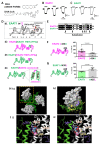
(A) The chemical structures of DHA and DHA-CoA. (B) Representative traces of L-glutamate (L-Glu, 50 μM for 2 min, black horizontal line)-induced current obtained from Xenopus oocytes overexpressing EAAT2 clamped at −50 mV in the absence or presence of DHA (100 μM for 2 min, grey horizontal line). When coapplied, DHA increased the L-Glu-induced EAAT2 current, and the effect disappeared after washout. (C) Representative traces of L-Glu-induced EAAT1 currents in the absence or presence of DHA. When the compounds were coapplied, DHA tended to decrease the EAAT1 current, and the effect disappeared after washout. (D) Topology of EAAT1, EAAT2 (d1). EAAT1/2 is organized into eight transmembrane (TM1-8) and two helical hairpins (HP1 and HP2), which are re-entrant loops. TM7b-HP2a sequence and connector sequence in the red square. d2-d4. EAAT1-EAAT2 hybrid chimeras: EAAT2 (EAAT1 TM7b-HP2a) (d2), EAAT1 (EAAT2 TM7b-HP2a) (d3), and EAAT1 (EAAT2 connector) (d4). (E) Amino acid alignment from TM7b to HP2a of EAAT2 and EAAT1. The common amino acids are shown on a black background. Single amino acid back-mutations were performed at the sites indicated by black arrowheads in the EAAT1 (EAAT2 TM7b-HP2a) chimaera. (F) Left, topology of EAAT2 L434A. Right, comparison of the effects of DHA on EAAT2 and EAAT2 L434A. Data are shown as rates of increase by DHA. (G) Left, topology of EAAT1 A435L. Right, comparison of the effects of DHA on EAAT1 and EAAT1 A435L. Data are shown as rates of increase by DHA. (H) Proposed binding conformation for DHA in the transport domain/scaffold domain interface of the EAAT2 homology model in the outward facing state (OFS). (h1). Extracellular view of the trimerized EAAT2 OFS homology model based on the EAAT1 crystal structure. The scaffold domain is shown as a green ribbon. The transport domain is shown as a grey surface. (h2). Magnified monomer in the hatched square in h1 in the presence of DHA. The lipid crevice calculated by SiteMap exists at the interface between the scaffold domain and the transport domain (yellow space). DHA is docked to the lipid crevice (carbon: purple spheres; hydrogen: white spheres). (I) Docking poses of DHA in the lipid pocket in the vicinity of HP2 according to the induced fit docking protocol. The scaffold domain and transport domain are shown in green and grey ribbons, respectively. The carbons in DHA and EAAT2 L434 are represented by purple and yellow sticks, respectively. The atoms in L-Glu are shown as follows: carbon, blue sphere; hydrogen, white sphere; oxygen, red sphere; nitrogen, hidden. Na+ is shown as a pink sphere. Two types of DHA conformations could be visualized according to the position of the carboxylic group, i.e., one with a carboxyl group on the upper side (i1) and the other with a carboxyl group on the lower side (i2). Both of them have similar U-shaped conformations. The inset shows the DHA conformations in each case. The three-dimensional position of DHA is in close proximity to the L-Glu binding site and Na+ binding site. Error bars represent the mean ±SD. The numbers written within parentheses in the respective figures represent the number of independent experiments. Statistical differences between groups were determined by two-tailed paired Student’s t test. p values are indicated in each figure panel [58].

Similar articles

Cited by

References

    1. Arriza J.L., Fairman W.A., Wadiche J.I., Murdoch G.H., Kavanaugh M.P., Amara S.G. Functional comparisons of three glutamate transporter subtypes cloned from human motor cortex. J. Neurosci. 1994;14:5559–5569. doi: 10.1523/JNEUROSCI.14-09-05559.1994. - DOI - PMC - PubMed
    1. Kanai Y., Hediger M.A. Primary structure and functional characterization of a high-affinity glutamate transporter. Nature. 1992;360:467–471. doi: 10.1038/360467a0. - DOI - PubMed
    1. Fairman W.A., Vandenberg R.J., Arriza J.L., Kavanaugh M.P., Amara S.G. An excitatory amino-acid transporter with properties of a ligand-gated chloride channel. Nature. 1995;375:599–603. doi: 10.1038/375599a0. - DOI - PubMed
    1. Arriza J.L., Eliasof S., Kavanaugh M.P., Amara S.G. Excitatory amino acid transporter 5, a retinal glutamate transporter coupled to a chloride conductance. Proc. Natl. Acad. Sci. USA. 1997;94:4155–4160. doi: 10.1073/pnas.94.8.4155. - DOI - PMC - PubMed
    1. Tanaka K., Watase K., Manabe T., Yamada K., Watanabe M., Takahashi K., Iwama H., Nishikawa T., Ichihara N., Kikuchi T., et al. Epilepsy and exacerbation of brain injury in mice lacking the glutamate transporter GLT-1. Science. 1997;276:1699–1702. doi: 10.1126/science.276.5319.1699. - DOI - PubMed

LinkOut - more resources