. 2024 Apr 20;14(4):238.
doi: 10.3390/metabo14040238.

Anaplerotic Therapy Using Triheptanoin in Two Brothers Suffering from Aconitase 2 Deficiency

Affiliations

Anaplerotic Therapy Using Triheptanoin in Two Brothers Suffering from Aconitase 2 Deficiency

Maximilian Penkl et al. Metabolites. .

Abstract

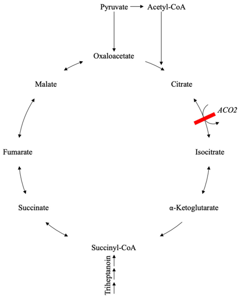
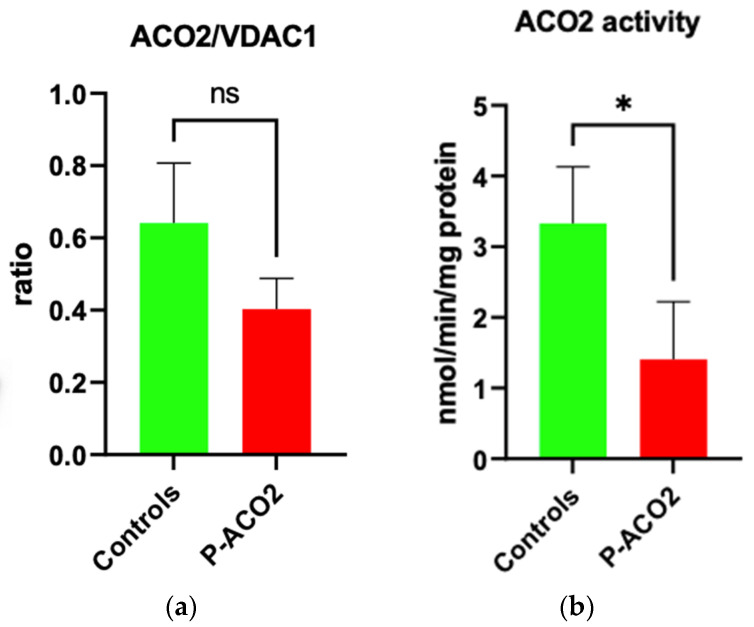
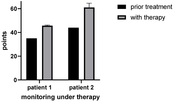
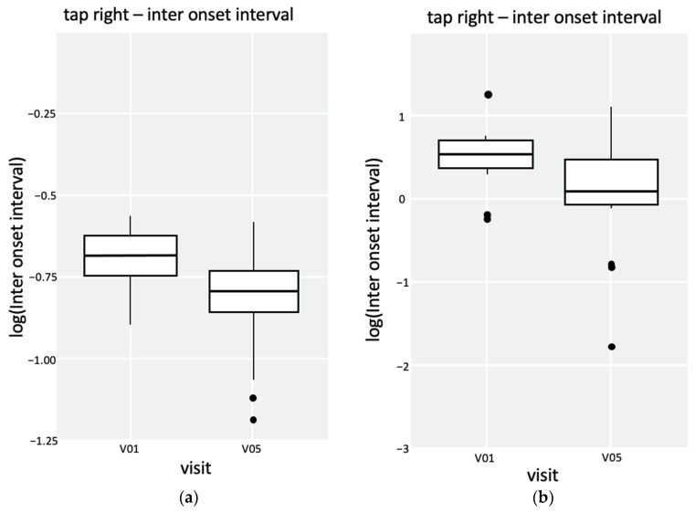
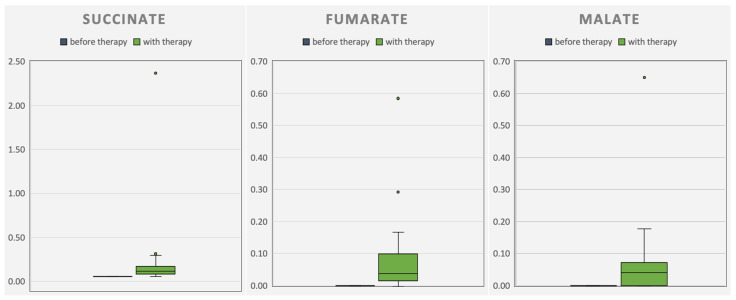
Citric acid cycle deficiencies are extremely rare due to their central role in energy metabolism. The ACO2 gene encodes the mitochondrial isoform of aconitase (aconitase 2), the second enzyme of the citric acid cycle. Approximately 100 patients with aconitase 2 deficiency have been reported with a variety of symptoms, including intellectual disability, hypotonia, optic nerve atrophy, cortical atrophy, cerebellar atrophy, and seizures. In this study, a homozygous deletion in the ACO2 gene in two brothers with reduced aconitase 2 activity in fibroblasts has been described with symptoms including truncal hypotonia, optic atrophy, hyperopia, astigmatism, and cerebellar atrophy. In an in vivo trial, triheptanoin was used to bypass the defective aconitase 2 and fill up the citric acid cycle. Motor abilities in both patients improved.

Keywords: ACO2 mutation; anaplerotic therapy; case report; citric acid cycle; triheptanoin.


Conflict of interest statement

The authors declare no conflicts of interest.

Figures

Figure 1
Figure 1
Scheme of the physiological and defective citric acid cycle in aconitase 2 deficiency with an overview of the CAC with its intermediates and bypassing Triheptanoin. The position of the aconitase 2 enzyme in CAC is illustrated as ACO2. Blocked pathway by defective ACO2 marked with a red line. Triheptanoin bypasses aconitase 2 and feeds the CAC as succinyl-CoA.
Figure 2
Figure 2
Determination of plasma C7 fatty acids for patient 1. Section of peak intensity chromatogram of plasma medium-chain free fatty acids by gas chromatography–single quadrupole mass spectrometry. The top image shows plasma C7 fatty acids prior to therapy, and the bottom image shows measurements during therapy.
Figure 3
Figure 3
Determination of plasma C7 fatty acids for patient 2. Section of peak intensity chromatogram of plasma medium-chain free fatty acids by gas chromatography–single quadrupole mass spectrometry. The top image shows plasma C7 fatty acids prior to therapy, and the bottom image shows measurements during therapy.
Figure 4
Figure 4
Western blot analysis of the ACO2 levels of the affected individual and four controls.
Figure 5
Figure 5
Determination of ACO2 activity in the affected individual and controls. (a) Densitometric analysis of the ACO2/VDAC1 ratio of the Western blot results. (b) Enzymatic activity of ACO2 in the affected individual and controls. VDAC1 was used as a mitochondrial loading control. A non-parametric Mann–Whitney U test was used for statistical analysis. The enzymatic measurement was repeated twice with the same controls used in Figure 3. For both experiments, isolated mitochondria from human primary skin fibroblasts were used. For the enzymatic determination, 50 µg mitochondria per 1 mL assay volume were taken. Values present mean ± SD. (a): p = 0.28; (b): * p = 0.0238.
Figure 6
Figure 6
Monitoring of motor ability with HFMS for patient 1 and CHOP for patient 2. Patient 1: Sum of calculated points in HFMS before (n = 1) and during therapy (n = 3). Patient 2: Sum of calculated points in CHOP before (n = 1) and during therapy (n = 3).
Figure 7
Figure 7
Q-Motor analysis of patient 1 with box plots of speeded tapping tests of the right hand and the left foot. (a) Speeded tapping performance of the right hand. The p-value in the Wilcoxon rank sum test is 9.813−9. (b) Speeded tapping performance of the left foot. The p-value in the Wilcoxon rank sum test is 0.003691.
Figure 8
Figure 8
Progression of the measured excretory organic acids succinate, malate, and fumarate in the urine of patient 1. The organic acids fumarate and malate before therapy (blue) were not detectable (n = 5). With therapy (green), a higher excretion of the organic acids was detected (n = 38).
Figure 9
Figure 9
Progression of the measured excretory organic acids succinate, malate, and fumarate in the urine of patient 2. Five samples before therapy (blue; n = 5). With therapy (green), a higher excretion of the organic acids fumarate and malate was detected (n = 38).

References

    1. Sadat R., Barca E., Masand R., Donti T.R., Naini A., De Vivo D.C., DiMauro S., Hanchard N.A., Graham B.H. Functional Cellular Analyses Reveal Energy Metabolism Defect and Mitochondrial DNA Depletion in a Case of Mitochondrial Aconitase Deficiency. Mol. Genet. Metab. 2016;118:28–34. doi: 10.1016/j.ymgme.2016.03.004. - DOI - PMC - PubMed
    1. Chen Y., Cai G.H., Xia B., Wang X., Zhang C.C., Xie B.C., Shi X.C., Liu H., Lu J.F., Zhang R.X., et al. Mitochondrial Aconitase Controls Adipogenesis through Mediation of Cellular ATP Production. FASEB J. 2020;34:6688–6702. doi: 10.1096/fj.201903224RR. - DOI - PubMed
    1. Neumann M.A.C., Grossmann D., Schimpf-Linzenbold S., Dayan D., Stingl K., Ben-Menachem R., Pines O., Massart F., Delcambre S., Ghelfi J., et al. Haploinsufficiency Due to a Novel ACO2 Deletion Causes Mitochondrial Dysfunction in Fibroblasts from a Patient with Dominant Optic Nerve Atrophy. Sci. Rep. 2020;10:16736. doi: 10.1038/s41598-020-73557-4. - DOI - PMC - PubMed
    1. Ciccarone F., Vegliante R., Di Leo L., Ciriolo M.R. The TCA Cycle as a Bridge between Oncometabolism and DNA Transactions in Cancer. Semin. Cancer Biol. 2017;47:50–56. doi: 10.1016/j.semcancer.2017.06.008. - DOI - PubMed
    1. Xin J.C., Wang X., Kaufman B.A., Butow R.A. Aconitase Couples Metabolic Regulation to Mitochondrial DNA Maintenance. Science. 2005;307:714–717. doi: 10.1126/science.1106391. - DOI - PubMed

LinkOut - more resources