. 2024 Apr 12;10(2):23.
doi: 10.3390/ncrna10020023.

Dynamic Localization of Paraspeckle Components under Osmotic Stress

Affiliations

Dynamic Localization of Paraspeckle Components under Osmotic Stress

Aysegul Yucel-Polat et al. Noncoding RNA. .

Abstract

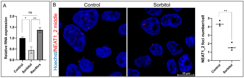
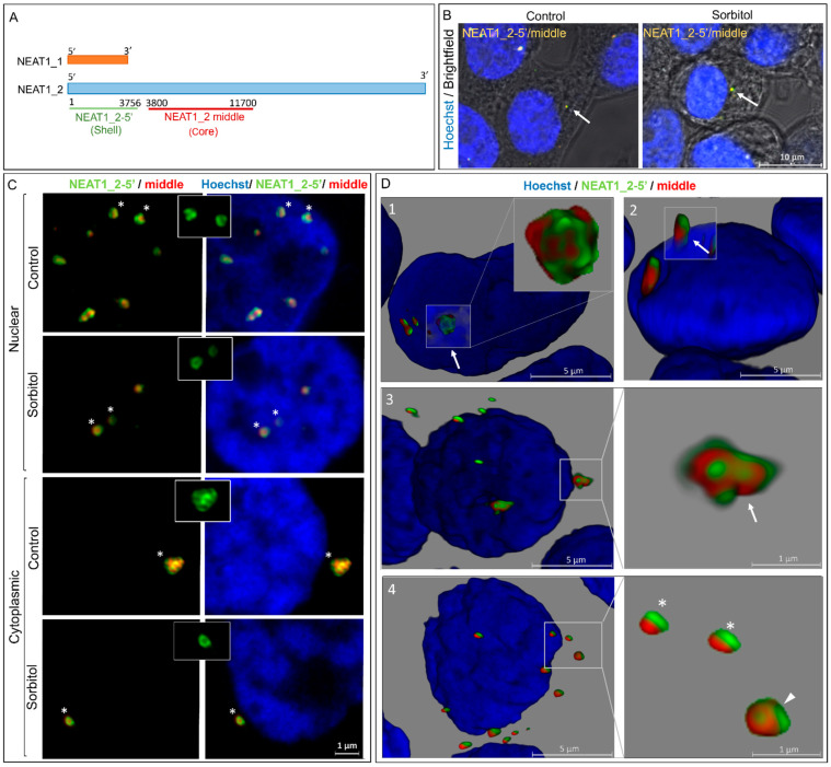
Paraspeckles are nuclear condensates formed by NEAT1_2 lncRNA and different RNA-binding proteins. In general, these membraneless organelles function in the regulation of gene expression and translation and in miRNA processing, and in doing this, they regulate cellular homeostasis and mediate pro-survival in the cell. Despite evidence showing the importance of paraspeckles in the stress response, the dynamics of paraspeckles and their components under conditions of osmotic stress remain unknown. We exposed HEK293T cells to sorbitol and examined NEAT1_2 expression using real-time PCR. Localization and quantification of the main paraspeckle components, NEAT1_2, PSPC1, NONO, and SFPQ, in different cellular compartments was performed using smFISH and immunofluorescence. Our findings showed a significant decrease in total NEAT1_2 expression in cells after osmotic stress. Sorbitol shifted the subcellular localization of NEAT1_2, PSPC1, NONO, and SFPQ from the nucleus to the cytoplasm and decreased the number and size of NEAT1_2 foci in the nucleus. PSPC1 formed immunoreactive cytoplasmic fibrils under conditions of osmotic stress, which slowly disassembled under recovery. Our study deepens the paraspeckle dynamics in response to stress, suggesting a novel role for NEAT1_2 in the cytoplasm in osmotic stress and physiological conditions.

Keywords: NEAT1_2; cytoplasmic aggregates; membraneless organelles; osmotic stress; paraspeckle proteins.


Conflict of interest statement

The authors declare no conflicts of interest.

Figures

Figure 1
Figure 1
NEAT1_2 expression decreases under the conditions of osmotic stress in HEK293T cells. (A) qPCR shows that total NEAT1_2 expression was significantly reduced under conditions of osmotic stress (sorbitol, 4 h) and returned to control levels after 4 h of recovery (Rec, 4 h) (ΔCt values were used for statistical analyses, one-way ANOVA, * p < 0.05, ** p < 0.01, n = 5, error bars indicate SEM). (B) Single-molecule FISH (smFISH) using Stellaris RNA probes against the NEAT1_2 middle segment shows that the number of NEAT1_2 foci in the cells decreased under conditions of osmotic stress (t-test, ** p < 0.01).
Figure 2
Figure 2
NEAT1_2 localizes in the cytoplasm of HEK293T cells forming typical shell–core structures. (A) Schematic representations of the positions of the smFISH probes used to identify distinct segments of NEAT1. (B) NEAT1_2 foci are observed in the cytoplasm at baseline and under conditions of osmotic stress (arrows). (C) The shell–core structure of NEAT1_2 (5′ segment in the shell and middle segment in the core) was detected in both cytoplasmic and nuclear NEAT1_2 foci in the control and sorbitol samples. Insets show the patchy pattern of the 5′ segment of NEAT1_2 foci. (asterisk used to demonstrate the granules in the insets) (D) 3D reconstruction shows NEAT1_2 foci of varying sizes and conformations in both the nuclear and cytosolic compartments. This includes clusters (arrows in 1 and 3) and single granules (arrowhead in 4) with shell–core structures in the nucleus (1) and cytoplasm (3 and 4) in the sorbitol samples. Small granules that, which lack the shell–core structure and showed the 5′ and middle segments of NEAT1_2 side by side, were also observed in the nucleus and cytosol under conditions of osmotic stress (asterisk in 4). Some NEAT1_2 clusters were observed partially residing within the nucleus and extending into the cytoplasm (arrow in 2).
Figure 3
Figure 3
The number and size of NEAT1_2 foci change in HEK293T cells under osmotic stress. (A) Quantification of smFISH experiments showed that the number of nuclear NEAT1_2 foci decreased under conditions of osmotic stress, while the cytoplasmic foci increased (100 cells per n, n = 3, one-way ANOVA, ** p < 0.01). (B) Violin plots show the distribution of NEAT1_2 foci size under different conditions. A decrease in NEAT1_2 foci size in the nucleus was observed under osmotic stress compared to the control and 4 h recovery (C: control; S: 4 h sorbitol; Rec: recovery-4 h; 100 cells per n, n = 3, One-way ANOVA, * p < 0.05, error bars indicate SEM). (C) Under conditions of osmotic stress, cytoplasmic NEAT1_2 foci were significantly larger than nuclear NEAT1_2 foci (100 cells per n, n = 3, t-test, ** p < 0.01, error bars indicate SEM).
Figure 4
Figure 4
Paraspeckle proteins colocalize in the cytoplasm of HEK293T cells after osmotic stress. (A) Under conditions of osmotic stress, PSPC1, NONO, and SFPQ redistributed from the nucleus to the cytoplasm where they colocalized. Pictures show the merging of the PSP staining. (B) After osmotic stress, PSPC1 immunoreactive fibrils were formed in the cytoplasm (arrows). After 8 h of recovery, cytoplasmic PSPC1(+) fibrils were observed partially assembled (arrowhead), and SFPQ and NONO returned to the nucleus. Nuclei were stained using Hoechst (blue).
Figure 5
Figure 5
Colocalization of NEAT1_2 with PSPs in the nucleus and cytoplasm of HEK293T cells. (A) smFISH/IF experiments showed that PSPC1, NONO, and NEAT1_2 form paraspeckles in the nucleus as expected in the baseline condition. After 30 min of incubation with sorbitol, PSPC1 and NONO redistributed to the cytosol, and paraspeckles began to disappear. After 4 h of sorbitol treatment, paraspeckles were barely visible in the nucleus. (B) NEAT1_2 localizes in small granules in the cytosol with (arrowheads) or without PSPC1, NONO, and SFPQ (arrows) under osmotic stress.
Figure 6
Figure 6
Paraspeckle components colocalize with stress granules in the cytoplasm under conditions of osmotic stress. (A) Colocalization images showed that cytoplasmic NEAT1_2 rarely colocalizes in G3BP1(+) SGs (I and II, asterisk). No extensive colocalization was observed (III and IV, arrowhead). (B) PSPC1, NONO, and SFPQ were present together in SGs after osmotic stress but only in a small subpopulation of the cells. Line profiles of the fluorescence intensities on the right show the colocalization of PSPs with TIA-1 and G3BP1 in areas in the squares.

References

    1. Yamazaki T., Yamamoto T., Hirose T. Micellization: A new principle in the formation of biomolecular condensates. Front. Mol. Biosci. 2022;9:974772. doi: 10.3389/fmolb.2022.974772. - DOI - PMC - PubMed
    1. Yamazaki T., Yamamoto T., Yoshino H., Souquere S., Nakagawa S., Pierron G., Hirose T. Paraspeckles are constructed as block copolymer micelles. EMBO J. 2021;40:e107270. doi: 10.15252/embj.2020107270. - DOI - PMC - PubMed
    1. Yamamoto T., Yamazaki T., Hirose T. Triblock copolymer micelle model of spherical paraspeckles. Front. Mol. Biosci. 2022;9:925058. doi: 10.3389/fmolb.2022.925058. - DOI - PMC - PubMed
    1. Chen L.L., Carmichael G.G. Altered nuclear retention of mRNAs containing inverted repeats in human embryonic stem cells: Functional role of a nuclear noncoding RNA. Mol. Cell. 2009;35:467–478. doi: 10.1016/j.molcel.2009.06.027. - DOI - PMC - PubMed
    1. Hirose T., Virnicchi G., Tanigawa A., Naganuma T., Li R., Kimura H., Yokoi T., Nakagawa S., Bénard M., Fox A.H., et al. NEAT1 long noncoding RNA regulates transcription via protein sequestration within subnuclear bodies. Mol. Biol. Cell. 2014;25:169–183. doi: 10.1091/mbc.e13-09-0558. - DOI - PMC - PubMed

LinkOut - more resources