. 2024 Sep;34(3):693-701.
doi: 10.1007/s00062-024-01418-y. Epub 2024 Apr 26.

Thalamo-mesencephalic Branches of the Posterior Cerebral Artery: a 3D Rotational Angiography Study

Affiliations

Thalamo-mesencephalic Branches of the Posterior Cerebral Artery: a 3D Rotational Angiography Study

Maximilian Rauch et al. Clin Neuroradiol. 2024 Sep.

Abstract

Purpose: The thalamo-mesencephalic (TM) branches of the posterior cerebral artery (PCA) supply critical structures. Previous descriptions of these vessels are inconsistent and almost exclusively rely on cadaver studies. We aimed to provide a neuroradiological description of TM vessels in vivo based on routine 3D rotational angiographies (3D-RA).

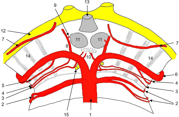
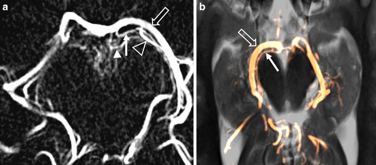
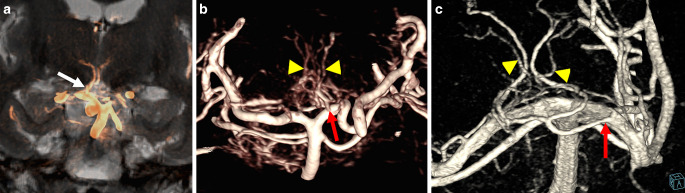
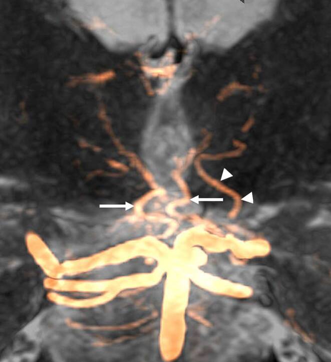
Methods: We analyzed 3D-RAs of 58 patients with pathologies remote from the PCA. PCA-origins were considered. Delineation, origin and number of branches of the collicular artery (CA), the accessory CA (ACA), the posterior thalamoperforating artery (PTA), the thalamogeniculate artery (TGA), and the posterior medial (PMCA) and lateral (PCLA) choroid arteries were assessed. The PTAs were categorized based on Percheron's suggested classification.

Results: A CA was identified in 84%, an ACA in 20%. The PTA was delineated in 100%. In 27%, PTA anatomy had features of several Percheron types (n = 7) or vessels emanating from a net like origin (n = 9). 26% had a type IIb PTA. A fetal type PCA origin with hypoplastic ipsilateral P1 was observed in 5 cases with type IIa (n = 2) or type IIb (n = 3) PTAs originating from contralateral P1. The TGA was identified in 85% of patients, with ≥ 2 branches in 67%. The PMCA was delineable in 41%, the PLCA in 100%.

Conclusion: The prevalence of a proper "Artery of Percheron" type IIb PTA seems to be higher than previously reported. A fetal type P1-origin may be predictive of a type IIa/b PTA emanating from contralateral P1. 3D-RA may be useful for planning PCA interventions, as impairment of TM branches is a severe risk.

Keywords: Digital subtraction angiography; Mesencephalon; Neuroanatomy; Posterior cerebral artery; Thalamus.


Conflict of interest statement

M. Rauch, J. Berkefeld, M. Klonowski, E. Hattingen and S. Weidauer declare that they have no competing interests.

Figures

Fig. 1
Fig. 1
Study flowchart describing the data procurement and patient selection process intended for retrospective analysis
Fig. 2
Fig. 2
a Sagittal 3D rotational angiography maximum intensity projection (MIP) of the intracranial posterior arterial circulation, red square delineating magnification in Fig. 2b. b sagittal MIP of the proximal posterior cerebral artery (PCA) and its branches. BA basilar artery; PLCA posterior lateral choroidal artery; PMCA posterior medial choroidal artery; PTA posterior thalamoperforating arteries; SCA superior cerebellar artery; TGA thalamogeniculate artery; P1–P3 segments of the PCA
Fig. 3
Fig. 3
Superficial course of the arteries of the upper pons and mesencephalon. 1: basilar artery; 2: superior cerebellar artery, lateral branches arises inferior, medial branches arises superior; 3: collicular artery; 4: accessory collicular artery; 5: posterior medial choroidal artery; 6: posterior cerebral artery; 7: anterior choroidal artery; 8: posterior communicating artery; 9: anterior thalamoperforating artery (tuberothalamic artery); 10: posterior thalamoperforating arteries; 11: mammillary bodies; 12: optic tract; 13: hypophyseal stalk; 14: cerebral peduncles; 15: oculomotor nerve
Fig. 4
Fig. 4
Percheron’s classification of the posterior thalamoperforating arteries (PTA). a type 1, bilateral branches (arrows) originating from the P1-segment of the posterior cerebral artery (PCA). b type 2a, unilateral branches (arrows). c type 2b, unilateral common trunk of the PTA, i.e. proper “artery of Percheron” (arrow). d bilateral branches with common anastomosing origin (arrow)
Fig. 5
Fig. 5
a 3D rotational digital subtraction angiography, axial maximum intensity projection (MIP) of the posterior cerebral artery (PCA, open arrow) with collicular artery (CA, arrow), accessory CA (arrowhead) and posterior medial choroidal artery (PMCA, open arrowhead). b axial MIP of the PCA (open arrow) and the CA (arrow) projected onto axial T2 weighted MR images
Fig. 6
Fig. 6
Artery of Percheron. a coronal maximum intensity projection (MIP) of the artery of Percheron (arrow) projected onto coronal T2 weighted MR image. b posterior thalamoperforating arteries (PTA, arrowheads) emanating from a netlike unilateral structure (arrow). c volume rendering of the basilar tip and the P1-segments showing the artery of Percheron (arrow) and accessory small penetrating branches (arrowheads) originating from a netlike structure surrounding the basilar tip and the right P1-segment
Fig. 7
Fig. 7
Transverse section of the upper mesencephalon and course of the collicular artery (CA) and lateral vascular territories of the midbrain. 1: posterior cerebral artery (PCA, P1-segment); 2: CA; 3, 4: accessory CA (ACA), left ACA originating from the P1-segment, right ACA originating from the CA; 5: anterior choroidal artery (ACHA); 6: substantia nigra; 7: tectal plate; green: anterolateral group (CA, ACHA, posterior medial choroidal artery [PMCA]); blue: lateral group (CA, PMCA, PCA); purple: posterior group (CA, PMCA)
Fig. 8
Fig. 8
Coronal maximum intensity projection (MIP) of the posterior thalamoperforating arteries (PTA, arrows) and the left anterior thalamoperforating artery (ATA, arrowheads) projected onto a coronal T2 weighted MR image

References

    1. Bogousslavsky J, Regli F, Uske A. Thalamic infarcts: clinical syndromes, etiology, and prognosis. Neurology. 1988;38:837–48. 10.1212/WNL.38.6.837 - DOI - PubMed
    1. Carrera E, Bogousslavsky J. The thalamus and behavior: effects of anatomically distinct strokes. Neurology. 2006;66:1817–23. 10.1212/01.wnl.0000219679.95223.4c - DOI - PubMed
    1. Weidauer S, Nichtweiß M, Zanella F, Lanfermann H. Assessment of paramedian thalamic infarcts: MR imaging, clinical features and prognosis. Eur Radiol. 2004;14:1615–26. 10.1007/s00330-004-2303-7 - DOI - PubMed
    1. Ruchalski K, Hathout GM. A medley of midbrain maladies: a brief review of midbrain anatomy and syndromology for radiologists. Radiol Res Pract. 2012;2012:258524. - PMC - PubMed
    1. Grochowski C, Maciejewski R. Diversity among posterior thalamoperforating branches originated from P1 segment: systematic review. Folia Morphol (warsz). 2017;76:335–9. 10.5603/FM.a2017.0012 - DOI - PubMed

LinkOut - more resources