

Estimation of the number of motor units in the human extensor digitorum brevis using MScanFit
- PMID: 38669263
- PMCID: PMC11051589
- DOI: 10.1371/journal.pone.0302214
Estimation of the number of motor units in the human extensor digitorum brevis using MScanFit
Abstract
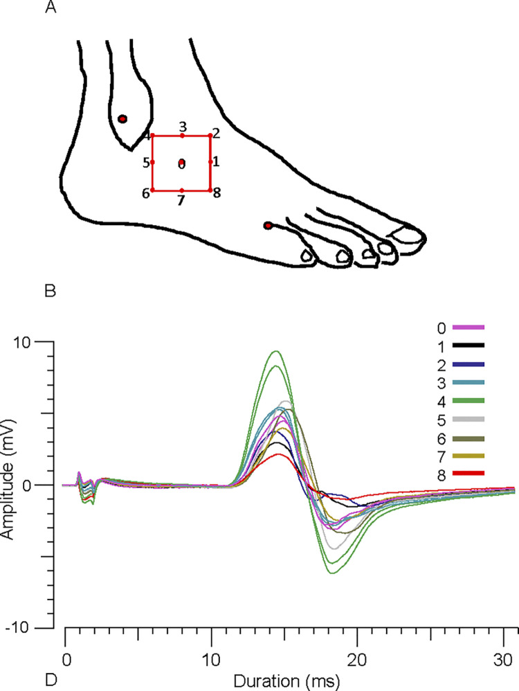
Objective: Our aim was to determine the number and size parameters of EDB motor units in healthy young adults using MScanFit, a novel approach to motor unit number estimation (MUNE). Since variability in MUNE is related to compound muscle action potential (CMAP) size, we employed a procedure to document the optimal EDB electromyographic (EMG) electrode position prior to recording MUNE, a neglected practice in MUNE.
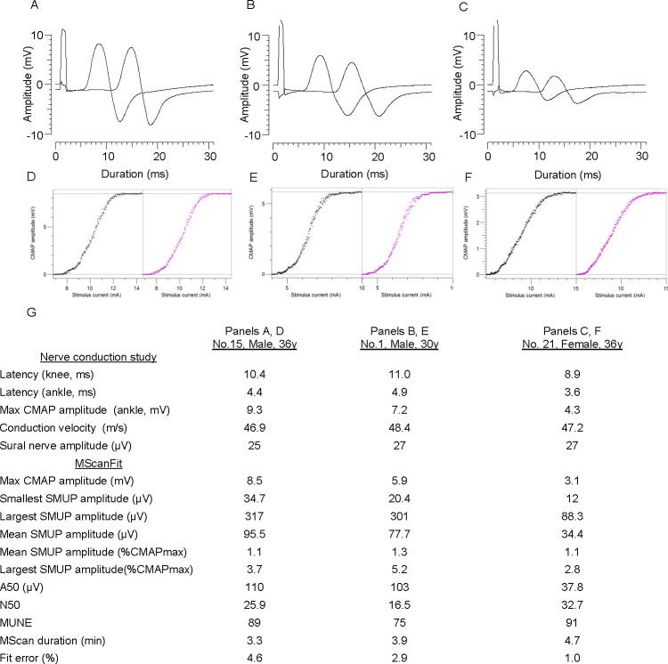
Methods: Subjects were 21 adults 21-44 y. Maximum CMAPs were recorded from 9 sites in a 4 cm2 region centered over the EDB and the site with the largest amplitude was used in the MUNE experiment. For MUNE, the peroneal nerve was stimulated at the fibular head to produce a detailed EDB stimulus-response curve or "MScan". Motor unit number and size parameters underlying the MScan were simulated using the MScanFit mathematical model.
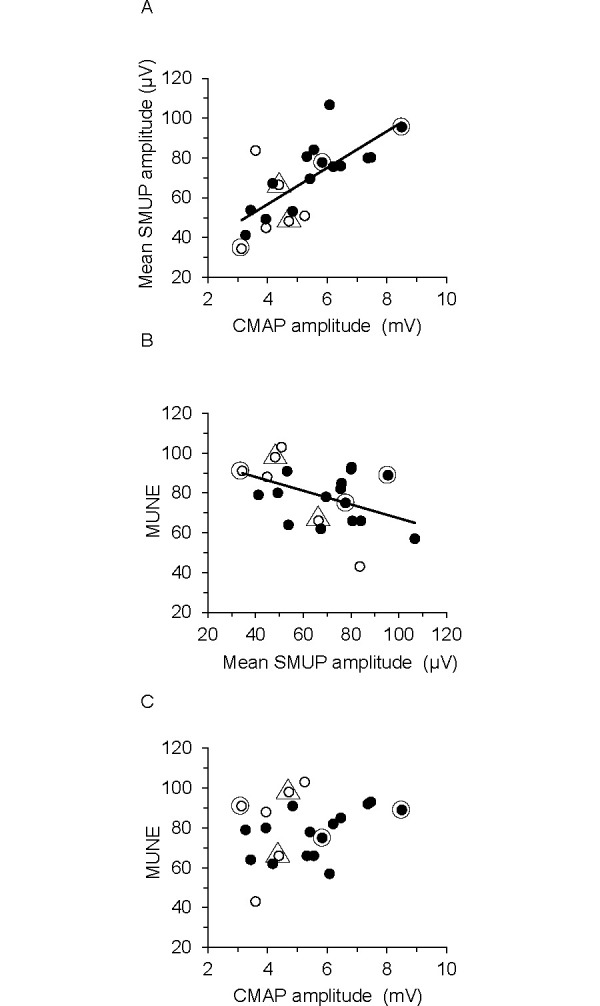
Results: In 19 persons, the optimal recording site was superior, superior and proximal, or superior and distal to the EDB mid-belly, whereas in 3 persons it was proximal to the mid-belly. Ranges of key MScanFit parameters were as follows: maximum CMAP amplitude (3.1-8.5 mV), mean SMUP amplitude (34.4-106.7 μV), mean normalized SMUP amplitude (%CMAP max, 0.95-2.3%), largest SMUP amplitude (82.7-348 μV), and MUNE (43-103). MUNE was not related to maximum CMAP amplitude (R2 = 0.09), but was related to mean SMUP amplitude (R2 = -0.19, P = 0.05).
Conclusion: The EDB CMAP was highly sensitive to electrode position, and the optimal position differed between subjects. Individual differences in EDB MUNE were not related to CMAP amplitude. Inter-subject variability of EDB MUNE (coefficient of variation) was much less than previously reported, possibly explained by better optimization of the EMG electrode and the unique approach of MScanFit MUNE.
Copyright: © 2024 Klein et al. This is an open access article distributed under the terms of the Creative Commons Attribution License, which permits unrestricted use, distribution, and reproduction in any medium, provided the original author and source are credited.
Conflict of interest statement
The authors have declared that no competing interests exist.
Figures
Similar articles
-
MScanFit and StairFit Motor Unit Number Estimation of the Extensor Digitorum Brevis and Abductor Digiti Minimi Muscles.Muscle Nerve. 2025 Mar;71(3):446-449. doi: 10.1002/mus.28341. Epub 2025 Jan 10. Muscle Nerve. 2025. PMID: 39789990
-
Feasibility and reliability of MScanFit motor unit number estimation in peroneus longus muscle.Muscle Nerve. 2022 Oct;66(4):503-507. doi: 10.1002/mus.27667. Epub 2022 Jul 13. Muscle Nerve. 2022. PMID: 35763284
-
Reliability of MScanFit Motor Unit Number Estimation in the Trapezius Muscle.Muscle Nerve. 2025 Feb;71(2):166-170. doi: 10.1002/mus.28303. Epub 2024 Nov 27. Muscle Nerve. 2025. PMID: 39601192
-
Motor unit number estimation (MUNE): Where are we now?Clin Neurophysiol. 2018 Aug;129(8):1507-1516. doi: 10.1016/j.clinph.2018.04.748. Epub 2018 May 24. Clin Neurophysiol. 2018. PMID: 29804042 Review.
-
Statistical motor unit number estimation: from theory to practice.Muscle Nerve. 2003 Sep;28(3):263-72. doi: 10.1002/mus.10351. Muscle Nerve. 2003. PMID: 12929186 Review.
References
Publication types
MeSH terms
LinkOut - more resources
Full Text Sources
