

Longitudinal brain volumetrics in glioma survivors
- PMID: 38669700
- PMCID: PMC11875424
- DOI: 10.3171/2024.1.JNS231980
Longitudinal brain volumetrics in glioma survivors
Abstract
Objective: Radiation therapy (RT) is used selectively for patients with low-grade glioma (LGG) given the concerns for potential cognitive effects in survivors, but prior cognitive outcome studies among LGG survivors have had inconsistent findings. Translational studies that characterize changes in brain anatomy and physiology after treatment of LGG may help to both contextualize cognitive findings and improve the overall understanding of radiation effects in normal brain tissue. This study aimed to investigate the hypothesis that patients with LGG who are treated with RT will experience greater brain volume loss than those who do not receive RT.
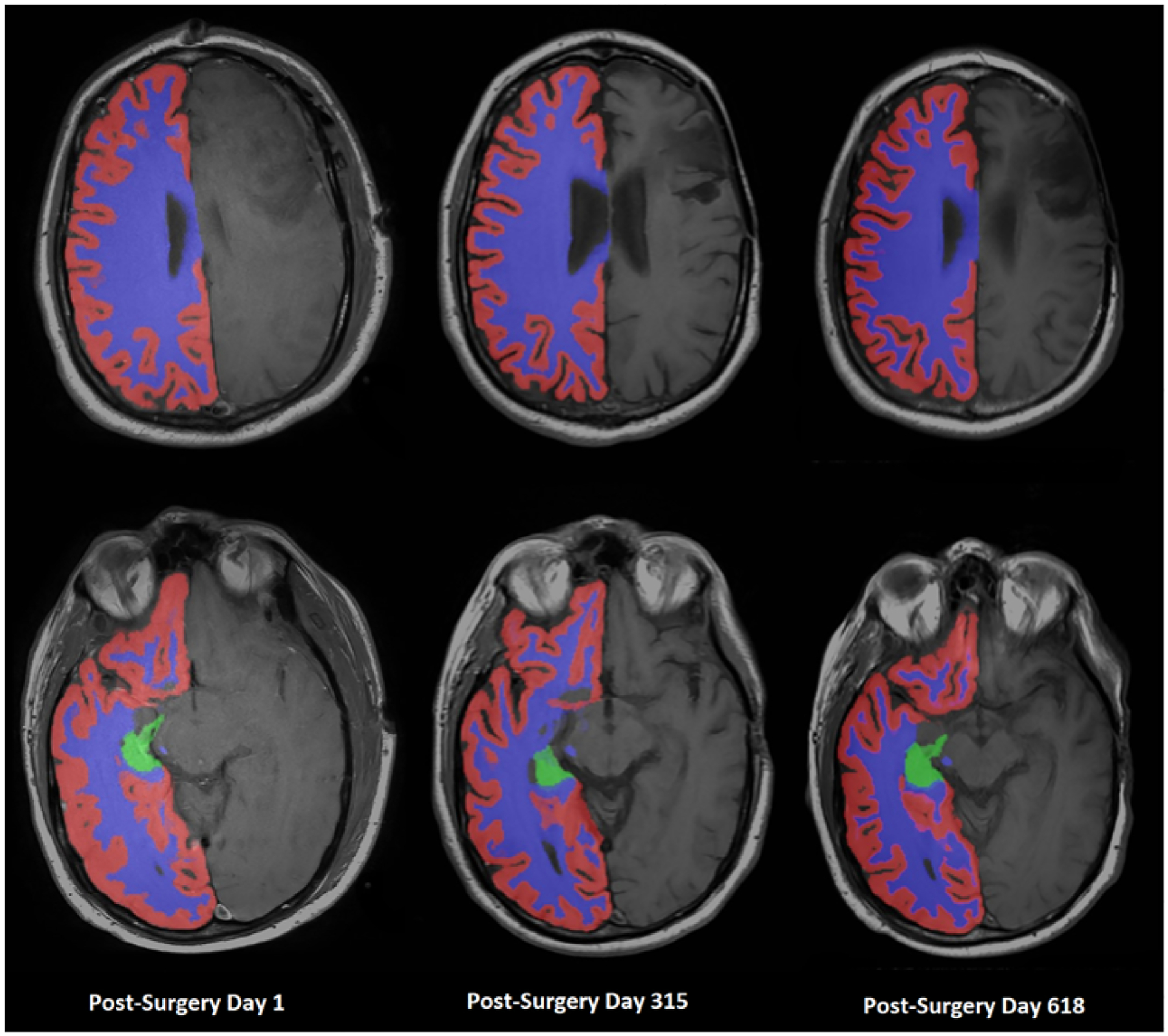
Methods: This retrospective longitudinal study included all patients with WHO grade 2 glioma who received posttreatment surveillance MRI at the University of Alabama at Birmingham. Volumetric analysis of contralateral cortical white matter (WM), cortical gray matter (GM), and hippocampus was performed on all posttreatment T1-weighted MRI sequences using the SynthSeg script. The effect of clinical and treatment variables on brain volumes was assessed using two-level hierarchical linear models.
Results: The final study cohort consisted of 105 patients with 1974 time points analyzed. The median length of imaging follow-up was 4.6 years (range 0.36-18.9 years), and the median number of time points analyzed per patient was 12 (range 2-40). Resection was performed in 79 (75.2%) patients, RT was administered to 61 (58.1%) patients, and chemotherapy was administered to 66 (62.9%) patients. Age at diagnosis (β = -0.06, p < 0.001) and use of RT (β = -1.12, p = 0.002) were associated with the slope of the contralateral cortical GM volume model (i.e., change in GM over time). Age at diagnosis (β = -0.08, p < 0.001), midline involvement (β = 1.31, p = 0.006), and use of RT (β = -1.45, p = 0.001) were associated with slope of the contralateral cortical WM volume model. Age (β = -0.0027, p = 0.001), tumor resection (β = -0.069, p < 0.001), use of chemotherapy (β = -0.0597, p = 0.003), and use of RT (β = -0.0589, p < 0.001) were associated with the slope of the contralateral hippocampus volume model.
Conclusions: This study demonstrated volume loss in contralateral brain structures among LGG survivors, and patients who received RT experienced greater volume loss than those who did not. The results of this study may help to provide context for cognitive outcome research in LGG survivors and inform the design of future strategies to preserve cognition.
Keywords: MRI; brain volumetry; hierarchical linear modeling; low-grade glioma; oncology; radiation therapy; tumor.
Figures


References
-
- Nabors LB, Portnow J, Ahluwalia M, et al. Central Nervous System Cancers, Version 3.2020, NCCN Clinical Practice Guidelines in Oncology. J Natl Compr Canc Netw. 2020; 18(11): 1537–1570. - PubMed
-
- van den Bent MJ, Afra D, de Witte O, et al. Long-term efficacy of early versus delayed radiotherapy for low-grade astrocytoma and oligodendroglioma in adults: the EORTC 22845 randomised trial. Lancet. 2005; 366(9490): 985–990. - PubMed
-
- Koutsarnakis C, Neromyliotis E, Komaitis S, et al. Effects of brain radiotherapy on cognitive performance in adult low-grade glioma patients: a systematic review. Radiother Oncol. 2021; 160: 202–211. - PubMed
-
- Teipel SJ, Grothe M, Lista S, Toschi N, Garaci FG, Hampel H. Relevance of magnetic resonance imaging for early detection and diagnosis of Alzheimer disease. Med Clin North Am. 2013; 97(3): 399–424. - PubMed
MeSH terms
Grants and funding
LinkOut - more resources
Full Text Sources
Medical
