Review
. 2024 Apr 25;187(9):2052-2078.
doi: 10.1016/j.cell.2024.03.037.

Principles and therapeutic applications of adaptive immunity

Affiliations
Review

Principles and therapeutic applications of adaptive immunity

Hongbo Chi et al. Cell. .

Abstract

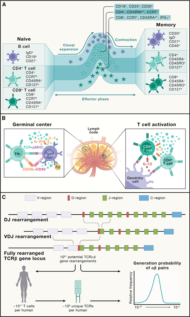
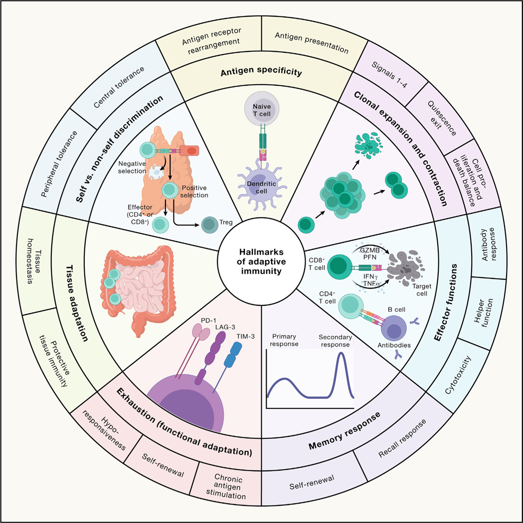
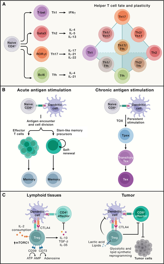
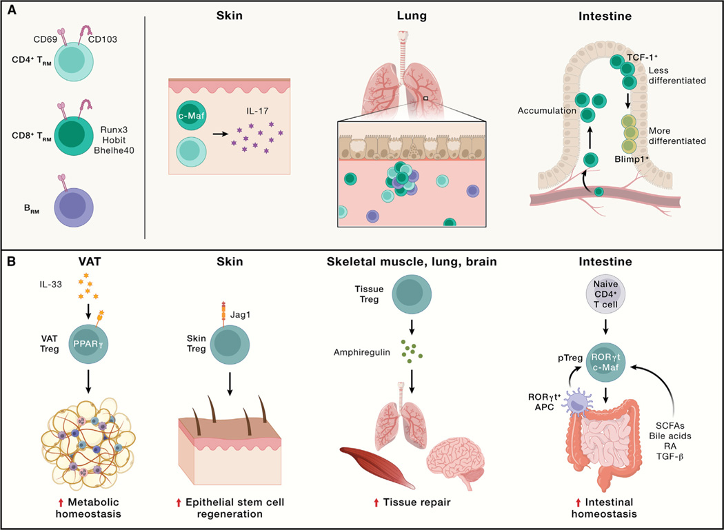
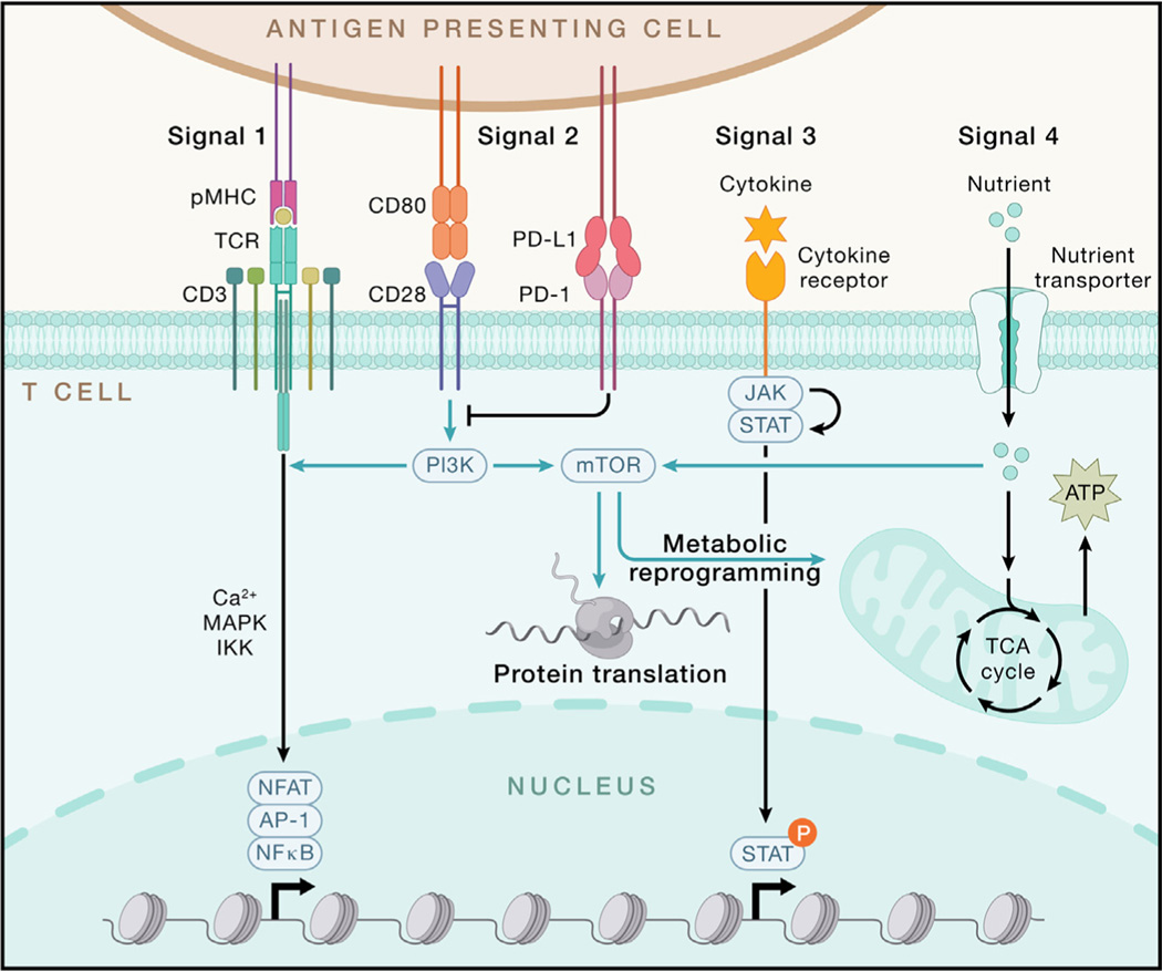
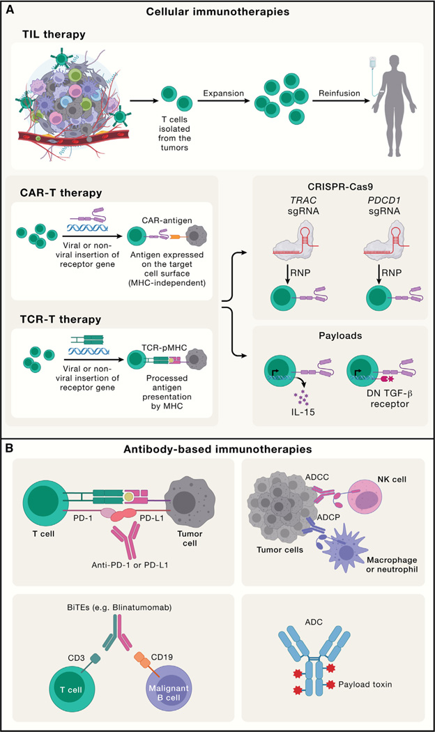
Adaptive immunity provides protection against infectious and malignant diseases. These effects are mediated by lymphocytes that sense and respond with targeted precision to perturbations induced by pathogens and tissue damage. Here, we review key principles underlying adaptive immunity orchestrated by distinct T cell and B cell populations and their extensions to disease therapies. We discuss the intracellular and intercellular processes shaping antigen specificity and recognition in immune activation and lymphocyte functions in mediating effector and memory responses. We also describe how lymphocytes balance protective immunity against autoimmunity and immunopathology, including during immune tolerance, response to chronic antigen stimulation, and adaptation to non-lymphoid tissues in coordinating tissue immunity and homeostasis. Finally, we discuss extracellular signals and cell-intrinsic programs underpinning adaptive immunity and conclude by summarizing key advances in vaccination and engineering adaptive immune responses for therapeutic interventions. A deeper understanding of these principles holds promise for uncovering new means to improve human health.


Conflict of interest statement

Declaration of interests H.C. has consulted for Kumquat Biosciences, Inc., Chugai Pharmaceutical Co., and ONO Pharmaceutical Co. and is a co-inventor on patents/patent applications in the fields of immunotherapy. M.P. serves on the scientific advisory board of Vaxart, Inc. P.G.T. has consulted or received honoraria and/or travel support from Illumina, J&J, Pfizer, Merck, and 10x Genomics, and his lab has received funding from ElevateBio. P.G.T. serves on the scientific advisory boards of ImmunoScape, Shennon Bio, and CytoAgents.

Figures

Figure 1.
Figure 1.. Adaptive immune cell states and the primary response
(A) B cells, CD4+ T cells, and CD8+ T cells acquire multiple differentiation fates during a normal immune response. Naive T cells and B cells are quiescent cells. Upon activation (not depicted; see B), these cells undergo clonal expansion and acquire effector function (effector phase). Following pathogen control, most effector cells undergo apoptosis (contraction), leaving long-lived memory cells to provide continued immune surveillance (memory). Shown are the major markers used to distinguish various differentiation states or subsets of human T cell and B cell populations discussed in this review. (B) Primary immune activation occurs in the lymph node. Lymph nodes are spatially organized into discrete B cell and T cell zones. Innate immune cells, especially DCs, process and present antigens to naive CD4+ and CD8+ T cells in the paracortical T cell zone, activating them and inducing clonal expansion. Here, the secretion of IL-12 by DCs drives type 1 differentiation in the responding T cells. IL-2 secretion by CD4+ T cells promotes CD8+ T cell expansion and effector function. Follicular CD4+ T cells also migrate to the B cell zone, where they interact with antigen-specific B cells in the germinal center. Here, B cell recognition of intact antigen via BCRs promotes B cell activation and antigen presentation to CD4+ T cells, which in turn provide activation signals to B cells by CD40L-CD40 interaction and cytokines (e.g., IL-4 and IL-21). (C) The generation of antigen receptor diversity. TCRs and BCRs are generated by the process of V(D)J recombination, which involves the fusion of separate V, D (on TCRβ and heavy chains), and J segments. Depicted is a TCRβ-chain recombination. Subsequent mRNA splicing brings the constant region together with the V(D)J fusion. This process can result in a vast potential diversity, which is only minimally represented in any individual organism. The distribution of probabilities for an individual TCR to recombine in an individual varies over 20 orders of magnitude.
Figure 2.
Figure 2.. The hallmarks of adaptive immune responses
The scheme illustrates the seven hallmarks of adaptive immunity described within the article: antigen specificity (contributed by antigen receptor rearrangement and antigen presentation); clonal expansion and contraction (contributed by antigen, co-stimulation, cytokines, and nutrients [signals 1–4, respectively]; quiescence exit; and a balance of cell proliferation and death); effector functions (mediated by B cell antibody responses, CD4+ T cell helper function, and CD8+ T cell cytotoxicity); memory response (contributed by recall response and self-renewal of memory cells); exhaustion or functional adaptation (contributed by chronic antigen stimulation, self-renewal, and hypo-responsiveness); tissue adaptation (presented as protective tissue immunity and tissue homeostasis); and self versus non-self discrimination (contributed by central and peripheral tolerance). PFN, perforin; GZMB, granzyme B.
Figure 3.
Figure 3.. Major types of adaptive immune responses, including effector, memory, exhausted T cell, and regulatory T cell responses
(A) Left: naive CD4+ T cells become activated via TCR activation combined with co-stimulation and further respond to cytokines in the microenvironment to differentiate into Th1, Th2, Th17, and Tfh cells. These effector T cells express the unique transcription factors T-bet, Gata3, RORγt, and Bcl6, respectively, and produce distinct cytokines. Specifically, Th1 cells produce IFNγ; Th2 cells secrete IL-4, IL-5, and IL-13; Th17 cells synthesize IL-17, IL-21, and IL-22; and Tfh cells generate IL-4 and IL-21. Right: the schematic highlights the representative interconnectedness in the plasticity between CD4+ helper T cell subsets. T helper cells with plasticity can display phenotypes from two distinct CD4+ T cell lineages. (B) Acute (left) and chronic (right) antigen stimulation results in the development of memory and exhausted CD8+ T cells, respectively. Left: memory CD8+ T cells can de-differentiate from a subset of effector cells or arise directly from stem-like memory precursors that are derived from antigen-stimulated naive T cells. Right: under persistent antigen stimulation, CD8+ T cells upregulate TOX expression and differentiate into Tpex cells, which further become Tex cells. In Tex cells, a transitory Tex population bridges the differentiation between Tpex and Tex cells, although Tex cells may also form without progression through a functional effector state (not depicted). (C) Left: in lymphoid tissues, thymus-derived Treg cells expressing the transcription factor Foxp3 exert immune suppressive function through multiple mechanisms. Treg cells secrete immunosuppressive cytokines (e.g., IL-10, TGF-β, and IL-35) and express co-inhibitory molecules such as CTLA4 to suppress effector T cells. They also express enzymes CD39 and CD73 that convert pro-inflammatory extracellular ATP to adenosine. The high expression of IL-2 receptors on Treg cells serves as an IL-2 “sink” to dampen IL-2-induced stimulatory effects on NK and CD8+ T cells (not depicted). Both IL-2 signaling and mTORC1 activity are required for Treg suppressive function and metabolic fitness in vivo. Right: Treg cells are a major component of the immunosuppressive tumor microenvironment. Intratumoral Treg cells have unique requirements for nutrients and metabolic programs and may co-localize with a specialized dendritic cell population called cDC1s to suppress the antitumor function of CD8+ T cells.
Figure 4.
Figure 4.. Functional importance of lymphocytes in non-lymphoid tissues in maintaining tissue immunity and homeostasis
(A) TRM and BRM cells in different non-lymphoid tissues or tumors. CD69 is a typical tissue-resident marker for TRM and BRM cells, while CD103 expression is more limited in lineage and tissue location compared with CD69. CD8+ TRM cells are more extensively studied, with transcriptional regulators such as Runx3, Hobit, and Bhelhe40 identified in certain tissues. In the skin, both CD4+ and CD8+ TRM cells producing IL-17 are present, with CD8+ TRM cells requiring c-Maf but not Runx3 for their generation. In the lung, CD4+ TRM, CD8+ TRM, and BRM cells are present during influenza infection. In the intestine, two distinct subsets of CD8+ TRM cells are present: one with enhanced memory potential and high TCF-1 expression, while the other expresses high levels of Blimp1. (B).Treg cells in different non-lymphoid tissues. Visceral adipose tissue (VAT) Treg cells, regulated by PPARγ and IL-33 signaling, orchestrate metabolic homeostasis and downmodulate tissue inflammation. In the skin, Treg cells expressing Notch ligand Jagged 1 (Jag1) localize to hair follicles and promote epithelial stem cell regeneration. Upon tissue injury, Treg cells produce amphiregulin to promote tissue repair in multiple tissues, including the skeletal muscle, lung, and brain. In the intestine, peripheral Treg (pTreg) cells are derived from naive T cells in response to short-chain fatty acids (SCFAs), secondary bile acids, retinoic acid (RA), or TGF-β and are marked by RORγt and c-Maf expression. RORγt+ APCs are crucial for intestinal pTreg cell generation.
Figure 5.
Figure 5.. Integration of extrinsic signals and intrinsic programs drives T cell responses
T cell activation largely relies on signals 1–3 (antigen, co-stimulation, and cytokines). TCR stimulation occurs upon recognition of the peptide presented by MHC molecule (pMHC), which is expressed on antigen-presenting cells (APCs). TCR-pMHC engagement initiates downstream signaling, leading to the activation of transcription factors NFAT, AP-1, and NF-κB mainly through Ca2+, mitogen-activated protein kinase (MAPK), and IKK-dependent signaling, respectively. CD28 serves as the major signal 2 for T cell activation, which induces downstream PI3K-AKT-mTOR signaling, while PD-1 delivers a co-inhibitory signal to block CD28-dependent signaling. Upon binding their receptors, signal 3 cytokines activate JAK-induced STAT phosphorylation, which is pivotal for potentiating T cell activation and differentiation. Nutrients function as signal 4 to license T cell immunity partly through fueling the tricarboxylic acid (TCA) cycle to generate ATP and activating mTOR to enhance metabolism and protein translation during T cell activation. P, phosphorylation.
Figure 6.
Figure 6.. Engineering adaptive immune responses for cell-and antibody-based therapies
(A).TIL, CAR-T, and TCR-T are three prominent cellular immunotherapies for cancer. For TIL therapy, T cells are isolated from the tumors for expansion and reinfusion into tumor patients; for CAR-T cells, CARs are engineered to recognize surface antigen independently of MHC-mediated antigen presentation; for TCR-T, a specific TCR gene recognizing the tumor antigen is ectopically expressed in T cells to expand tumor-specific T cells for tumor killing, which requires peptide-MHC (pMHC) expression on tumor cells. For CAR-T or TCR-T therapies, new strategies have been developed to modify intracellular signaling pathways in these tumor-specific T cells to enhance tumor killing, such as CRISPR-mediated genome editing of the TRAC or PDCD1 locus or introduction of payloads (e.g., cytokines like IL-15 and dominant-negative [DN] TGF-β receptor) expressed on T cells. These strategies increase the antitumor function in these cellular therapies. RNP, Cas9-expressing ribonucleoprotein. (B).Antibody-based immunotherapies include ICB targeting co-inhibitory molecules (e.g., PD-1 or PD-L1), which unleash endogenous T cell-mediated antitumor responses; antibody-dependent cellular cytotoxicity (ADCC; mainly mediated by NK cells) or antibody-dependent cellular phagocytosis (ADCP; mainly mediated by macrophages and neutrophils); a BiTE that bridges T cells and tumor cells by targeting the CD3 chain of TCRs on the T cells and CD19 on malignant B cells to promote T cell-mediated tumor killing; and ADCs, which are composed of a tumor antigen-targeting antibody, a linker, and a payload toxin.

Similar articles

Cited by

References

    1. Cooper MD (2015). The early history of B cells. Nat. Rev. Immunol. 15, 191–197. 10.1038/nri3801. - DOI - PubMed
    1. Miller JFAP (2020). The function of the thymus and its impact on modern medicine. Science 369, eaba2429. 10.1126/science.aba2429. - DOI - PubMed
    1. Lu LL, Suscovich TJ, Fortune SM, and Alter G. (2018). Beyond binding: antibody effector functions in infectious diseases. Nat. Rev. Immunol. 18, 46–61. 10.1038/nri.2017.106. - DOI - PMC - PubMed
    1. Dupic T, Marcou Q, Walczak AM, and Mora T. (2019). Genesis of the αβ T-cell receptor. PLoS Comput. Biol. 15, e1006874. 10.1371/journal.pcbi.1006874. - DOI - PMC - PubMed
    1. Farber DL, Netea MG, Radbruch A, Rajewsky K, and Zinkernagel RM (2016). Immunological memory: lessons from the past and a look to the future. Nat. Rev. Immunol. 16, 124–128. 10.1038/nri.2016.13. - DOI - PubMed

Publication types

LinkOut - more resources