

Oxytocin and women's health in midlife
- PMID: 38670161
- PMCID: PMC11404667
- DOI: 10.1530/JOE-23-0396
Oxytocin and women's health in midlife
Abstract
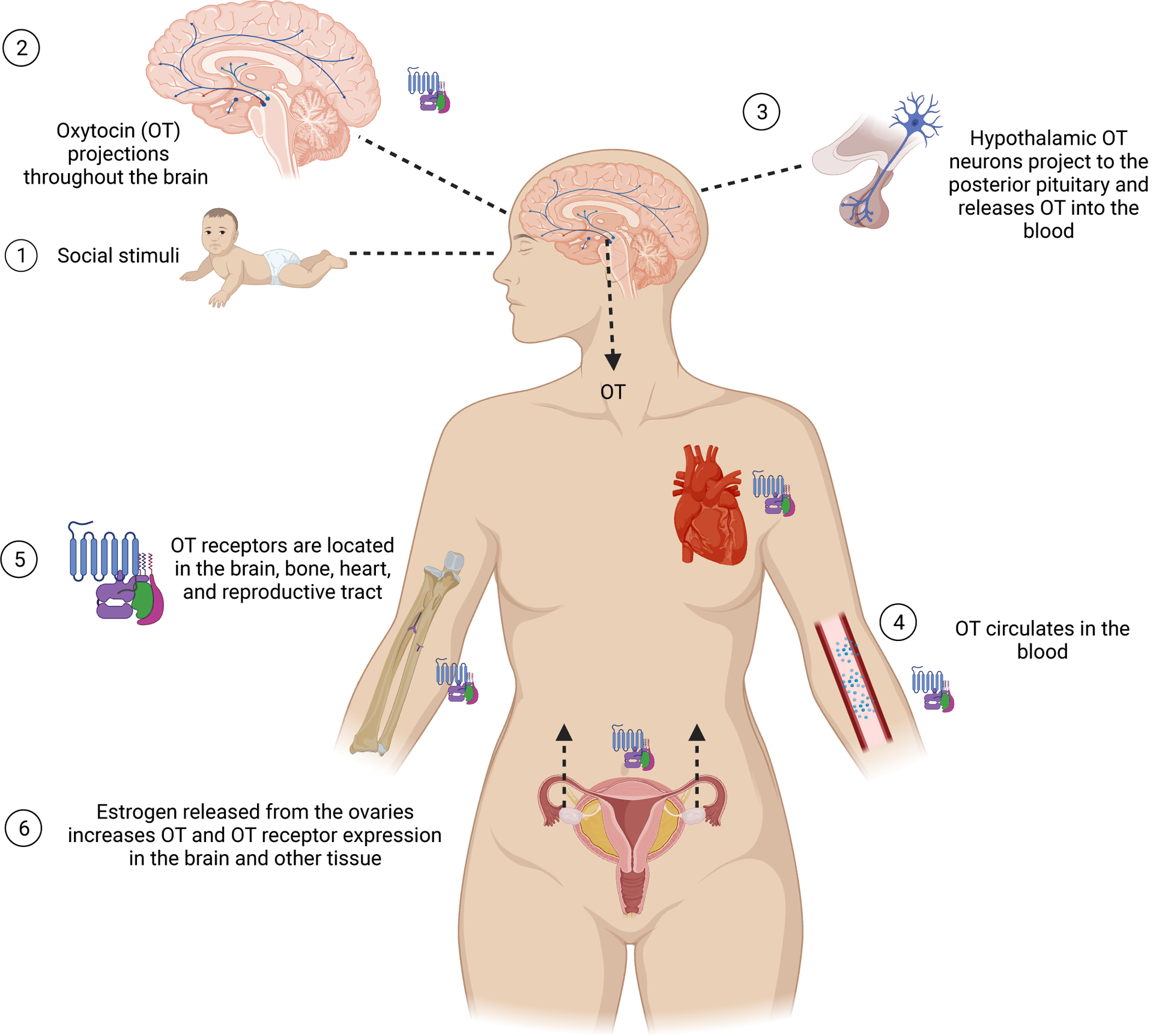
Menopause marks the cessation of fertility and the transition to post-reproductive years. Nearly 1 million US women experience menopause annually, but despite the significant impact it has on their physical and mental health, menopause has been insufficiently studied. Oxytocin is a neurohormone that regulates emotionality, social behaviors, and fundamental physiological systems. Localization of oxytocin receptors in the brain, reproductive tissues, bone, and heart support their role in mental health and potentially sleep, along with reproductive and cardiovascular functions. While experimental data linking oxytocin to behavior and physiology in animals are largely consistent, human data are correlative and inconclusive. As women transition into menopause, oxytocin levels decrease while their susceptibility to mood disorders, poor sleep, osteoporosis, and cardiovascular diseases increases. These concurrent changes highlight oxytocin as a potential influence on the health and mood of women along their reproductive life span. Here, we summarize experimental rodent and non-human primate studies that link oxytocin to reproductive aging and metabolic health and highlight the inconclusive findings in studies of women. Most human studies relied on a single oxytocin assessment in plasma or on intranasal oxytocin administration. The pulsatile release and short half-life of plasma oxytocin limit the validity of these methods. We discuss the need for oxytocin assessments in stable bio-samples, such as urine, and to use valid assays for assessment of associations between changing oxytocin levels and well-being across the reproductive life span. This work has the potential to guide therapeutic strategies that will one day alleviate adverse health outcomes for many women.
Keywords: cardiometabolic disease; menopause; mood disorders; reproductive aging; sleep.
Conflict of interest statement
Dr. Chervin reports research funded by the NIH; royalties as an author and editor for UpToDate; consulting for Eli Lilly & Company through a contract with the University of Michigan; and service on the board of the International Pediatric Sleep Association and advisory board for the non-profit Pajama Program.
Figures
References
-
- AMICO JA, ULBRECHT JS and ROBINSON AG (1987). “Clearance Studies of Oxytocin in Humans Using Radioimmunoassay Measurements of the Hormone in Plasma and Urine*.” The Journal of Clinical Endocrinology & Metabolism 64(2): 340–345. - PubMed
-
- Arnauld E, Bibene V, Meynard J, Rodriguez F and Vincent JD (1989). “Effects of chronic icv infusion of vasopressin on sleep-waking cycle of rats.” Am J Physiol 256(3 Pt 2): R674–684. - PubMed
-
- Arsenijevic Y, Dreifuss JJ, Vallet P, Marguerat A and Tribollet E (1995). “Reduced binding of oxytocin in the rat brain during aging.” Brain Res 698(1–2): 275–279. - PubMed
Publication types
MeSH terms
Substances
Grants and funding
LinkOut - more resources
Full Text Sources
Medical
Research Materials
