Review
. 2024 Jun;167(1):4-22.
doi: 10.1053/j.gastro.2024.03.042. Epub 2024 Apr 24.

New Insights on Genes, Gluten, and Immunopathogenesis of Celiac Disease

Affiliations
Review

New Insights on Genes, Gluten, and Immunopathogenesis of Celiac Disease

Valérie Abadie et al. Gastroenterology. 2024 Jun.

Abstract

Celiac disease (CeD) is a gluten-induced enteropathy that develops in genetically susceptible individuals upon consumption of cereal gluten proteins. It is a unique and complex immune disorder to study as the driving antigen is known and the tissue targeted by the immune reaction can be interrogated. This review integrates findings gained from genetic, biochemical, and immunologic studies, which together have revealed mechanisms of gluten peptide modification and HLA binding, thereby enabling a maladapted anti-gluten immune response. Observations in human samples combined with experimental mouse models have revealed that the gluten-induced immune response involves CD4+ T cells, cytotoxic CD8+ T cells, and B cells; their cross-talks are critical for the tissue-damaging response. The emergence of high-throughput technologies is increasing our understanding of the phenotype, location, and presumably function of the gluten-specific cells, which are all required to identify novel therapeutic targets and strategies for CeD.

Keywords: B Cells; CD4(+) T Cells; Celiac Disease; Gluten; HLA; Intraepithelial CD8(+) T Cells; Transglutaminase 2; Villous Atrophy.


Figures

Figure 1:
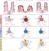
Figure 1:. Environmental, genetic, immunological, and histological features of celiac disease.
Celiac disease develops in genetically susceptible individuals carrying the HLA-DQ2 or HLA-DQ8 molecules. Although HLA is the key genetic determinant, additional non-HLA genes are thought to contribute to predisposing to disease development. Cereal gluten proteins are the driving antigens. Gluten-induced immune responses requires transglutaminase 2 (TG2) to be active, the presence of epithelial stress with upregulation of IL-15 and expression of non-classical MHC class I molecules on intestinal epithelial cells, and encompasses the differentiation of inflammatory IFN-γ and IL-21-producing CD4+ T cells, the expansion of TCRαβ and TCRγδ intraepithelial lymphocytes with cytotoxic properties, the expansion of TG2-specific and gluten-specific plasma cells, and the production and release of IgA and IgG antibodies against TG2 and deamidated gluten peptides (DGP). This pathogenic immune reaction results in the formation of a celiac lesion characterized by plasmacytosis in the lamina propria, an accumulation of intraepithelial lymphocytes, crypt hyperplasia and a destruction of epithelial cells resulting in the blunting of the intestinal villi.
Figure 2:
Figure 2:. TG2-mediated uptake, deamidation, and presentation of gluten peptides.
Left panel, TG2-reactive B cells can endocytose TG2-gluten complexes that serve as a hapten-carrier where TG2 is the hapten and the gluten peptide the carrier. TG2-gluten complex bound to the BCR is endocytosed. Deamidated gluten peptides are then released into the endosomal compartment where they are loaded onto MHC class II (HLA-DQ2 or HLA-DQ8) and presented to gluten-specific CD4+ T cells. Right panel, in myeloid cells, including dendritic cells and macrophages, endocytosis of ternary gluten-TG2- α2-macroglobulin complex is mediated by the endocytic low density lipoprotein receptor-related protein 1 (LRP-1) leading to the release of deamidated gluten in endosomes for MHC class II binding and ensuing presentation to gluten-specific T cell. In both these processes the cellular uptake and posttranslational modification of the gluten peptides are mechanistically coupled by involvement of TG2.
Figure 3:
Figure 3:. Interplay between gluten, HLA, TG2, CD4+ T cells, B cells, and T-IELs for the development of celiac disease.
Immune response to dietary occurs sequentially in inductive sites including mesenteric lymph nodes, Peyer’s patches and isolated lymphoid follicles (not shown here) and effector sites of the gut lamina propria. Upon gluten exposure, TG2-gluten complexes are formed, endocytosed and deamidated in the endosomal compartment of antigen-presenting cells before being loaded onto HLA-DQ2 or HLA-DQ8 molecules. Mucosal dendritic cells acting as sentinel cells migrate to the mesenteric lymph nodes and present deamidated gluten peptides to naïve T cells. In the presence of transforming growth factor (TGF)-β, retinoic acid, which is a vitamin A metabolite abundant in the gut, and inflammatory cytokines upregulated in the mucosa of celiac patients such as IL-15 or type-I interferons, dendritic cells acquire an inflammatory phenotype. Inflammatory dendritic cells secrete IL-12 promoting the differentiation of TH1 CD4+ T cells producing IFN-γ and IL-21. Gluten-specific CD4+ T cells can provide help to both gluten- and TG2-reactive B cells likely in Peyer’s patches where TG2 released from shed enterocytes and gluten peptides have penetrated through M cells. T cell-B cell interactions lead to the differentiation of plasma cells secreting anti-TG2 and anti-deamidated gluten peptides antibodies as well as effector CD4+ T cells that accumulate in the lamina propria. A pathogenic CD4+ T cell response of a certain magnitude is required for tissue destruction, but how CD4+ T cells directly impact on the activation of cytotoxic intraepithelial lymphocytes is still unknown. Intraepithelial CD8+ lymphocytes (T-IELs) expressing activating NK receptors including NKG2D and CD94-NKG2C recognize stress-induced ligands HLA-E and MHC class I polypeptide-related molecules (MIC)A/MICB on intestinal epithelial cells, hence acquiring cytotoxic properties and the ability to lyse the enterocytes.
Figure 4:
Figure 4:. Different subsets of mucosal T cells are associated with tissue destruction or tissue healing in healthy, untreated celiac disease, and treated celiac disease patients.
In healthy intestinal epithelium, the predominant population of CD8+ αβ T-IELs (light red) express the inhibitory NK receptor CD94/NKG2A and CD161. The predominant population of γδ T-IELs (grey) is a population of γδ T cells with innate cytotoxic properties with semi-invariant usage of Vγ4Vδ1. In untreated celiac mucosa, NKG2D/CD94-NKG2C-expressing CD8+ αβ red) and γδ (blue) T cells are induced to exhibit a highly cytotoxic phenotype and secrete granzymes and perforins. Gluten-specific CD4+ T cells (yellow) accumulate in the lamina propria where they have an activated phenotype expressing an array of activation markers and gut homing receptors. Within treated celiac mucosa, innate cytotoxic Vγ4Vδ1 diminish and NKG2D/CD94-NKG2C-expressing γδ (dark blue) and CD8+ αβ red) T-IELs increase in number, whereas gluten-specific CD4+ T cells downregulate many of the activation markers and express CD127.

References

    1. Abadie V, Sollid LM, Barreiro LB, Jabri B. Integration of genetic and immunological insights into a model of celiac disease pathogenesis. Annu Rev Immunol 2011;29:493–525. - PubMed
    1. Sollid LM, Markussen G, Ek J, et al. Evidence for a primary association of celiac disease to a particular HLA-DQ alpha/beta heterodimer. J Exp Med 1989;169:345–50. - PMC - PubMed
    1. Singh P, Arora A, Strand TA, et al. Global Prevalence of Celiac Disease: Systematic Review and Meta-analysis. Clin Gastroenterol Hepatol 2018;16:823–836 e2. - PubMed
    1. Lebwohl B, Sanders DS, Green PHR. Coeliac disease. Lancet 2018;391:70–81. - PubMed
    1. Lindfors K, Ciacci C, Kurppa K, et al. Coeliac disease. Nat Rev Dis Primers 2019;5:3. - PubMed