

Radiographical bony lesions after discontinuation of immunosuppressant therapy: bone involvement in sarcoidosis
- PMID: 38670568
- PMCID: PMC11057273
- DOI: 10.1136/bcr-2023-255611
Radiographical bony lesions after discontinuation of immunosuppressant therapy: bone involvement in sarcoidosis
Abstract
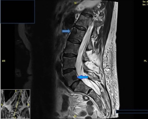
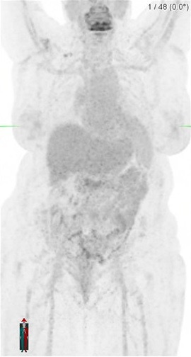
We describe a patient who had failed renal transplant after 13 years, eventually requiring a graft nephrectomy and discontinuation of immunosuppressive therapy, including antithymocyte globulin, tacrolimus and mycophenolate while on steroid avoidance protocol. Within a few months of complete discontinuation of the immunosuppressive medications, she developed lower back pain associated with numbness in her right anterolateral thigh. The radiological imaging demonstrated multiple bony lesions throughout her axial and appendicular skeleton with normal pulmonary findings. A computerised tomography-guided bone biopsy from the left iliac crest revealed fragments of bone with granulomatous inflammation, thus making the diagnosis of extrapulmonary sarcoidosis. Initiating treatment with prednisone resulted in near-complete resolution of symptoms. Long-term immunosuppressive therapy is administered to all renal transplant recipients to help prevent acute rejection and loss of renal allograft. This case highlights that immunosuppressants can conceal the presence of underlying conditions in transplant patients.
Keywords: General practice / family medicine; Radiology; Rheumatology.
© BMJ Publishing Group Limited 2024. Re-use permitted under CC BY-NC. No commercial re-use. See rights and permissions. Published by BMJ.
Conflict of interest statement
Competing interests: None declared.
Figures


References
Publication types
MeSH terms
Substances
LinkOut - more resources
Full Text Sources
Medical