. 2024 Apr 26;21(3):305-315.
doi: 10.21873/cgp.20449.

The Important Role of GPX1 and NF-κB Signaling Pathway in Human Gastric Cancer: Implications for Cell Proliferation and Invasion

Affiliations

The Important Role of GPX1 and NF-κB Signaling Pathway in Human Gastric Cancer: Implications for Cell Proliferation and Invasion

Byeong Il Jang et al. Cancer Genomics Proteomics. .

Abstract

Background/aim: Glutathione peroxidases (GPXs) are crucial antioxidant enzymes, counteracting reactive oxygen species (ROS). GPX overexpression promotes proliferation and invasion in cancer cells. Glutathione peroxidase-1 (GPX1), the most abundant isoform, contributes to invasion, migration, cisplatin resistance, and proliferation in various cancers. Nuclear factor-kappa B (NF-[Formula: see text]B) participates in cell proliferation, apoptosis, and tumor progression. The inhibition of NF-[Formula: see text]B expression reduces the malignancy of esophageal squamous cell carcinoma. This study aimed to explore the GPX1 and NF-[Formula: see text]B signaling pathways and their correlation with gastric cancer cell proliferation and invasion.

Materials and methods: Cell culture, complementary DNA microarray analysis, western blotting, reverse transcription-polymerase chain reaction, zymography, 3-(4,5-dimethylthiazol-2-yl)-2,5-diphenyltetrazolium bromide assay, GPX1 knock-down with short hairpin RNA (shRNA), standard two-chamber invasion assay, chromatin immunoprecipitation assay.

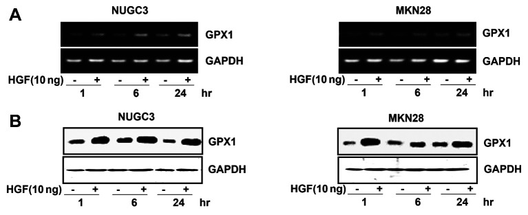
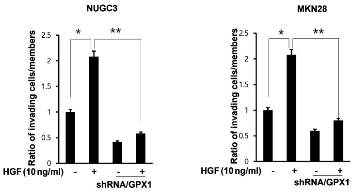
Results: Hepatocyte growth factor (HGF) up-regulated GPX1 expression in gastric cancer cells. The NF-[Formula: see text]B inhibitor, pyrrolidine dithiocarbamate down-regulated HGF-induced GPX1 protein levels. Furthermore, NF-[Formula: see text]B and urokinase-type plasminogen activators were down-regulated in GPX1-shRNA-treated cells. Treatment with an Akt pathway inhibitor (LY294002) led to the down-regulation of GPX1 and NF-[Formula: see text]B gastric cancer cells. GPX1 knockdown resulted in decreased HGF-mediated in vitro cell proliferation and invasion. The study identified the putative binding site of the GPX1 promoter containing the NF-[Formula: see text]B binding site, confirmed through chromatin immunoprecipitation.

Conclusion: HGF induced GPX1 expression through the NF-[Formula: see text]B and Akt pathways, suggesting a central role in gastric cell proliferation and invasion. Hence, GPX1 emerges as a potential therapeutic target for gastric cancer.

Keywords: GPX1; NF-B; gastric cancer; uPA.


Conflict of interest statement

The Authors have no conflicting interests.

Figures

Figure 1
Figure 1. The impact of HGF on GPX1 expression in NGUC3 and MKN28 cells was assessed. Cells (5×10⁵) were serum-starved for 24 h, treated with or without HGF (10 ng/ml) for the specified durations, and subsequently harvested. GPX1 RNA expression levels were validated via reverse transcription-polymerase chain reaction (A). GPX1 protein expression levels were confirmed using western blot analysis (B). The presented data represent typical results obtained from three independent experiments. HGF, Hepatocyte growth factor; NGUC3, poorly differentiated adenocarcinoma; MKN28, moderately differentiated tubular adenocarcinoma; GPX1, glutathione peroxidase-1.
Figure 2
Figure 2. HGF up-regulated GPX1 expression in NGUC-3 and MKN-28 cells. Cells (5×10⁵) were serum-starved for 24 h and treated with varying concentrations of HGF (0, 10, and 40 ng/ml) for 1 h before harvesting. Expression levels of GPX1 RNA were validated using RT-PCR (A). Concurrently, the expression levels of GPX1 protein were confirmed using western blot analysis (B). Presented data are representative results from three independent experiments. HGF, Hepatocyte growth factor; NGUC3, poorly differentiated adenocarcinoma; MKN28, moderately differentiated tubular adenocarcinoma; GPX1, glutathione peroxidase-1; RT-PCR, reverse transcriptase polymerase chain reaction.
Figure 3
Figure 3. Effect of PDTC, an NF-ĸB inhibitor, on HGF-induced GPX1. Serum-starved cells were pre-treated with varying concentrations of PDTC (50, 100, and 200 μM) and then exposed to 10 ng/ml of HGF. The expression levels of GPX1 and NF-ĸB were assessed using western blot analysis. Presented data are representative results from three independent experiments. PDTC, Pyrrolidine dithiocarbamate; HGF, hepatocyte growth factor; GPX1, glutathione peroxidase-1; NF-ĸB, nuclear factor-kappa B; NGUC3, poorly differentiated adenocarcinoma; MKN28, moderately differentiated tubular adenocarcinoma.
Figure 4
Figure 4. The levels of NF-ĸB and GPX1 proteins in response to HGF were reduced in cells expressing GPX1-shRNA. Control cells and stable GPX1-shRNA cells (5×10⁵/well) were plated overnight in a complete medium, starved for 24 h, then treated with or without HGF (10 ng/ml) for 1 h before harvesting. Expression levels of NF-ĸB and GPX1 were analyzed using western blot analysis. Presented data are representative results from three independent experiments. NF-ĸB, Nuclear factor-kappa B; GPX1, glutathione peroxidase-1; shRNA, short hairpin RNA; NGUC3, poorly differentiated adenocarcinoma; MKN28, moderately differentiated tubular adenocarcinoma.
Figure 5
Figure 5. Expression levels and enzyme activity of the uPA protein in response to HGF were down-regulated in cells expressing GPX1-shRNA. Control cells and stable GPX1-shRNA cells (5×10⁵) were treated with or without HGF (10 ng/ml) and subsequently harvested. The levels of uPA proteins in the culture media were analyzed using western blot analysis (A). Furthermore, the enzyme activity levels of uPA proteins in the culture media were assessed by zymography (B). Presented data are representative results from three independent experiments. uPA, Urokinase-type plasminogen activator; HGF, hepatocyte growth factor; GPX1, glutathione peroxidase-1; shRNA, short hairpin RNA; NGUC3, poorly differentiated adenocarcinoma; MKN28, moderately differentiated tubular adenocarcinoma.
Figure 6
Figure 6. Down-regulation of GPX1, NF-ĸB, and pAkt expression was observed, and was influenced by LY294002, acting as a pAkt inhibitor. Cells (5×10⁵) were pre-treated with different concentrations of LY294002 (1, 5, and 10 μM) and then treated with 10 ng/ml of HGF. The expression levels of GPX1, NF-ĸB, and pAkt proteins were evaluated by western blotting. Presented data are representative results from three independent experiments. GPX1, Glutathione peroxidase-1; NF-ĸB, nuclear factor-kappa B, pAkt; phosphor-protein kinase; NGUC3, poorly differentiated adenocarcinoma; MKN28, moderately differentiated tubular adenocarcinoma.
Figure 7
Figure 7. The effect of NF-ĸB knockdown on HGF regulation of GPX1 was investigated. ChIP assay results demonstrate the amplification of a fragment from the proximal GPX1 promoter, which contains the NF-ĸB binding site (A). Immunoprecipitation was conducted using an anti-NF-ĸB antibody (B). Presented data are representative results from three independent experiments. NF-ĸB, Nuclear factor-kappa B; HGF, hepatocyte growth factor; GPX1, glutathione peroxidase-1; CHIP; chromatin immunoprecipitation assay; NGUC3, poorly differentiated adenocarcinoma; MKN28, moderately differentiated tubular adenocarcinoma.
Figure 8
Figure 8. The effect of GPX1 on cell proliferation in the presence of HGF was investigated. Control cells (1×10³/well) and stable GPX1-shRNA cells were seeded in 96-well plates with DMEM supplemented with 5% FBS and incubated for 24 h. After serum starvation for an additional 24 h, cells were treated or untreated with HGF (10 ng/ml) for 72 h. Cell proliferation was measured using 3-(4,5-dimethylthiazol-2-yl)-2,5-diphenyltetrazolium bromide assays and expressed as a percentage of HGF-untreated control cells. The values represent means±standard deviation of three independent experiments. GPX1, Glutathione peroxidase-1; HGF, hepatocyte growth factor; shRNA, short hairpin RNA; DMEM, Dulbecco's Modified Eagle Medium, FBS; fetal bovine serum; NGUC3, poorly differentiated adenocarcinoma; MKN28, moderately differentiated tubular adenocarcinoma.
Figure 9
Figure 9. Impact of GPX1 on HGF-mediated cell invasion. Stable GPX1-shRNA cells and control cells (1×10³) were treated with or without HGF (10 ng/ml) for 48 h. Cell invasion capacity was measured using the standard two-chamber invasion assay with Matrigel migration chambers. The presented values represent means±standard deviation of three independent experiments. GPX1, Glutathione peroxidase-1; HGF, hepatocyte growth factor; shRNA, short hairpin RNA; NGUC3, poorly differentiated adenocarcinoma; MKN28, moderately differentiated tubular adenocarcinoma.
Figure 10
Figure 10. Study flow-chart. After cell culture and stimulation with HGF, the expression of GPX1 was analyzed at both mRNA and protein levels. Following inhibition of the NF-ĸB and Akt pathways, the levels of GPX1 were analyzed. Subsequently, to identify downstream genes, GPX1 knockdown was performed and analyzed. Then, using MTT assay and Two-chamber invasion assay, differences in cell proliferation and invasion were confirmed based on GPX1 knockdown. GPX1, Glutathione peroxidase-1; HGF, hepatocyte growth factor; shRNA, short hairpin RNA; NF-ĸB, nuclear factor-kappa B.
Figure 11
Figure 11. Schematic diagram of HGF-mediated GPX1 up-regulation, HGF mediated GPX1 up-regulation is regulated through the PI3K/Akt pathway and NF-ĸB, which was confirmed by pretreatment with LY294002 and PDTC, HGF-mediated GPX1 up-regulation increased expression uPA. Increased uPA mediates cell proliferation and invasion. GPX1, Glutathione peroxidase-1; HGF, hepatocyte growth factor; shRNA, short hairpin RNA; NF-ĸB, nuclear factor-kappa B; pAkt, phosphorprotein kinase; uPA, urokinase-type plasminogen activator; PDTC, pyrrolidine dithiocarbamate.
Figure 12
Figure 12. Schematic diagram of HGF-mediated GPX1 up-regulation, HGF mediated GPX1 up-regulation is regulated through the PI3K/Akt pathway and NF-ĸB, which was confirmed by pretreatment with LY294002 and PDTC, HGF-mediated GPX1 up-regulation increased expression uPA. Increased uPA mediates cell proliferation and invasion. GPX1, Glutathione peroxidase-1; HGF, hepatocyte growth factor; shRNA, short hairpin RNA; NF-ĸB, nuclear factor-kappa B; pAkt, phosphor-protein kinase; uPA, urokinase-type plasminogen activator; PDTC, pyrrolidine dithiocarbamate.

References

    1. Smyth EC, Nilsson M, Grabsch HI, van Grieken NC, Lordick F. Gastric cancer. Lancet. 2020;396(10251):635–648. doi: 10.1016/S0140-6736(20)31288-5. - DOI - PubMed
    1. Karimi P, Islami F, Anandasabapathy S, Freedman ND, Kamangar F. Gastric cancer: descriptive epidemiology, risk factors, screening, and prevention. Cancer Epidemiol Biomarkers Prev. 2014;23(5):700–713. doi: 10.1158/1055-9965.EPI-13-1057. - DOI - PMC - PubMed
    1. Correa P. Gastric cancer: overview. Gastroenterol Clin North Am. 2013;42(2):211–217. doi: 10.1016/j.gtc.2013.01.002. - DOI - PMC - PubMed
    1. Vlasova-St Louis I, Bohjanen PR. Post-transcriptional regulation of cytokine and growth factor signaling in cancer. Cytokine Growth Factor Rev. 2017;33:83–93. doi: 10.1016/j.cytogfr.2016.11.004. - DOI - PMC - PubMed
    1. Lee KH, Choi EY, Kim MK, Hyun MS, Eun JR, Jang BI, Kim TN, Kim SW, Song SK, Kim JH, Kim JR. Hepatocyte growth factor promotes cell survival by phosphorylation of BAD in gastric cancer cells. Oncol Res. 2008;17(1):23–32. doi: 10.3727/096504008784046072. - DOI - PubMed