. 2024 Apr 26;15(1):3553.
doi: 10.1038/s41467-024-47784-6.

Mucosal prime-boost immunization with live murine pneumonia virus-vectored SARS-CoV-2 vaccine is protective in macaques

Affiliations

Mucosal prime-boost immunization with live murine pneumonia virus-vectored SARS-CoV-2 vaccine is protective in macaques

Jaclyn A Kaiser et al. Nat Commun. .

Abstract

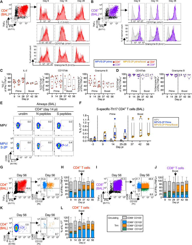
Immunization via the respiratory route is predicted to increase the effectiveness of a SARS-CoV-2 vaccine. Here, we evaluate the immunogenicity and protective efficacy of one or two doses of a live-attenuated murine pneumonia virus vector expressing SARS-CoV-2 prefusion-stabilized spike protein (MPV/S-2P), delivered intranasally/intratracheally to male rhesus macaques. A single dose of MPV/S-2P is highly immunogenic, and a second dose increases the magnitude and breadth of the mucosal and systemic anti-S antibody responses and increases levels of dimeric anti-S IgA in the airways. MPV/S-2P also induces S-specific CD4+ and CD8+ T-cells in the airways that differentiate into large populations of tissue-resident memory cells within a month after the boost. One dose induces substantial protection against SARS-CoV-2 challenge, and two doses of MPV/S-2P are fully protective against SARS-CoV-2 challenge virus replication in the airways. A prime/boost immunization with a mucosally-administered live-attenuated MPV vector could thus be highly effective in preventing SARS-CoV-2 infection and replication.


Conflict of interest statement

J.A.K., C.L., S.M., C.L.N. and U.J.B. are inventors on the provisional patent application number 63/502,829 entitled “Recombinant murine pneumonia virus expressing severe acute respiratory syndrome coronavirus 2 (SARS-COV-2) spike protein” filed by the United States, Department of Health and Human Services. The remaining authors declare no competing interests.

Figures

Fig. 1
Fig. 1. Evaluation of the replication, immunogenicity, and protective efficacy of one or two doses of MPV/S-2P in rhesus macaques.
A Timeline of the evaluation of MPV/S-2P in rhesus macaques. Three groups (n = 4 per group) were immunized by the IN/IT route. Group 1 received a single dose of MPV (empty-vector control, gray), and groups 2 (blue) and 3 (gold) received MPV/S-2P. Twenty-eight days after the first dose, macaques of group 3 received a second dose of MPV/S-2P (boost, 6.3 log10 PFU). Thirty-one to 32 days after a single dose (groups 1 and 2) or day 30 after the second dose (group 3, study day 58), macaques were challenged by the IN/IT route with the vaccine-matched WA1/2020 SARS-CoV-2 strain. Vital signs were documented for the duration of the study (Fig. S1). Macaques were euthanized on day 6 post-challenge. B Replication of MPV and MPV/S-2P in the upper and lower airways was evaluated by immunoplaque assay from nasopharyngeal swabs (NS) and tracheal lavage samples (TL), respectively, collected at the indicated days post immunization (pi) from MPV primed (n = 4), MPV/S-2P primed (n = 8) and boosted (n = 4) macaques. Medians (lines), min and max values (whiskers), 25th to 75th quartiles (boxes), and individual values are shown. The limit of detection was 0.7 log10 PFU/ml and 1 log10 PFU/ml for NS and TL, respectively (dotted line). The number of animals per group of macaques with detectable virus replication is indicated. Two-way ANOVA with Sidak post-test; exact p values are indicated for levels of significance p < 0.05 unless p < 0.0001 (****). Source data are provided in the Source Data file.
Fig. 2
Fig. 2. Immunogenicity of MPV/S-2P in the airways.
Immunogenicity in the upper airways (UA) and lower airways (LA) was evaluated from nasal washes (NW) (AC) and bronchoalveolar lavages (BAL) (DG), collected at the indicated day pi. Spike (S)-specific and receptor binding domain (RBD)-specific IgG (A, D) and IgA (B, E) was measured using ELISA and dissociation-enhanced lanthanide fluorescent (DELFIA) assays, respectively (limit of detection: 1.6 log10, dotted line). Dimeric anti-S IgA in the UA and LA was also evaluated by DELFIA assay (C, F; limit of detection is 1.0 log10, dotted line). G BAL samples were analyzed for their ability to block binding of tagged, soluble ACE2 to purified S protein from the vaccine-matched SARS-CoV-2 S protein (Wuhan strain) or variants of concern. ACE2 binding inhibition is expressed as % inhibition relative to a no-sample control (see also Fig. S2A). AG Medians (lines), min and max values (whiskers), 25th to 75th quartile (boxes), and individual values are shown for MPV primed (n = 4), MPV/S-2P primed (n = 8) and boosted (n = 4) macaques; two-way ANOVA with Sidak post-test; exact p values are indicated for levels of significance p < 0.05. Source data are provided in the Source Data file.
Fig. 3
Fig. 3. Prime/boost regimen of MPV/S-2P induced high level of serum anti-S antibodies with increased avidity, breadth and ADCP activity.
Anti-spike (S) and receptor binding domain (RBD)-specific serum IgG (A) and IgA (B) measured by ELISA or DELFIA (limit of detection: 3.0 log10, dotted line). Antibody avidity to S, determined by ELISA with or without NaSCN, which strips low-affinity antibodies; avidity index (AI), calculated as ratio of NaSCN-treated vs. PBS-treated serum IgG or IgA titers. C Antibody-dependent cellular phagocytosis (ADCP), determined by incubating biotinylated S protein complexed to neutravidin-labeled fluorescent beads with macaque sera, followed by measurement of the phagocytic activity of THP-1 monocytes [ref. , modified as described in Supplementary Methods 2]. Data are expressed as the 50% inhibitory concentration (IC50) corresponding to the dilution of serum that resulted in 50% of THP-1 cells being FITC positive for binding and/or phagocytosing the S protein. D 50% SARS-CoV-2 serum neutralizing-antibody titers (ND50) against the WA1/2020, B.1.1.7, and B.1.351 isolates; serum MPV neutralizing-antibody titers, determined by 60% plaque reduction neutralization tests (PRNT60). E Inhibition of binding of soluble, tagged angiotensin converting enzyme 2 receptor (ACE2) to indicated purified S proteins by serum antibodies, expressed as % inhibition relative to no-serum control (see also Fig. S2B). AE Medians (lines), min and max values (whiskers), 25th to 75th quartile (boxes), and individual values are shown for MPV primed (n = 4), MPV/S-2P primed (n = 8) and boosted (n = 4) macaques; two-way ANOVA with Sidak post-test; exact p values are indicated for levels of significance p < 0.05 unless p < 0.0001 (****). Source data are provided in the Source Data file.
Fig. 4
Fig. 4. MPV/S-2P immunization induced S-specific B cells in the blood that are restimulated after boosting.
Peripheral blood monocytic cells (PBMC) were stained with fluorochrome-labeled monoclonal antibodies and fluorochrome-labeled SARS-CoV-2 receptor binding domain (RBD) and S-2P probes to identify spike (S)-specific B cells (RBD+/S-2P+ or RBD/S-2P+, see Fig. S3A for gating strategy). A, D, F Dot plots from representative MPV/S-2P- or MPV (A only) immunized macaques showing the frequency of RBD+ and RBD S-specific B cells. B Frequency of S-specific B cells from MPV primed (n = 4), MPV/S-2P primed (n = 8) and boosted (n = 4) macaques [medians (lines), min and max (whiskers), 25th to 75th quartiles (boxes) on indicated days]. C Median frequencies with ranges of S-specific B cells in the blood of MPV/S-2P immunized macaques that are RBD+ (dark purple) or RBD (light purple) on indicated days (prime n = 8; boost n = 4 macaques). Isotype class of RBD+ and RBD S-specific B cells. D Representative dot plots from an MPV/S-2P-immunized macaque. E Median frequencies with ranges of IgM (gray), IgA (red) and IgG (blue) RBD+ (left) and RBD (right) S-specific B cells at the indicated day pi (prime n = 8; boost n = 4 macaques). F, G Activated memory (AM; CD21/CD27+; yellow), resident memory (RM; CD21+/CD27+; green), CD27 RM (CD21+/CD27; brown) or tissue-resident like memory (TLM; CD21/CD27; white) phenotypes of S-specific IgM+, IgA+, and IgG+ B cells of MPV/S-2P-immunized macaques. G Median frequencies with ranges (prime n = 8; boost n = 4 macaques). Source data are provided in the Source Data file.
Fig. 5
Fig. 5. IN/IT immunization with MPV/S-2P induces S-specific CD4+ and CD8+ T-cell responses in the blood and lower airways that are restimulated after boosting.
S-specific CD4+ and CD8+ T-cells in blood (AC) or bronchoalveolar lavage (BAL) (DF). Peripheral blood monocytic cells (PBMC) and bronchoalveolar lavage (BAL) cells from indicated days post-immunization (pi) or post-challenge (pc) were left unstimulated or stimulated with overlapping SARS-CoV-2 spike (S) or (BAL only) nucleoprotein (N) peptides, and processed for flow cytometry (see Fig. S3B for gating). Dot plots showing interferon gamma (IFNγ) and tumor necrosis factor alpha (TNFα) expression by CD4+ or CD8+ T-cells from PBMC (A) or BAL (D) of representative MPV (top) or MPV/S-2P-immunized (bottom) macaques. Background-corrected frequencies of S-specific IFNγ+/TNFα+ CD4+ (B, E) or CD8+ T-cells (C, F) from PBMC (B, C) or BAL (E, F) on indicated days. Expression of proliferation marker Ki-67 by IFNγ+/TNFα+ S-specific CD4+ (red) or CD8+ (purple) T-cells from blood (G, H) or BAL (I, J) of MPV/S-2P-immunized macaques. Gating and histograms showing evolution of Ki-67 expression by IFNγ+/TNFα+ CD4+ or CD8+ S-specific T-cells in blood (G) or BAL (I) from a representative MPV/S-2P-immunized macaque. IFNγ/TNFα T-cells (gray) are shown for comparison. Frequencies of Ki-67+ T-cells in IFNγ+/TNFα+ T-cells from blood (H) or BAL (J). B, C, E, F, H, J Medians (lines), min and max values (whiskers), 25th to 75th quartiles (boxes), and individual values are shown for MPV primed (n = 4), MPV/S-2P primed (n = 8) and boosted (n = 4) macaques. MPV/S-2P-primed and primed/boosted macaques are represented by circles or triangles. Source data are provided in the Source Data file.
Fig. 6
Fig. 6. MPV/S-2P induced S-specific Th1 and Th17 CD4+ and granzyme B expressing CD8+ T-cells in lower airways that remained functional after boost and transitioned to tissue-resident memory phenotype.
AL Bronchoalveolar lavage (BAL)-derived T-cells were stimulated with overlapping SARS-CoV-2 spike (S) or nucleoprotein (N) peptides or kept unstimulated and processed for flow cytometry. Dot plots, histograms (A, B), and frequencies (C, D) of S-specific interferon gamma/tumor necrosis factor alpha (IFNγ+/TNFα+) CD4+ (A, C, red), CD8+ (B, D, purple), and IFNγ/TNFα (A, B only, gray) T-cells expressing interleukin 2 (IL-2) (CD4+ T-cells only), CD107ab and granzyme B in BAL from MPV/S-2P-primed (C, D, circles, n = 8) and -boosted (C, D, triangles, n = 4) macaques on indicated days (C, D, medians (lines), min and max values (whiskers), 25th to 75th quartiles (boxes), and individual values are shown; see Fig. S3B for full gating). E, F MPV/S-2P induced a small population of S-specific T helper 17 (Th-17) CD4+ T-cells in airways that are restimulated after boost. E Dot plots from representative macaques and (F) frequencies of S-specific CD95/IL-17+ CD4+ T-cells from BAL on indicated days; medians (lines), min and max values (whiskers), 25th to 75th quartiles (boxes), and individual values are shown for MPV primed (n = 4), MPV/S-2P primed (n = 8) and boosted (n = 4) macaques. GL S-specific T-cells from BAL transition to circulating (CD69/CD103, gray) and tissue-resident memory [Trm; CD69+/CD103 (blue), CD69+/CD103+ (orange), CD69/CD103+ (green)] phenotypes. Dot plots (G, I, K) and frequencies (H, J, L) of Trm phenotypes in S-specific IFNγ+/TNFα+ CD4+ (G, H), CD8+ (I, J) and CD95+/IL-17+ CD4+ T-cells (K, L) from BAL on indicated days. H, J, L Median frequencies, stacked, with ranges, from MPV/S-2P primed (n = 8) and boosted (n = 4) macaques. Figure S4 shows Trm markers in PBMC-derived T-cells. Source data are provided in the Source Data file.
Fig. 7
Fig. 7. Efficacy of one or two doses of MPV/S-2P against SARS-CoV-2 challenge virus replication.
On days 31 or 32 post-prime [MPV- (gray) and MPV/S-2P-immunized macaques, blue] or on day 58 (corresponding to day 30 post-boost; MPV/S-2P-immunized macaques, gold) (n = 4 animals per group), macaques were challenged IN/IT with 6.3 log10 TCID50 of SARS-CoV-2, strain WA1/2020 (Fig. 1A). SARS-CoV-2 subgenomic E (sgE) RNA in the upper (A) and lower airways (B) following challenge. Nasopharyngeal swabs (NS) and bronchoalveolar lavages (BAL) were collected, and SARS-CoV-2 sgE mRNA, indicative of transcription/de-novo RNA synthesis and challenge virus replication, was quantified by RT-qPCR. C SARS-CoV-2 replication in lung tissues. On day 6 pc, SARS-CoV-2 sgE was quantified by RT-qPCR in six regions of lung tissues from each macaque. In each graph, the number of macaques with detectable sgE is indicated. Limit of detection: 2.85 log10 copies/ml for NS and BAL; 3.6 log10 copies/g for lung tissue. AC Medians (lines), min and max values (whiskers), 25th to 75th quartiles (boxes), and individual values are shown two-way ANOVA with Sidak post-test; exact p values are indicated for levels of significance p < 0.05. Source data are provided in the Source Data file.

Update of

References

    1. WHO. COVID-19 epidemiological update. in Emergency Situational Updates 164 edn (World Health Organization, 2024).
    1. Morens DM, Taubenberger JK, Fauci AS. Rethinking next-generation vaccines for coronaviruses, influenzaviruses, and other respiratory viruses. Cell Host Microbe. 2023;31:146–157. doi: 10.1016/j.chom.2022.11.016. - DOI - PMC - PubMed
    1. Shrestha LB, Foster C, Rawlinson W, Tedla N, Bull RA. Evolution of the SARS-CoV-2 omicron variants BA.1 to BA.5: implications for immune escape and transmission. Rev. Med. Virol. 2022;32:e2381. doi: 10.1002/rmv.2381. - DOI - PMC - PubMed
    1. Brock LG, et al. Murine pneumonia virus expressing the fusion glycoprotein of human respiratory syncytial virus from an added gene is highly attenuated and immunogenic in rhesus macaques. J. Virol. 2018;92:e00723–18. doi: 10.1128/JVI.00723-18. - DOI - PMC - PubMed
    1. Brock LG, Karron RA, Krempl CD, Collins PL, Buchholz UJ. Evaluation of pneumonia virus of mice as a possible human pathogen. J. Virol. 2012;86:5829–5843. doi: 10.1128/JVI.00163-12. - DOI - PMC - PubMed

Publication types

MeSH terms