. 2024 Apr 26;14(1):9573.
doi: 10.1038/s41598-024-59635-x.

P2X7 receptor knockout does not alter renal function or prevent angiotensin II-induced kidney injury in F344 rats

Affiliations

P2X7 receptor knockout does not alter renal function or prevent angiotensin II-induced kidney injury in F344 rats

Josselin Nespoux et al. Sci Rep. .

Abstract

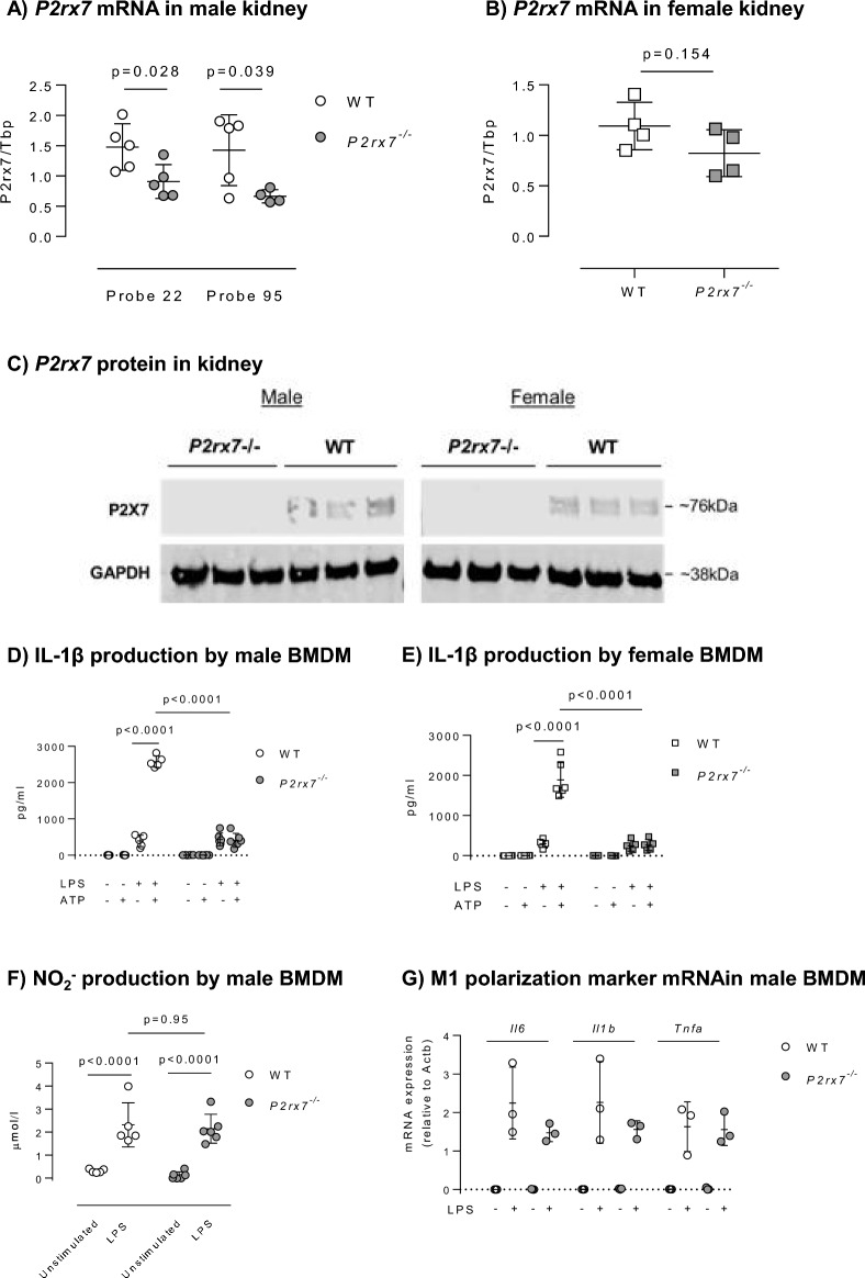
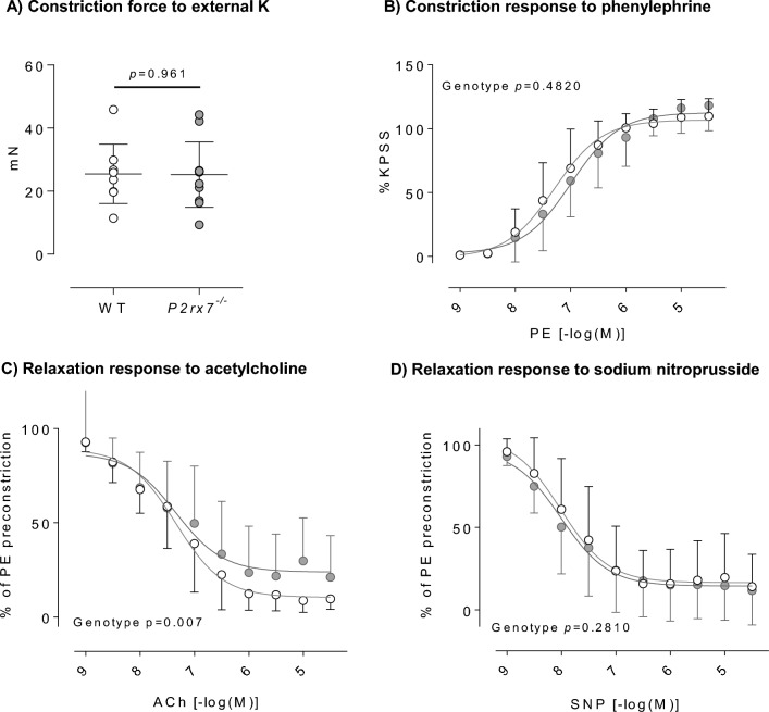
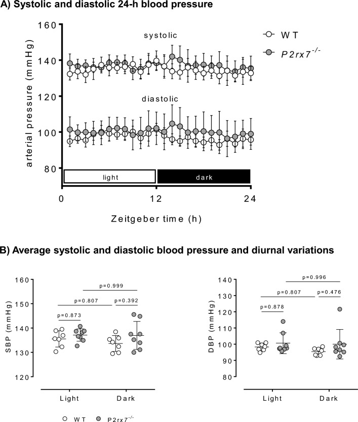
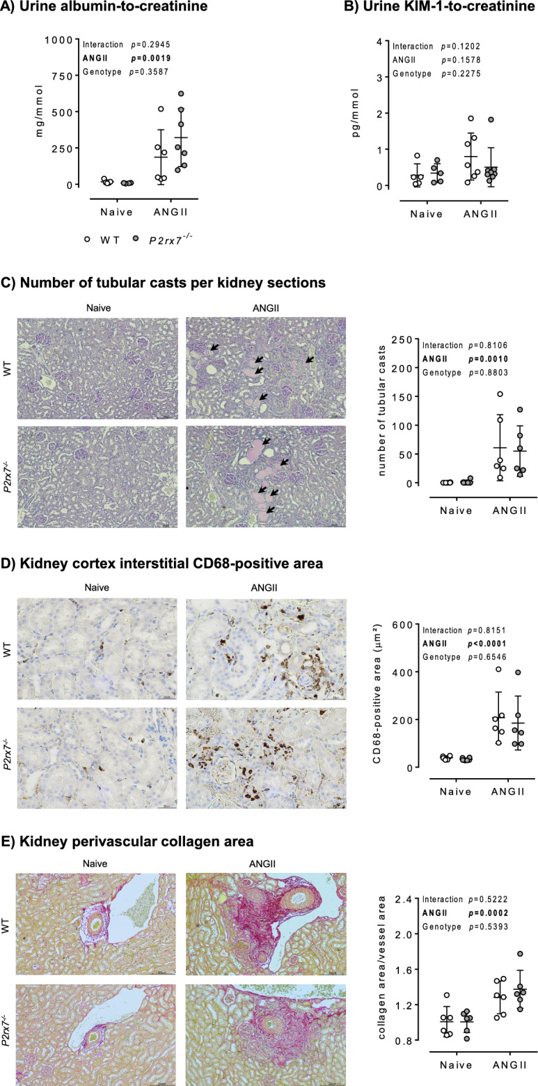
P2X7 receptors mediate immune and endothelial cell responses to extracellular ATP. Acute pharmacological blockade increases renal blood flow and filtration rate, suggesting that receptor activation promotes tonic vasoconstriction. P2X7 expression is increased in kidney disease and blockade/knockout is renoprotective. We generated a P2X7 knockout rat on F344 background, hypothesising enhanced renal blood flow and protection from angiotensin-II-induced renal injury. CRISPR/Cas9 introduced an early stop codon into exon 2 of P2rx7, abolishing P2X7 protein in kidney and reducing P2rx7 mRNA abundance by ~ 60% in bone-marrow derived macrophages. The M1 polarisation response to lipopolysaccharide was unaffected but P2X7 receptor knockout suppressed ATP-induced IL-1β release. In male knockout rats, acetylcholine-induced dilation of the renal artery ex vivo was diminished but not the response to nitroprusside. Renal function in male and female knockout rats was not different from wild-type. Finally, in male rats infused with angiotensin-II for 6 weeks, P2X7 knockout did not reduce albuminuria, tubular injury, renal macrophage accrual, and renal perivascular fibrosis. Contrary to our hypothesis, global P2X7 knockout had no impact on in vivo renal hemodynamics. Our study does not indicate a major role for P2X7 receptor activation in renal vascular injury.


Conflict of interest statement

The authors declare no competing interests. R.I. M. has since joined AstraZeneca and may own stock or stock options.

Figures

Figure 1
Figure 1
The role of P2X7 receptors in the hypertensive kidney. P2X7 receptors are non-selective cation channel receptors activated by extracellular adenosine triphosphate (ATP). P2X7 receptors are abundantly expressed by immune cells, such as macrophages. ATP released by damaged or activated cells activates P2X7 receptors on macrophages to mediate NOD−, LRR− and pyrin domain-containing protein 3 (NLRP3), Apoptosis-associated speck-like protein containing a caspase recruitment domain (CARD) (ASC) and caspase-1 assembly leading to inflammasome activation, and the subsequent maturation and release of pro-inflammatory cytokines such as interleukin (IL)-1β. These cytokines promote renal interstitial inflammation and promote sodium retention by tubular epithelial cells, thereby contributing to salt-sensitivity and hypertension. P2X7 receptors are also expressed in renal vascular endothelial cells, particularly in the pre-glomerular vasculature and vasa recta. ATP, released by endothelial cells, can activate endothelial P2X7 receptors in an autocrine/paracrine fashion to promote vasoconstriction, ultimately decreasing renal blood flow (RBF) and glomerular filtration rate (GFR). Figure contains a modified illustration from Servier Medical Art. Servier Medical Art by Servier is licensed under a Creative Commons Attribution 4.0 Unported License (https://creativecommons.org/licenses/by/4.0/).
Figure 2
Figure 2
Validation of P2X7 deletion in P2rx7−/− rats. (A) Partial decrease in P2rx7 mRNA abundance in the kidney of male P2rx7−/− vs. WT using two PCR primer sets (n = 5 rats/group). (B) Trend for a partial decrease in P2rx7 mRNA abundance in the kidney of female P2rx7−/− vs. WT (n = 4 rats/group). (C) Western blot demonstrating the absence of P2X7 protein expression in kidney samples of male and female P2rx7−/− rats using an antibody directed against the C‐terminus of P2X7. GAPDH expression was used as a loading control (n = 3 rats/group). (D) Suppressed IL‐1β production by BMDM from male rats primed for 4 h with 1 μg/mL of LPS, then stimulated for 1 h with 3 mM ATP (n = 5–6 rats/group). (E) Suppressed IL‐1β production by BMDM from female rats primed for 4 h with 1 μg/mL of LPS, then stimulated for 1 h with 3 mM ATP (n = 5–6 rats/group). (F) Similar NO2 production by male WT and P2rx7−/− BMDM following stimulation with LPS for 4 h (n = 5–6 rats/group). (G) Similar induction of M1 polarisation marker genes by male WT and P2rx7−/− BMDM following stimulation with LPS for 4 h (n = 3 rats/group). Data are means ± SD and statistical analysis performed using t-test (A,B) or 2-way ANOVA with Holm–Sidak post hoc correction (DG). For all analyses, P < 0.05 was considered significant.
Figure 3
Figure 3
Ex vivo renal artery contractility in male P2rx7−/− rats. (A) Similar external K+-evoked constriction force in male WT and P2rx7−/− rat renal artery. (B) Similar vasoconstriction of male WT and P2rx7−/− rat renal arteries to increasing phenylephrine (PE) concentrations. (C) Impaired vasodilation of male P2rx7−/− rat renal arteries to increasing acetylcholine (ACh) concentrations. (D) Similar vasodilation of male WT and P2rx7−/− rat renal arteries to increasing sodium nitroprusside (SNP) concentrations. For all, n = 9–11 rats/group. Data are means ± SD and statistical analysis performed using t-test (A) or 2-way ANOVA (BD). For all analyses, P < 0.05 was considered significant.
Figure 4
Figure 4
In vivo renal hemodynamics and the pressure natriuresis relationship in male P2rx7−/− rats. (A) Change in mean arterial pressure (MAP) following ligation of coeliac, superior mesenteric, and distal aorta ligation. (B) Change in renal artery blood flow (RBF) as measured using a Transonic Doppler flow probe placed around the right main renal artery. (C) Change in renal vascular resistance (RVR). (D) Change in glomerular filtration rate (GFR). (E) Change in urinary sodium excretion rate (UNaV). (F) Change in urine flow rate (UV). (G) Urinary nitrite/nitrate excretion rate (UNOxV). For all, n = 9–10 rats/group. Data are means ± SD and statistical analysis performed using t-test (AF) or 2-way ANOVA with Holm–Sidak post hoc correction (G). For all analyses, P < 0.05 was considered significant.
Figure 5
Figure 5
Radiotelemetry blood pressure in healthy male P2rx7−/− rats. Blood pressure was recorded for 5 consecutive days using radiotelemetry devices. Data from each 24 h period were averaged. (A) 24 h systolic and diastolic blood pressure profiles in WT and P2rx7−/− rats. (B) Average systolic and diastolic blood pressure during light (inactive) and dark (active) 12 h phases in WT and P2rx7−/− rats. n = 7–8 rats/group. Data are means ± SD and statistical analysis performed using 2-way ANOVA with Holm–Sidak post hoc correction (B). For all analyses, P < 0.05 was considered significant.
Figure 6
Figure 6
Kidney injury, inflammation, and fibrosis in male P2rx7−/− rats following chronic ANGII infusion. Male WT and P2rx7−/− rats underwent 5–6 week ANGII infusion. Urine and kidneys were harvested at end of infusion for the quantification of kidney injury, inflammation and fibrosis. (A) Quantification of albuminuria following ANGII infusion (n = 4–7 rats/group). (B) Quantification of the tubular injury biomarker KIM-1 in urine following ANGII infusion (n = 5–8 rats/group). (C) Number of tubular casts in kidney sections stained with PAS (n = 6 rats/group). (D) Quantification of the macrophage marker CD68-positive area in kidney sections (n = 6 rats/group). (E) Quantification of perivascular collagen area in kidney sections (n = 6 rats/group). Data are means ± SD and statistical analysis performed using 2-way ANOVA with Holm–Sidak post hoc correction to test for the effect of angiotensin II (ANGII), P2rx7 knockout (Genotype), and the interaction. For all analyses, P < 0.05 was considered significant.

References

    1. Burnstock, G. Purines and purinoceptors: Molecular biology overview☆. In Reference Module in Biomedical Sciences B9780128012383047413 (Elsevier, 2014). 10.1016/B978-0-12-801238-3.04741-3.
    1. Burnstock G. Purine and purinergic receptors. Brain Neurosci. Adv. 2018;2:2398212818817494. doi: 10.1177/2398212818817494. - DOI - PMC - PubMed
    1. Burnstock G, Evans LC, Bailey MA. Purinergic signalling in the kidney in health and disease. Purinergic Signal. 2014;10:71–101. doi: 10.1007/s11302-013-9400-5. - DOI - PMC - PubMed
    1. Vallon V, Unwin RJ, Inscho EW, Leipziger J, Kishore BK. Extracellular nucleotides and P2 receptors in renal function. Physiol. Rev. 2019 doi: 10.1152/physrev.00038.2018. - DOI - PMC - PubMed
    1. Menzies RI, Unwin RJ, Bailey MA. Renal P2 receptors and hypertension. Acta Physiol. 2015;213:232–241. doi: 10.1111/apha.12412. - DOI - PubMed

Publication types