. 2024 Apr 26;14(1):9571.
doi: 10.1038/s41598-024-60169-5.

Hyper-N-glycosylated SEL1L3 as auto-antigenic B-cell receptor target of primary vitreoretinal lymphomas

Affiliations

Hyper-N-glycosylated SEL1L3 as auto-antigenic B-cell receptor target of primary vitreoretinal lymphomas

Michelle Elbert et al. Sci Rep. .

Abstract

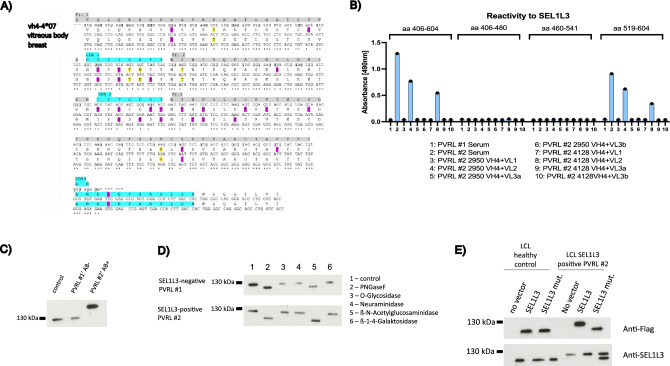
Primary vitreoretinal lymphoma (PVRL) is a rare subtype of DLBCL and can progress into primary central nervous system lymphoma (PCNSL). To investigate the role of chronic antigenic stimulation in PVRL, we cloned and expressed B-cell receptors (BCR) from PVRL patients and tested for binding against human auto-antigens. SEL1L3, a protein with multiple glycosylation sites, was identified as the BCR target in 3/20 PVRL cases. SEL1L3 induces proliferation and BCR pathway activation in aggressive lymphoma cell lines. Moreover, SEL1L3 conjugated to a toxin killed exclusively lymphoma cells with respective BCR-reactivity. Western Blot analysis indicates the occurrence of hyper-N-glycosylation of SEL1L3 at aa 527 in PVRL patients with SEL1L3-reactive BCRs. The BCR of a PVRL patient with serum antibodies against SEL1L3 was cloned from a vitreous body biopsy at diagnosis and of a systemic manifestation at relapse. VH4-04*07 was used in both lymphoma manifestations with highly conserved CDR3 regions. Both BCRs showed binding to SEL1L3, suggesting continued dependence of lymphoma cells on antigen stimulation. These results indicate an important role of antigenic stimulation by post-translationally modified auto-antigens in the genesis of PVRL. They also provide the basis for a new treatment approach targeting unique lymphoma BCRs with ultimate specificity.

Keywords: Auto-antigens; B-cell receptor antigens; Primary CNS lymphoma; Primary vitreoretinal lymphoma; SAMD14/neurabin-I; SEL1L3.


Conflict of interest statement

The authors declare no competing interests.

Figures

Figure 1
Figure 1
(A) ELISA to evaluate the affinity of 20 Fabs derived from published PCNSL cases (Belhouachi et al.) against the antigens SAMD14/Neurabin-I, LRPAP1, RpoC, and Galectin-3. Among tested Fabs, two showed reactivity to SAMD14/Neurabin-I. No discernible binding was observed between PCNSL Fabs and LRPAP1, RpoC, or Galectin-3. (B) Twenty PVRL-BCR sequences (Belhouachi et al.) were cloned and produced as Fab-antibodies in E. coli and screened for binding to human antigens on a UniPEx human cDNA expression library protein macroarray, identifying SEL1L3 as possible BCR target. ELISA assays showed binding of 3/20 PVRL Fabs (PVRLs #22, #30 and #39) to SEL1L3. (C) Reactivity of PVRL patient sera and recombinant Fabs #15, #22, #30 to SEL1L3 as it was expressed on the UniPEx membrane (aa 406–604, termed as full-length) and SEL1L3 fragments (Fr1: aa 406–480; Fr2: aa 460–541; Fr3: aa 519–604; Fr3-3: aa 564–604; Fr3-2: aa 537–578; Fr3-2: aa 519–559), assessed by ELISA. Patient serum #1 and recombinant Fab #15 show no reactivity to full-length SEL1L3 or its’ fragments. PVRL patient serum #2 and recombinant Fabs #22 and #30 are reactive to full-length SEL1L3. Binding properties to SEL1L3 fragments suggest an epitope region spanning approximately from aa 560 to aa 580. ELISAs of figure were repeated multiple times and figures (AC) are exemplary. ELISAs of Fig. 2B were performed simultaneously in two wells, which is represented by two dots on top of the bars.
Figure 2
Figure 2
(A) DLBCL cell lines TMD8 and Oci-Ly3 were transfected with BCRs of PVRL cases #22 and #30. Both BCRs were previously identified as reactive to SEL1L3. TMD8 cells express BCRs of unknown reactivity as natural BCR. For Oci-Ly3 cells, Ars2 has been identified as cognate auto-antigen of their BCR. Expression of BCRs from PVRL cases #22 and #30 was induced with doxycycline ( +). Oci-Ly3 and TMD8 cells were incubated for 45 min with medium, Ars2-Flag or SEL1L3-Flag. After washing steps, FITC-A coupled anti-Flag antibody was used as secondary system. Ars2 shows binding to OCI-Ly3 cells but not to TMD8 cells regardless of the expression of SEL1L3-reactive BCRs (A and B). SEL1L3 shows binding to TMD8 and OCI-Ly3 cells after the expression of SEL1L3-reactive BCRs of PVRL cases #22 and #30 is switched on using doxycycline. (B) Without induction of BCR expression, no binding of SEL1L3 to either cell line is observed. (C) 12% SDS-Page Western blots of DLBCL cell line lysates. TMD8 and Oci-Ly3 cells transfected with an inducible SEL1L3-reactive BCR were incubated over night with SEL1L3, LRPAP1, Ars2, anti-IgM or medium. Expression of SEL1L3-reactive BCRs derived from PVRL patients #22 and #30 was activated the day before with doxycycline (induction of BCR expression is marked with +). In OCI-Ly3 cells, BCR pathway proteins are upregulated after incubation with their cognate BCR antigen Ars2 (3) and the positive control anti-IgM (4). For SEL1L3 (1) this effect was observed only after induction of a SEL1L3-reactive BCR. Medium (5) only or LRPAP1 (2) had no influence on the activation status of the BCR pathway of OCI-Ly3 cells. Similar results were observed for TMD8 cells with the exception that Ars2 had no effect on the upregulation of BCR signaling pathway proteins since TMD8 cells do not express an Ars2-reactive BCR. (D) Incubation with SEL1L3 epitope induces proliferation of DLBCL cell lines only after expression of patient-derived, SEL1L3-reactive BCRs (induction of BCR expression is marked with +) on TMD8 and Oci-Ly3 cells. Ars2 as cognate antigen of Oci-Ly3 BCRs was used as positive control. Proliferation experiments shown in 2D were performed in triplicate, i.e. simultaneously in three separate wells.
Figure 3
Figure 3
(A) DLBCL cell lines TMD8 and Oci-Ly3 were transfected with BCRs of PVRL cases #22 and #30. Both PVRL cases were previously identified as reactive to SEL1L3. TMD8 cells express BCRs of unknown reactivity as their natural BCR. For Oci-Ly3 cells, Ars2 has been identified as cognate auto-antigen of their BCR. The BCR binding epitope of SEL1L3 (aa 560–585) conjugated to a truncated form of Pseudomonas exotoxin A (ETA) reduces proliferation of lymphoma cells expressing the respective BCR. Ars2-ETA was used as positive control on Ars2-reactive OCI-Ly3 cells. Rpoc, an irrelevant antigen, conjugated to ETA’ was used as negative control. Expression of BCR was induced with doxycycline ( +). (B) Flow cytometric annexin assays using the Annexin V-FITC Apoptosis Detection Kit (Sigma-Aldrich) were performed to elucidate the mechanisms of cell death after treatment with SEL1L3 immunotoxins. OCI-LY3 and TMD8 cells transfected to express a BCR with reactivity against SEL1L3 were treated with Ars2-ETA, RpoC-ETA, SEL1L3-ETA (all at 0.5 µg/mL), staurosporine (1 µg/mL), or medium. After incubation with SEL1L3 immunotoxins both OCI-Ly3 and TMD8 cells undergo mainly apoptosis if expression of SEL1L3-reactive BCRs is switched on by doxycycline (upper row) and has no effect on both cell lines without prior induction of respective BCRs (bottom row). Quantitative measurements are shown in Suppl. Table 2. (C) LDH cytotoxicity assays of SEL1L3-ETA immunotoxins on Oci-Ly3 as well as TMD8 cells expressing SEL1L3-reactive BCRs derived from PVRL patients #22 and #30. Ars2-ETA constructs served as positive control as Ars2 is the cognate antigen of the constitutively expressed BCR in Oci-Ly3 cells. TMD8 cells express BCRs of unknown reactivity as their natural BCR. Bars are depicted in pairs, left bar in light blue (expression of SEL1L3-reactive BCRs turned on) and right bar in red (no expression of SEL1L3-reactive BCRs). Cells were incubated with medium, SEL1L3 or Ars2. Starting at doses of 0.125 µg/ml, SEL1L3-ETA constructs mediated dose-dependent and specific lysis of lymphoma cells (TMD8 and Oci-Ly3) only after doxycycline-induced expression of SEL1L3-reactive BCRs. Ars2-ETA constructs mediate cytotoxicity to Oci-Ly3 cells regardless of PVRL BCR expression and had no effect on TMD8 cells. Proliferation assays in Fig. 3A and LDH assays in Fig. 3C were performed in triplicate, i.e. simultaneously in three separate wells. M medium, S SEL1L3-ETA, A Ars2-ETA.
Figure 4
Figure 4
(A) DNA sequences of a PVRL case at first diagnosis and at relapse (obtained from a breast punch biopsy). VH4-04*07 was used in both lymphoma manifestations. Amino acids marked in pink and yellow (silent mutations) differ from the germline sequence. CDR regions are marked in light blue. Ig DNA sequences of the primary tumor and at relapse are largely consistent and show a striking conservation of the CDR3 region, indicating a continued dependence of lymphoma cells on BCR stimulation. (B) Lymphoma BCRs were cloned and expressed from both biopsies (BCR cloned from the eye at initial diagnosis: PVRL #2 2950, BCR cloned from breast biopsy at relapse: PVRL #2 4128) and showed binding to SEL1L3 in ELISA. Both BCRs use the same heavy and light chain (VH4 + VL2). Serum of respective patient (PVRL #2 Serum) was reactive to SEL1L3, serum of a control PVRL patient showed no binding to SEL1L3 (PVRL #1 Serum). (C) 10% SDS-PAGE Western blot analysis was performed on LCL lysates generated from a healthy control, an SEL1L3 antibody-negative PVRL patient, and an antibody-positive PVRL patient. SEL1L3 protein from the SEL1L3-positive PVRL patient exhibited a higher molecular mass (> 130 kDa) in comparison to SEL1L3 in the antibody-negative PVRL patient and the healthy control (< 130 kDa). (D) Upon treatment with N-acetyl-β-D-glucosaminidase and PNGase F, the difference in molecular weight vanishes, implicating post translational hyper-N-glycosylation of SEL1L3 as trigger of an auto-immune reaction in some PVRL patients. (E) 8% SDS-PAGE Western blot of LCL cells derived from a healthy donor and a PVRL patient with serum anti-SEL1L3 antibodies using Anti-Flag antibody and Anti-SEL1L3 antibodies. LCL cells were transfected with either wild type SEL1L3 or SEL1L3 mutated at amino acid 527, the suspected site of hyper-N-glycosylation. Western blots developed using the Anti-Flag antibody exclusively detect the transfected SEL1L3 protein, whereas Western blots developed with commercially available anti-SEL1L3 antibodies can detect both transfected and naturally expressed SEL1L3. In LCLs derived from a PVRL patient positive for SEL1L3 antibodies, the constitutively expressed SEL1L3 exhibits a higher molecular weight compared to the mutated SEL1L3 and SEL1L3 expressed in LCLs derived from a healthy control. Mutation of amino acid 527 leads to a lower molecular weight of transfected SEL1L3 in PVRL patient-derived LCLs compared to SEL1L3 expressed in the SEL1L3 auto-antibody positive PVRL patient. These findings suggest that the observed molecular weight difference in SEL1L3 between healthy individuals and certain PVRL patients can be attributed to hyper-N-glycosylation at aa 527.
Figure 5
Figure 5
(A) PVRL LCL #2 cells (LCLs generated from a PVRL patient with SEL1L3-reactive lymphoma BCR and anti-SEL1L3 serum titer) were incubated over night with SEL1L3 (1), LRPAP1 (2), Ars2 (3), anti-IgM (4) or medium (5). Western blot analysis shows strong activation and upregulation of BCR pathway proteins after incubation with SEL1L3 and anti-IgM (positive control). LRPAP1, Ars2 and medium had no effect on BCR pathway activation. (B) In line with these results, more than 50% of PVRL LCL #2 cells also showed strong binding to SEL1L3 as assessed by flow cytometry. PVRL LCL #2 cells were incubated for 45 min with medium, Ars2-Flag or SEL1L3-Flag. After washing steps, FITC-A coupled anti-Flag antibody was used as secondary system. Quantitative measurements are shown in Suppl. Table 3. (C) Results of the immunoglobulin variable region gene PCR of PVRL patient-derived LCL cells with a SEL1L3-reactive BCR (PVRL LCL #2). VH1-2*02 and VK1-39*01 were identified as monoclonal heavy and light chain, respectively. Primers used in (1) and (2) in lanes 1–14 are shown in Suppl. Table 4. This is in contrast to the heavy chain variable region VH4-04*07, that was found in both lymphoma manifestations of this patient. (D) Epitope mapping for BCRs generated from PVRL LCL #2 and from biopsy at relapse was performed. LCLs were generated of the PVRL patient from whom the biopsy at relapse was taken. This patients lymphoma cells express BCRs with SEL1L3 reactivity. SEL1L3 epitopes differed between both BCRs indicating their different clonal origin.

References

    1. Alaggio R, Amador C, Anagnostopoulos I, et al. The 5th edition of the World Health Organization classification of haematolymphoid tumours: Lymphoid neoplasms. Leukemia. 2022;36(7):1720–1748. doi: 10.1038/s41375-022-01620-2. - DOI - PMC - PubMed
    1. Fend F, Ferreri AJM, Coupland SE. How we diagnose and treat vitreoretinal lymphoma. Br. J. Haematol. 2016;173(5):680–692. doi: 10.1111/BJH.14025. - DOI - PubMed
    1. Deckert M, Montesinos-Rongen M, Brunn A, Siebert R. Systems biology of primary CNS lymphoma: From genetic aberrations to modeling in mice. Acta Neuropathol. 2014;127(2):175–188. doi: 10.1007/s00401-013-1202-x. - DOI - PubMed
    1. Montesinos-Rongen M, Brunn A, Bentink S, et al. Gene expression profiling suggests primary central nervous system lymphomas to be derived from a late germinal center B cell. Leukemia. 2008;22(2):400–405. doi: 10.1038/SJ.LEU.2405019. - DOI - PMC - PubMed
    1. Coupland SE, Chan CC, Smith J. Pathophysiology of retinal lymphoma. Ocul. Immunol. Inflam. 2009;17(4):227–237. doi: 10.1080/09273940903168696. - DOI - PMC - PubMed

Publication types

MeSH terms