

Effects on gait kinematics, pedobarography, functional and subjective results after isolated chopart injury
- PMID: 38671405
- PMCID: PMC11046766
- DOI: 10.1186/s12891-024-07467-1
Effects on gait kinematics, pedobarography, functional and subjective results after isolated chopart injury
Abstract
Background: This study analysed changes in gait and pedobarography and subjective and functional outcomes after isolated Chopart joint injury.
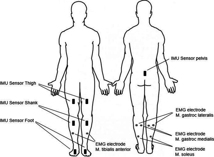
Methods: The results of 14 patients were reviewed. Kinematic 3D gait analysis, comparative bilateral electromyography (EMG) and pedobarography were performed.
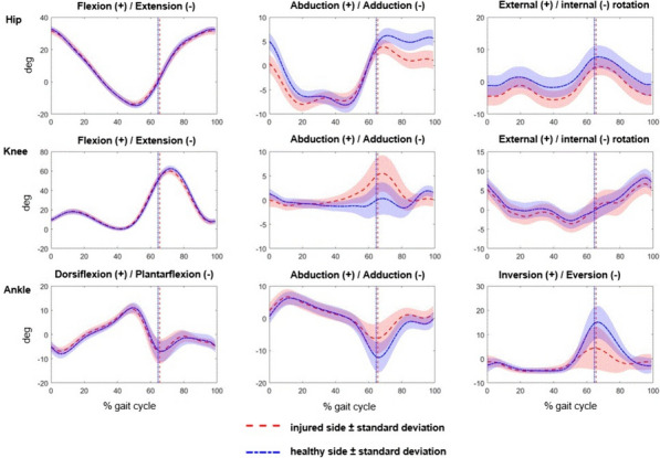
Results: On the injured side, the 3D gait analysis showed a significantly increased internal rotation and decreased external rotation of the hip and significantly decreased adduction and decreased range of motion (ROM) for the ankle. On the healthy side, the pedobarography revealed a significantly increased mean force in the forefoot, an increased peak maximum force and an increased maximum pressure in the metatarsal. When standing, significantly more weight was placed on the healthy side. The EMG measurements showed no significant differences between the healthy and injured legs.
Conclusions: After isolated Chopart injuries, significant changes in gait and pedobarography can be seen over the long term.
Keywords: Chopart injury; Fracture; Gait analysis; Midfoot; Pressure distribution.
© 2024. The Author(s).
Conflict of interest statement
The authors declare no competing interests.
Figures
Similar articles
-
Combined three-dimensional gait and plantar pressure analyses detecting significant functional deficits in children with juvenile idiopathic arthritis.Gait Posture. 2018 Oct;66:247-254. doi: 10.1016/j.gaitpost.2018.08.041. Epub 2018 Sep 9. Gait Posture. 2018. PMID: 30218839
-
A comparative study of pedobarography and ankle kinematics between children with idiopathic clubfoot after a soft tissue release procedure and controls.Int Orthop. 2020 Feb;44(2):319-327. doi: 10.1007/s00264-019-04447-2. Epub 2019 Dec 3. Int Orthop. 2020. PMID: 31796992
-
Optomechanical Analysis of Gait in Patients with Ankylosing Spondylitis.Sensors (Basel). 2025 Mar 14;25(6):1797. doi: 10.3390/s25061797. Sensors (Basel). 2025. PMID: 40292924 Free PMC article.
-
Loss of Mechanical Ankle Function Is Not Compensated by the Distal Foot Joints in Patients with Ankle Osteoarthritis.Clin Orthop Relat Res. 2021 Jan 1;479(1):105-115. doi: 10.1097/CORR.0000000000001443. Clin Orthop Relat Res. 2021. PMID: 32947288 Free PMC article.
-
Pedobarography and ankle-foot kinematics in children with symptomatic flexible flatfoot after medialising calcaneal osteotomy and controls: a comparative study.Int Orthop. 2024 Nov;48(11):2873-2879. doi: 10.1007/s00264-024-06290-6. Epub 2024 Sep 5. Int Orthop. 2024. PMID: 39235617
References
-
- Court-Brown CM, Zinna S, Ekrol I. Classification and epidemiology of mid-foot fractures. Foot. 2006;16:138–141. doi: 10.1016/j.foot.2006.03.003. - DOI
MeSH terms
LinkOut - more resources
Full Text Sources
