. 2024 Apr 26;22(1):245.
doi: 10.1186/s12964-024-01614-w.

DEC1 is involved in circadian rhythm disruption-exacerbated pulmonary fibrosis

Affiliations

DEC1 is involved in circadian rhythm disruption-exacerbated pulmonary fibrosis

Shuai-Jun Chen et al. Cell Commun Signal. .

Abstract

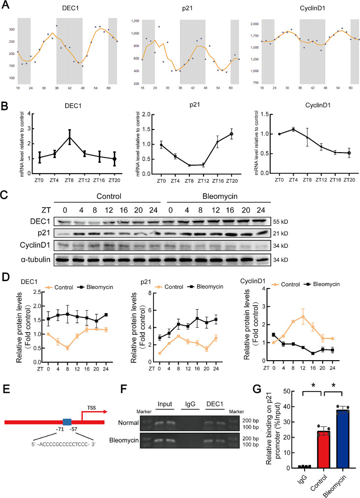
Background: The alveolar epithelial type II cell (AT2) and its senescence play a pivotal role in alveolar damage and pulmonary fibrosis. Cell circadian rhythm is strongly associated with cell senescence. Differentiated embryonic chondrocyte expressed gene 1 (DEC1) is a very important circadian clock gene. However, the role of DEC1 in AT2 senescence and pulmonary fibrosis was still unclear.

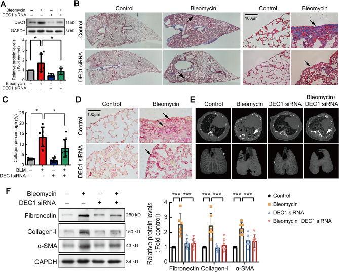
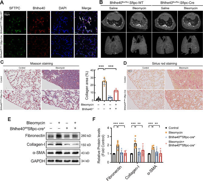
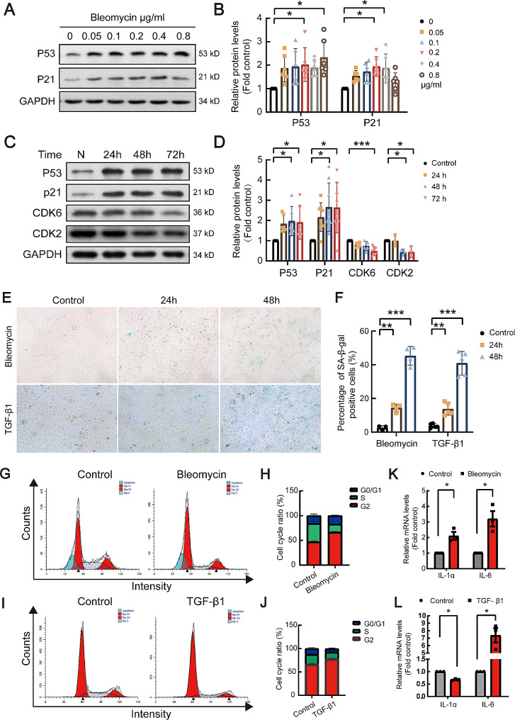
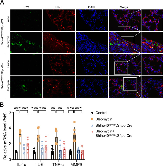
Results: In this study, a circadian disruption model of light intervention was used. It was found that circadian disruption exacerbated pulmonary fibrosis in mice. To understand the underlying mechanism, DEC1 levels were investigated. Results showed that DEC1 levels increased in lung tissues of IPF patients and in bleomycin-induced mouse fibrotic lungs. In vitro study revealed that bleomycin and TGF-β1 increased the expressions of DEC1, collagen-I, and fibronectin in AT2 cells. Inhibition of DEC1 mitigated bleomycin-induced fibrotic changes in vitro and in vivo. After that, cell senescence was observed in bleomycin-treated AT2 cells and mouse models, but these were prevented by DEC1 inhibition. At last, p21 was confirmed having circadian rhythm followed DEC1 in normal conditions. But bleomycin disrupted the circadian rhythm and increased DEC1 which promoted p21 expression, increased p21 mediated AT2 senescence and pulmonary fibrosis.

Conclusions: Taken together, circadian clock protein DEC1 mediated pulmonary fibrosis via p21 and cell senescence in alveolar epithelial type II cells.

Keywords: Alveolar epithelial type II cell; DEC1; Pulmonary fibrosis; p21.


Conflict of interest statement

The authors declare that no conflict of interest exists.

Figures

Fig. 1
Fig. 1
Circadian rhythm disruption exacerbated pulmonary fibrosis in C57BL/6J mice. All C57BL/6J mice were synchronized to a light dark cycle with 12 h of light and 12 h of darkness (LD) for 2 weeks. Then the CJL group was treated by advancing light onset 8 h in every 48 h, 5 days in rhythm disruption was exactly a cycle. The LD group was maintained at normal circadian rhythms. (A) Schematic diagram of animal model for disrupted circadian rhythms. (B, C) BMAL1 and DEC1 proteins were measured by western blotting, changes in relative density of BMAL1 and DEC1 were presented. n = 6. (D) Survival analysis of mice in each group. (E) Representative images of lung tissue with Masson staining. Original magnification, ×400. (F) Collagen deposition area (%) of lung tissue in each group. n = 6. (G) Representative immunoblots of collagen-I, DEC1 and α-SMA proteins in lung lysates. (H) Changes in protein levels of collagen-I, DEC1 and α-SMA (n = 5). Data are shown as mean ± SEM of n individual experiments. *P < 0.05, P values were determined by Student’s t-test (C) or one-way ANOVA followed by the Bonferroni’s test (F, H)
Fig. 2
Fig. 2
DEC1 expression increased in IPF patient′s lung tissues and bleomycin induced C57BL/6J mouse fibrotic lungs. (A) Representative images of Masson staining and DEC1 protein immunohistochemistry of lung tissue from IPF patients. The control was adjacent normal lung tissues of lung cancer. Original magnification, ×400. (B) C57BL/6J mouse pulmonary fibrosis model, representative images of DEC1 protein immunofluorescence staining and Masson staining of lung tissues. Original magnification, ×400
Fig. 3
Fig. 3
Bleomycin and TGF-β1 increased expressions of collagen-I, fibronectin and DEC1 in rat alveolar epithelial cells. (A, B) Primary alveolar epithelial cells were treated with bleomycin (0.2 µg/ml) for 48 h, and Western blot was performed to detect collagen-I (n = 6), fibronectin (n = 7), α-SMA (n = 10), BMAL1 (n = 8) and DEC1 (n = 8) proteins. (C-F) Serial concentrations of bleomycin or TGF-β1 (2 ng/ml) were used to treat RLE-6TN cells. The expression of related proteins, collagen-I, fibronectin and DEC1, was detected by Western blot. The representative images of immunoblots and statistical analysis were shown. n = 4. *P < 0.05, **P < 0.01, ***P < 0.001. P values were determined by Student’s t-test (B) or one-way ANOVA followed by the Dunnett’s test (D, F)
Fig. 4
Fig. 4
DEC1-siRNA suppressed bleomycin-induced mouse pulmonary fibrosis. C57BL/6J mice were subjected to intraperitoneal injection of DEC1 siRNA lentivirus to knock down DEC1 gene, and then a pulmonary fibrosis model was established by intraperitoneal injection of bleomycin. (A) Efficiency validation of DEC1 interference in mouse lung tissue (n = 5). (B) A panoramic and 400-times magnified representative images of Masson staining of mouse lung tissue. (C) Quantitative analysis of collagen deposition area in mouse lung tissue (n = 6). (D) Sirius red staining of mouse lung tissues. (E) Micro-CT scan image and 3D reconstruction of mouse lung. (F) Western blot analysis of fibronectin, collagen-I, and α-SMA in mouse lung tissue (n = 6), quantitative analysis of fibronectin, collagen-I, and α-SMA protein blots. *P < 0.05, **P < 0.01, ***P < 0.001. P values were determined by the one-way ANOVA followed by the Bonferroni’s test
Fig. 5
Fig. 5
Mouse type II alveolar epithelial cell specific knockout of DEC1 attenuated bleomycin-induced mouse pulmonary fibrosis. (A) Representative images of DEC1 gene knockout in mouse alveolar epithelial cells were verified by immunofluorescence. Original magnification, ×400. (B) DEC1 conditional knockout mice were used to construct a pulmonary fibrosis model. The figure showed CT scan and 3D reconstruction images of mouse lung. (C) Masson staining of lung tissues. Original magnification, ×200. (D) Sirius red staining of lung tissues. Original magnification, ×200. (E, F) Representative blots showing the expression level of related proteins in lung tissue of mice, detected by Western blot analysis (n = 6). *P < 0.05, **P < 0.01, ***P < 0.001. P values were determined by the one-way ANOVA followed by the Bonferroni’s test
Fig. 6
Fig. 6
Bleomycin and TGF-β1 induced cell senescence in type II alveolar epithelial cells. (A, B) RLE-6TN cells were treated with a serial concentration of bleomycin, and Western blot analysis was performed to detect protein levels of p21 and p53. (A) representative blots. (B) Statistical analysis, n = 6. (C, D) After treatment with bleomycin for a serial of time points, Western blot was used to detect the protein expression of p53, p21, CDK6, and CDK2 in RLE-6TN. Statistical analysis was conducted. (E, F) Cells were treated with bleomycin (0.2 µg/ml) or TGF-β1 (2 ng/ml), and cell senescence levels were assessed by β-galactosidase assay. (G-J) Bleomycin or TGF-β1 was used to treat cells for 24 h, changes in cell cycle were detected by flow cytometry. (K, L) Senescence-related gene miRNA levels were tested using qRT-PCR. *P < 0.05, **P < 0.01, ***P < 0.001. P values were determined by the one-way ANOVA followed by the Bonferroni’s test
Fig. 7
Fig. 7
Mouse alveolar epithelial cell specific knockout of DEC1 attenuated ATII cell senescence in bleomycin-induced mouse pulmonary fibrosis. (A) Representative immunofluorescence images of p21 protein expression in pulmonary fibrosis models constructed in mice with or without DEC1 knockout. Original magnification, ×400. Green: p21, red: SFTPC. (B) qRT-PCR was performed to detect mRNA levels of the senescence-associated secretory phenotype (SASP) factors, including IL-1α, IL-6, TNF-α, and MMP-9 in mouse lung tissues (n = 6). *P < 0.05, **P < 0.01, ***P < 0.001. P values were determined by the one-way ANOVA followed by the Bonferroni’s test
Fig. 8
Fig. 8
Knockdown of DEC1 with DEC1 siRNA attenuated bleomycin or TGF-β1 induced ATII cell senescencein vitro. (A) RLE-6TN cells were treated with bleomycin and DEC1 siRNA, and the expression levels of senescence-associated proteins DEC1, p53, p21, and CDK2 were detected by Western blot analysis. (B) Statistical analysis of the Western blot bands corresponding to the senescence-associated proteins detected. DEC1: n = 12, p53: n = 8, p21: n = 6, and CDK2: n = 3. (C, D) After treating RLE-6TN cells with bleomycin or TGF-β1 with or without DEC1 siRNA, cellular senescence was detected by staining with β-galactosidase. (E, F) Statistical analysis of senescent cells in according to C and D, n = 3. (G, H) SASP (IL-1α and IL-6) in epithelial cells was detected by qRT-PCR after treating RLE-6TN cells with bleomycin with or without DEC1 siRNA (G: n = 4, H: n = 6). (I, J) RLE-6TN cells were treated with bleomycin or TGF-β1 with or without DEC1 siRNA, and changes in the cell cycle were detected by flow cytometry, n = 3. *P < 0.05, **P < 0.01, ***P < 0.001, ns means nonsignificant. P values were determined by the one-way ANOVA followed by the Bonferroni’s test
Fig. 9
Fig. 9
The cell cycle is rhythmical, and bleomycin disrupts the cell cycle rhythm in alveolar epithelial cells. (A) The website predicted the rhythms of circadian clock proteins and related cell cycle protein (Circadian Expression Profiles Data Base: http://circadb.hogeneschlab.org/). (B) The rhythms of the mRNA levels of circadian clock genes and cell cycle genes were detected in C57 mouse lung tissue by qRT-PCR. n = 6. (C, D) After treat RLE-6TN cells with bleomycin, Western blot analysis was performed to detect the rhythmic change of related proteins, and statistical analysis of blots was conducted. n = 3. (E) Schematic illustration of the binding sites between DEC1 and p21 promoter regions. (F) DNA electrophoresis gels showed PCR products obtained after the reaction with ChIP-enriched DNA. (G) ChIP-qPCR was used to quantitatively detect the binding of DEC1 to the p21 promoter region. n = 4. *P < 0.05. P values were determined by the one-way ANOVA followed by the Bonferroni’s test

References

    1. Raghu G, Remy-Jardin M, Richeldi L, Thomson CC, Inoue Y, Johkoh T, et al. Idiopathic pulmonary fibrosis (an update) and progressive pulmonary fibrosis in adults: an Official ATS/ERS/JRS/ALAT Clinical Practice Guideline. Am J Respir Crit Care Med. 2022;205:e18–47. doi: 10.1164/rccm.202202-0399ST. - DOI - PMC - PubMed
    1. Parimon T, Yao C, Stripp BR, Noble PW, Chen P. Alveolar epithelial type II cells as drivers of lung fibrosis in idiopathic pulmonary fibrosis. Int J Mol Sci. 2020; 21. - PMC - PubMed
    1. Mora AL, Rojas M, Pardo A, Selman M. Emerging therapies for idiopathic pulmonary fibrosis, a progressive age-related disease. Nat Rev Drug Discov. 2017;16:755–72. doi: 10.1038/nrd.2017.170. - DOI - PubMed
    1. Reyfman PA, Walter JM, Joshi N, Anekalla KR, McQuattie-Pimentel AC, Chiu S, et al. Single-cell transcriptomic analysis of human lung provides insights into the Pathobiology of Pulmonary Fibrosis. Am J Respir Crit Care Med. 2019;199:1517–36. doi: 10.1164/rccm.201712-2410OC. - DOI - PMC - PubMed
    1. Yao C, Guan X, Carraro G, Parimon T, Liu X, Huang G et al. Senescence of alveolar type 2 cells Drives Progressive Pulmonary Fibrosis. Am J Respir Crit Care Med. 2019. - PMC - PubMed

Publication types

MeSH terms

Substances