. 2024 Mar 24;13(4):389.
doi: 10.3390/antiox13040389.

Effects of L-Type Voltage-Gated Calcium Channel (LTCC) Inhibition on Hippocampal Neuronal Death after Pilocarpine-Induced Seizure

Affiliations

Effects of L-Type Voltage-Gated Calcium Channel (LTCC) Inhibition on Hippocampal Neuronal Death after Pilocarpine-Induced Seizure

Chang-Jun Lee et al. Antioxidants (Basel). .

Abstract

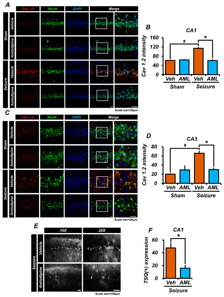
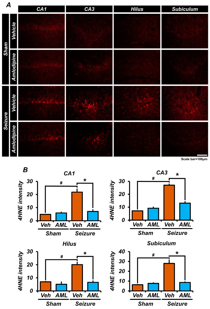
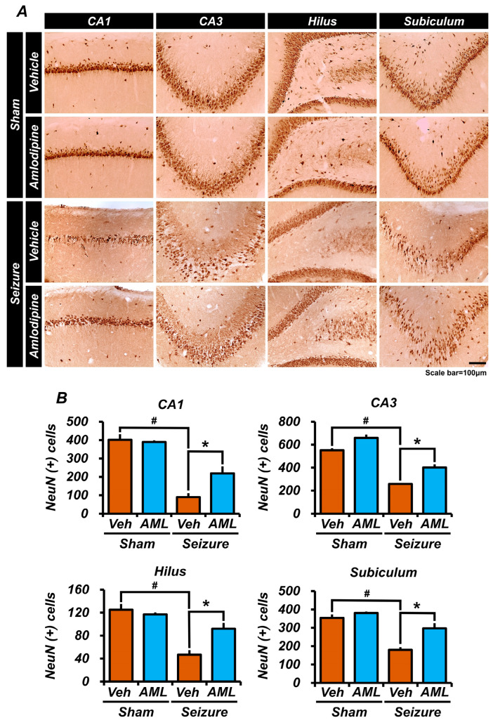
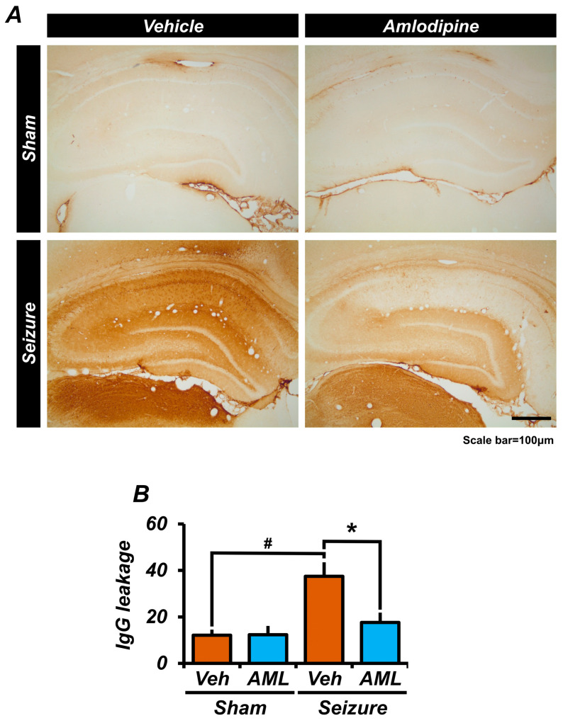
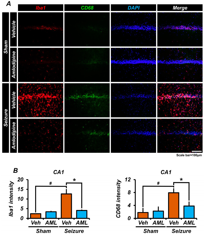
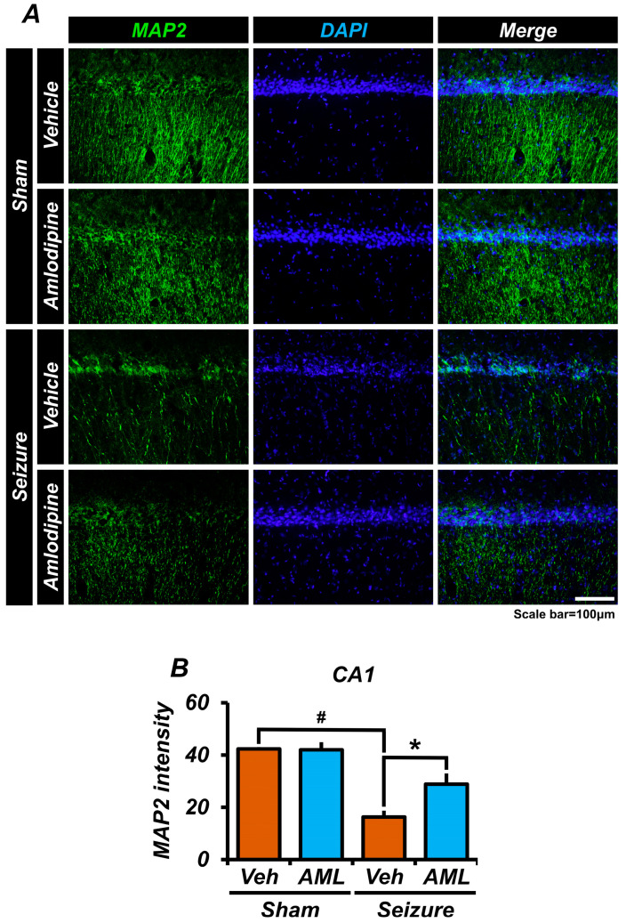
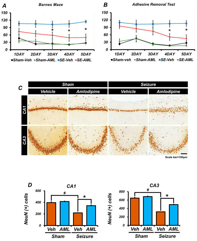
Epilepsy, marked by abnormal and excessive brain neuronal activity, is linked to the activation of L-type voltage-gated calcium channels (LTCCs) in neuronal membranes. LTCCs facilitate the entry of calcium (Ca2+) and other metal ions, such as zinc (Zn2+) and magnesium (Mg2+), into the cytosol. This Ca2+ influx at the presynaptic terminal triggers the release of Zn2+ and glutamate to the postsynaptic terminal. Zn2+ is then transported to the postsynaptic neuron via LTCCs. The resulting Zn2+ accumulation in neurons significantly increases the expression of nicotinamide adenine dinucleotide phosphate (NADPH) oxidase subunits, contributing to reactive oxygen species (ROS) generation and neuronal death. Amlodipine (AML), typically used for hypertension and coronary artery disease, works by inhibiting LTCCs. We explored whether AML could mitigate Zn2+ translocation and accumulation in neurons, potentially offering protection against seizure-induced hippocampal neuronal death. We tested this by establishing a rat epilepsy model with pilocarpine and administering AML (10 mg/kg, orally, daily for 7 days) post-epilepsy onset. We assessed cognitive function through behavioral tests and conducted histological analyses for Zn2+ accumulation, oxidative stress, and neuronal death. Our findings show that AML's LTCC inhibition decreased excessive Zn2+ accumulation, reactive oxygen species (ROS) production, and hippocampal neuronal death following seizures. These results suggest amlodipine's potential as a therapeutic agent in seizure management and mitigating seizures' detrimental effects.

Keywords: L-type voltage-gated calcium channel; amlodipine; neuronal death; oxidative stress; seizure; zinc.


Conflict of interest statement

The authors declare no competing interests.

Figures

Figure 1
Figure 1
Amlodipine administration reduced Cav 1.2 activation and zinc accumulation in neurons following seizure. (A,C) Confocal micrographs of Cav1.2 (red), NeuN (green), and DAPI (blue) in the hippocampal CA1 and CA3 regions. Scale bar = 20 µm. (B,D) The bar graph shows the intensity of Cav1.2 in the hippocampal regions CA1 and CA3. Both graphs indicate that the intensity of Cav1.2 in the seizure-amlodipine group is similar to that of the sham groups and approximately half of that in the seizure-vehicle group, with significant differences. Additionally, there was no significant difference between the sham-vehicle group and the sham-amlodipine group. Animals were sacrificed on day 7. Cav 1.2 expression decreased in the seizure-amlodipine group, compared with seizure-vehicle group, by about 46% in theCA1 region (seizure-vehicle group, 113.9 ± 5.9; seizure-amlodipine group, 61.0 ± 7.2; sham-vehicle group, 61.6 ± 2.7; sham-amlodipine group, 64.0 ± 3.3), and by 54% in the CA3 region (seizure-vehicle group, 65.7 ± 5.5; seizure-amlodipine group, 30.0 ± 4.2; sham-vehicle group, 20.0 ± 1.7; sham-amlodipine group, 29.3 ± 9.7). The sample sizes were n = 3 animals for each sham group and n = 5 animals for each seizure group. (Bonferroni post hoc test after Kruskal–Wallis test, CA1: chi-squared = 10.212, df = 3, p = 0.017; CA3: chi-squared = 10.471, df = 3, p = 0.015.) (E) Difference in the number of TSQ (+) cells between the seizure-vehicle and seizure-amlodipine groups in the CA1 region one day after the seizure. (F) The seizure-amlodipine group shows a significant reduction in TSQ-positive neuron cells, with a decrease of about 66%, compared to the seizure-vehicle group. Thus, amlodipine treatment after a seizure reduces Cav1.2 over-activation and zinc accumulation, compared to vehicle treatment after a seizure. Animals were sacrificed on day 1. TSQ (+) neuron cells decreased in the seizure-amlodipine group, compared with seizure-vehicle group, by about 66% in the CA1 region (seizure-vehicle group, 47.5 ± 8.0; seizure-amlodipine group, 16.0 ± 5.0). Scale bar = 100 µm (the sample sizes were n = 5 animals for the seizure-amlodipine group, n = 6 animals for the seizure-vehicle group), (Mann–Whitney U test measurement results, whole brain region z = 2.373, p = 0.018). # indicates significant difference between sham vehicle and seizure vehicle groups, * Significantly different from the vehicle group, p < 0.05.
Figure 2
Figure 2
Administration of amlodipine decreased reactive oxidative stress (ROS) after seizure. ROS was detected using 4-hydroxy-2-nonenal (4HNE) (red) staining in the hippocampal CA1, CA3, dentate gyrus (DG), and subiculum (Sub) regions, 7 days after the seizure. (A) Micrographs showing 4HNE (red) in the hippocampal CA1, CA3, DG, and Sub regions. (B) In every region, there was no significant difference between the sham groups. There were differences in intensity between the seizure-vehicle and seizure-amlodipine groups in every region, including the CA3 region. In every region, the intensity was similar between the sham groups and the seizure-amlodipine-treated group, and there was also a significant difference between the sham-vehicle group and the seizure-vehicle group. Therefore, administering amlodipine after a seizure significantly reduced ROS, compared to administering the vehicle after a seizure. Animals were sacrificed on day 7. The expression of 4HNE decreased in the seizure-amlodipine group, compared with seizure-vehicle group, by about 68% in the CA1 region (seizure-vehicle group, 21.8 ± 3.1; seizure-amlodipine group, 7.0 ± 2.0; sham-vehicle group, 4.8 ± 0.3; sham-amlodipine group, 5.9 ± 1.0), by 52% in the CA3 region (seizure-vehicle group, 27.0 ± 2.4; seizure-amlodipine group, 13.1 ± 1.6; sham-vehicle group, 7.1 ± 0.5; sham-amlodipine group, 29.2 ± 1.3), by 69% in the hilus (seizure-vehicle group, 20.1 ± 3.4; seizure-amlodipine group, 6.6 ± 1.8; sham-vehicle group, 7.0 ± 0.6; sham-amlodipine group, 5.1 ± 2.1), and by 67% in the subiculum (seizure-vehicle group, 28.0 ± 4.0; seizure-amlodipine group, 8.6 ± 1.2; sham-vehicle group, 6.3 ± 0.7; sham-amlodipine group, 7.6 ± 1.1). Scale bar = 100 µm (the sample sizes were n = 5 animals for each sham group, n = 6 animals for the seizure-vehicle group, and n = 8 animals for the seizure-amlodipine group). (Bonferroni post hoc test after Kruskal–Wallis test, CA1: chi-squared = 11.085, df = 3, p = 0.011; CA3: chi-squared = 14.367, df = 3, p = 0.002; subiculum: chi-squared = 13.485, df = 3, p = 0.004; dentate gyrus: chi-squared = 12.686, df = 3, p = 0.005). # indicates significant difference between sham vehicle and seizure vehicle groups, * Significantly different from the vehicle group, p < 0.05.
Figure 3
Figure 3
Amlodipine administration increased neuron survival after seizure. (A) Micrographs of NeuN (+) cells representing surviving cells in the hippocampal CA1, CA3, hilus, and subiculum (Sub) regions. The number of surviving neurons appears to be greater in the seizure-amlodipine group than in the seizure-vehicle group. (B) Cell counting in the micrographs of every region shows a significant difference between the sham-vehicle group and the seizure-vehicle group. In the CA1 and hilus regions, cell counting showed that the seizure-amlodipine group had approximately twice the number of NeuN (+) cells as the seizure-vehicle group. In the CA3 and Sub regions, the seizure-amlodipine group had more NeuN (+) cells, which was a noticeable difference from the seizure-vehicle group. Additionally, in every region, there is no significant difference in NeuN (+) cells between the sham-vehicle group and the sham-amlodipine group. Therefore, administering amlodipine after a seizure significantly increases neuron survival compared to treating with a vehicle after a seizure. Animals were sacrificed on day 7. The number of NeuN (+) cells was higher in the seizure-amlodipine group than the seizure-vehicle group by about 58% in the CA1 region (seizure-vehicle group, 89.5 ± 21.5; seizure-amlodipine group, 215.4 ± 32.3; sham-vehicle group, 401.6 ± 22.3; sham-amlodipine group, 389.5 ± 6.9), by 34.3% in the CA3 region (seizure-vehicle group, 257.9 ± 8.7; seizure-amlodipine group, 392.8 ± 24.7; sham-vehicle group, 551.1 ± 28.3; sham-amlodipine group, 659.0 ± 19.4), by 49% in the hilus region (seizure-vehicle group, 46.7 ± 8.0; seizure-amlodipine group, 92.1 ± 10.0; sham-vehicle group, 125.2 ± 11.0; sham-amlodipine group, 117.0 ± 2.8), and by 39% in the subiculum (seizure-vehicle group, 179.9 ± 14.1; seizure-amlodipine group, 297.0 ± 25.4; sham-vehicle group, 353.8 ± 14.9; sham-amlodipine group, 380.3 ± 6.6). Scale bar = 100 µm (the sample sizes were n = 5 animals for each sham group, n = 6 animals for the seizure-vehicle group, and n = 8 animals for the seizure-amlodipine group). (Bonferroni post hoc test after Kruskal–Wallis test, CA1: chi-squared = 17.649, df = 3, p = 0.001; CA3: chi-squared = 18.543, df = 3, p < 0.001; subiculum: chi-squared =15.535, df = 3, p = 0.001; dentate gyrus: chi-squared = 15.649, df = 3, p < 0.001.) # indicates significant difference between sham vehicle and seizure vehicle groups, * Significantly different from the vehicle group, p < 0.05.
Figure 4
Figure 4
Amlodipine administration reduced blood–brain barrier (BBB) breakdown after seizure. (A,B) The micrographs show the amount of IgG leakage in a specific region of the whole brain. We found a significant difference between the sham-vehicle group and the seizure-vehicle group. There is also a twofold decrease in IgG leakage between the seizure-amlodipine group and the seizure-vehicle group. However, the IgG leakage in the seizure-amlodipine group is similar to that of the sham groups. Also, the sham-vehicle group shows no significant difference when compared with the sham-amlodipine group. This indicates that administering an amlodipine treatment after a seizure significantly reduces IgG leakage. The scale bar represents a distance of 100 µm. The data are presented as mean values with the standard error of the mean (SEM) indicated. The sample sizes were n = 3 animals for each sham group, n = 5 animals for the seizure-vehicle group, and six for the seizure-amlodipine group. The difference between the seizure-amlodipine group and the seizure-vehicle group is considered statistically significant, with a p-value less than 0.05. Animals were sacrificed on day 7. IgG leakage decreased in the seizure-amlodipine group, compared with the seizure-vehicle group, by about 53% in the hippocampal area (seizure-vehicle group, 37.4 ± 6.0; seizure-amlodipine group, 17.6 ± 4.3; sham-vehicle group, 12.1 ± 2.4; sham-amlodipine group, 12.3 ± 3.8). (Bonferroni post hoc test after Kruskal–Wallis test of IgG, CA1: chi-squared = 7.763, df = 3, p = 0.051.) # indicates significant difference between sham vehicle and seizure vehicle groups, * Significantly different from the vehicle group, p < 0.05.
Figure 5
Figure 5
Amlodipine administration reduced astrocyte over-activation after seizure. (A) The micrographs show the staining of GFAP (green), C3 (red), and DAPI (blue) in the hippocampal CA1 region. (B) The bar graph illustrates the intensity of GFAP, which is significantly different in the sham-vehicle group compared with the seizure-vehicle group; there is also a significant difference between the seizure-amlodipine group and the seizure-vehicle group. The intensity of C3 in the seizure-amlodipine group is comparable to each sham group and is approximately three times lower than that of the seizure-vehicle group. There is also a significant difference between the sham-vehicle group and the seizure-vehicle group. Also, there is no significant difference in GFAP and C3 intensity among the various sham groups. This indicates that administering an amlodipine treatment after a seizure reduces the over-activation of astrocytes. The scale bar represents a distance of 100 µm. The data are presented as mean values, with the standard error of the mean (SEM) indicated. The sample sizes were five animals for each sham group, six animals for the seizure-vehicle group, and eight animals for the seizure-amlodipine group. The difference between the seizure-amlodipine group and the seizure-vehicle group is considered statistically significant, with a p-value less than 0.05. Animals were sacrificed on day 7. GFAP and C3 intensity decreased in the seizure-amlodipine group, compared with the seizure-vehicle group, by about 33% in the CA1 region for GFAP (seizure-vehicle group, 17.3 ± 1.8; seizure-amlodipine group, 11.6 ± 1.0; sham-vehicle group, 5.1 ± 0.4; sham-amlodipine group, 3.7 ± 0.8) and by 64% in the CA1 region for C3 (seizure-vehicle group, 17.3 ± 2.5; seizure-amlodipine group, 6.2 ± 1.4; sham-vehicle group, 5.5 ± 1.9; sham-amlodipine group, 12.9 ± 0.8). (Bonferroni post hoc test after Kruskal–Wallis test for GFAP, CA1: chi-squared = 12.894, df = 3, p = 0.005; Bonferroni post hoc test after Kruskal–Wallis test for C3, CA1: chi-squared = 12.246, df = 3, p = 0.007.) # indicates significant difference between sham vehicle and seizure vehicle groups, * Significantly different from the vehicle group, p < 0.05.
Figure 6
Figure 6
Amlodipine administration reduced microglia over-activation after seizure. (A) The micrographs depict the staining of Iba1 (red), CD68 (green), and DAPI (blue). There is a high presence of detected cells positive for both Iba1 and CD68 in the seizure-vehicle group. (B) The bar graph displays the intensity of Iba1 staining, which is found to be twice as high in the seizure-vehicle group compared to the other groups. The intensity of Iba1 in both the seizure-amlodipine group and each sham group is almost similar. Additionally, there is a significant difference in C3 intensity between the seizure-vehicle group and the seizure-amlodipine group, with the latter showing a slightly higher intensity compared to each sham group. Additionally, both graphs illustrate a significant difference between the sham-vehicle group and the seizure-vehicle group, while showing no significant difference among the sham groups. This indicates that administering amlodipine after a seizure reduces the over-activation of astrocytes, in comparison to the seizure-vehicle group. The scale bar represents a distance of 100 µm. The data are presented as mean values with the standard error of the mean (SEM) indicated. The sample sizes were five animals for each sham group, six animals for the seizure-vehicle group, and eight animals for the seizure-amlodipine group. The difference between the seizure-amlodipine group and the seizure-vehicle group is considered statistically significant, with a p-value less than 0.05. Animals were sacrificed on day 7. Iba1 and CD68 intensity decreased in the seizure-amlodipine group, compared with the seizure-vehicle group, by about 68% in the CA1 region for Iba1 (seizure-vehicle group, 12.6 ± 2.5; seizure-amlodipine group, 4.1 ± 0.5; sham-vehicle group, 2.4 ± 0.1; sham-amlodipine group, 3.4 ± 0.7) and by 52% in the CA1 region for CD68 (seizure-vehicle group, 7.9 ± 1.6; seizure-amlodipine group, 3.8 ± 0.7; sham-vehicle group, 1.8 ± 0.3; sham-amlodipine group, 2.3 ± 0.5). (Bonferroni post hoc test after Kruskal–Wallis test for Iba1, CA1: chi-squared = 16.884, df = 3, p = 0.001; Bonferroni post hoc test after Kruskal–Wallis test for CD68, CA1: chi-squared = 12.379, df = 3, p = 0.006.) # indicates significant difference between sham vehicle and seizure vehicle groups, * Significantly different from the vehicle group, p < 0.05.
Figure 7
Figure 7
Amlodipine administration reduced microtubule disruption after seizure. (A) The micrographs illustrate the staining of MAP2 (green) and DAPI (blue). The intensity of MAP2 staining in the seizure groups is lower than in the sham groups. (B) The bar graph demonstrates that each sham group exhibits a similar intensity of MAP2 staining. There is a significant difference between the sham-vehicle group and the seizure-vehicle group, and the seizure-amlodipine group has a lower intensity than the sham groups but a higher intensity than the seizure-vehicle group, with a significant difference observed. Additionally, there was no significant difference between the sham-vehicle group and the sham-amlodipine group. These results indicate that administering amlodipine after a seizure provides protection to microtubules. The scale bar represents a distance of 100 µm. The data are presented as mean values, with the standard error of the mean (SEM) indicated. The sample sizes were five animals for each sham group, six animals for the seizure-vehicle group, and eight animals for the seizure-amlodipine group. The difference between the seizure-amlodipine group and the seizure-vehicle group is considered statistically significant, with a p-value less than 0.05. Animals were sacrificed on day 7. Microtubule intensity increased in the seizure-amlodipine group, compared with the seizure-vehicle group, by about 43% in the CA1 area (seizure-vehicle group, 16.3 ± 2.4; seizure-amlodipine group, 28.9 ± 4.1; sham-vehicle group, 42.4 ± 0.7; sham-amlodipine group, 42.0 ± 2.9). (Bonferroni post hoc test after Kruskal–Wallis test, CA1: chi-squared =10.452, df = 3, p = 0.015.) # indicates significant difference between sham vehicle and seizure vehicle groups, * Significantly different from the vehicle group, p < 0.05.
Figure 8
Figure 8
Amlodipine administration improved spatial cognitive, memory, and cognitive function recovery after seizure. One week following the seizures for the seizure groups, and after a recovery period for the sham groups, we conducted several tests, and the results were as follows: (A) Barnes maze test: This test assessed spatial cognitive ability recovery. The sham groups outperformed the seizure groups. Notably, the seizure-amlodipine group performed better than the seizure-vehicle group, with the most significant differences observed between the third and fifth day. Additionally, there was no significant difference between the sham-vehicle group and the sham-amlodipine group on any day. Statistical analyses (a repeated measures test, followed by an ANOVA) revealed significant differences, especially in group interaction effects. (Barnes maze: time x group: F = 3.268, p < 0.021.) (B) Adhesive removal test: This measured cognitive function recovery, and the seizure-amlodipine group demonstrated progressive improvement from day one to five. In contrast, the seizure-vehicle group exhibited negligible progression, with stark differences apparent on the fourth and fifth days. Additionally, there was no significant difference between the sham groups on any day. Statistical analyses (a repeated measures test, followed by an ANOVA) revealed significant differences, especially in group interaction effects (adhesive removal: time * group: F = 12.005, p < 0.006). (C) We analyzed micrographs for neuronal nuclei (NeuN)-positive cells in the hippocampal CA1 and CA3 areas. (D) Bar graphs showed the sham groups had similar NeuN (+) cell counts in the hippocampal regions. In contrast, there was a noticeable discrepancy in the seizure groups, with a significant difference compared to the sham-vehicle group, as the amlodipine treatment seemed to bolster recovery in spatial cognition and general cognitive functions. Also, there were no differences in the CA1 and CA3 regions between the various sham groups. Each sham group consisted of five animals, while the seizure groups had six. Statistical significance was achieved between the seizure-amlodipine and seizure-vehicle groups, with a p-value below 0.05. Animals were sacrificed on day 12. The number of NeuN (+) cells was higher in the seizure-amlodipine group, compared with the seizure-vehicle group, by about 36% in the CA1 region (seizure-vehicle group, 218.2 ± 12.1; seizure-amlodipine group, 342.7 ± 21.1; sham-vehicle group, 395.8 ± 23.1; sham-amlodipine group, 414.9 ± 14.4) and by 35% in the CA3 region (seizure-vehicle group, 320.4 ± 5.3; seizure-amlodipine group, 492.5 ± 18.8; sham-vehicle group, 646.6 ± 33.5; sham-amlodipine group, 682.3 ± 24.2). (Bonferroni post hoc test after Kruskal–Wallis test: CA1: p = 0.006; CA3: p = 0.004.) This underscores the neuroprotective potential of amlodipine post-seizure. # indicates significant difference between sham vehicle and seizure vehicle groups, * Significantly different from the vehicle group, p < 0.05.

Similar articles

Cited by

References

    1. Arnold S.T., Dodson W.E. Epilepsy in children. Baillieres Clin. Neurol. 1996;5:783–802. - PubMed
    1. Goldberg E.M., Coulter D.A. Mechanisms of epileptogenesis: A convergence on neural circuit dysfunction. Nat. Rev. Neurosci. 2013;14:337–349. doi: 10.1038/nrn3482. - DOI - PMC - PubMed
    1. Shen Z., Haragopal H., Li Y.V. Zinc modulates synaptic transmission by differentially regulating synaptic glutamate homeostasis in hippocampus. Eur. J. Neurosci. 2020;52:3710–3722. doi: 10.1111/ejn.14749. - DOI - PubMed
    1. Stefan H. Pathophysiology of human epilepsy: Imaging and physiologic studies. Curr. Opin. Neurol. 2000;13:177–181. doi: 10.1097/00019052-200004000-00011. - DOI - PubMed
    1. Jeong J.H., Lee S.H., Kho A.R., Hong D.K., Kang D.H., Kang B.S., Park M.K., Choi B.Y., Choi H.C., Lim M.S., et al. The Transient Receptor Potential Melastatin 7 (TRPM7) Inhibitors Suppress Seizure-Induced Neuron Death by Inhibiting Zinc Neurotoxicity. Int. J. Mol. Sci. 2020;21:7897. doi: 10.3390/ijms21217897. - DOI - PMC - PubMed

LinkOut - more resources