. 2024 Apr 12;13(4):454.
doi: 10.3390/antiox13040454.

Nrf2 Plays a Key Role in Erythropoiesis during Aging

Affiliations

Nrf2 Plays a Key Role in Erythropoiesis during Aging

Serge Cedrick Toya Mbiandjeu et al. Antioxidants (Basel). .

Abstract

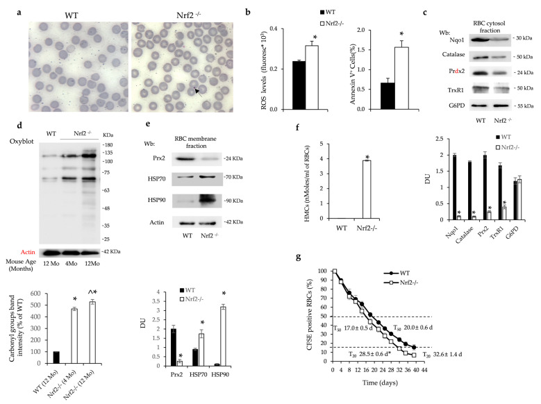
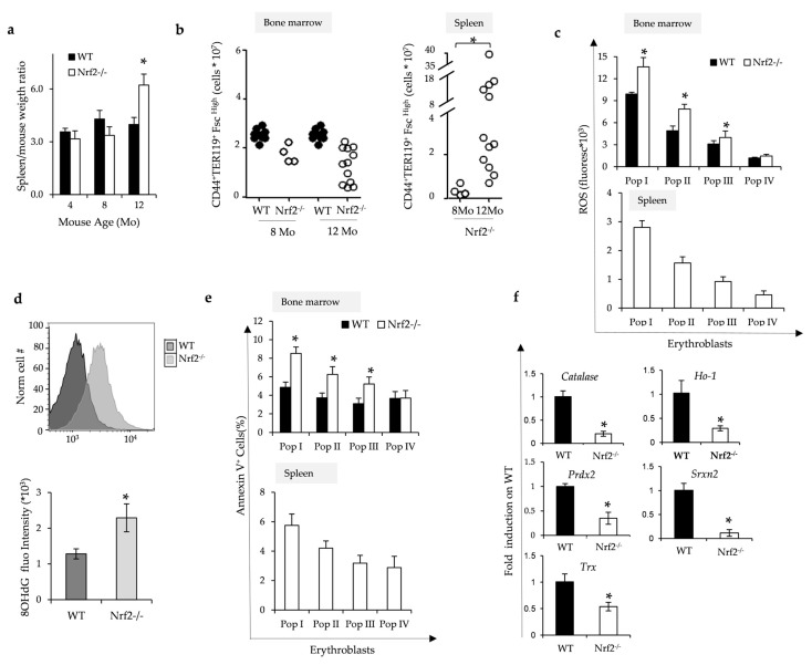
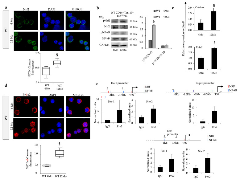
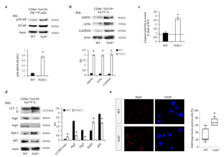
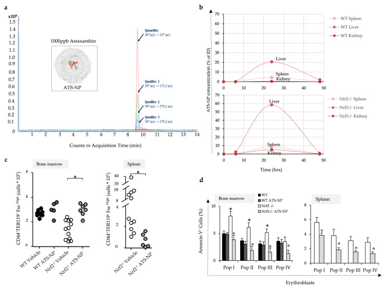
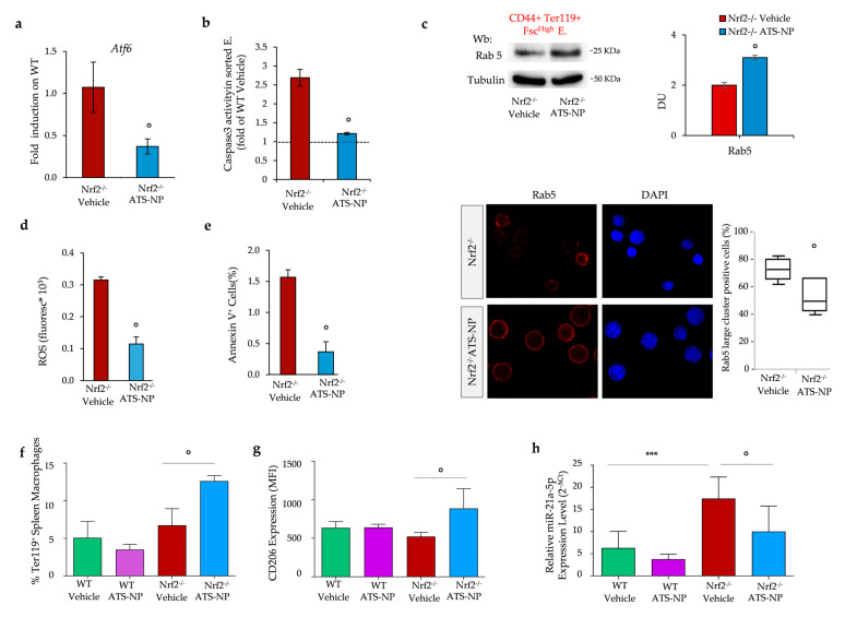
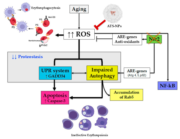
Aging is characterized by increased oxidation and reduced efficiency of cytoprotective mechanisms. Nuclear factor erythroid-2-related factor (Nrf2) is a key transcription factor, controlling the expression of multiple antioxidant proteins. Here, we show that Nrf2-/- mice displayed an age-dependent anemia, due to the combined contributions of reduced red cell lifespan and ineffective erythropoiesis, suggesting a role of Nrf2 in erythroid biology during aging. Mechanistically, we found that the expression of antioxidants during aging is mediated by activation of Nrf2 function by peroxiredoxin-2. The absence of Nrf2 resulted in persistent oxidation and overactivation of adaptive systems such as the unfolded protein response (UPR) system and autophagy in Nrf2-/- mouse erythroblasts. As Nrf2 is involved in the expression of autophagy-related proteins such as autophagy-related protein (Atg) 4-5 and p62, we found impairment of late phase of autophagy in Nrf2-/- mouse erythroblasts. The overactivation of the UPR system and impaired autophagy drove apoptosis of Nrf2-/- mouse erythroblasts via caspase-3 activation. As a proof of concept for the role of oxidation, we treated Nrf2-/- mice with astaxanthin, an antioxidant, in the form of poly (lactic-co-glycolic acid) (PLGA)-loaded nanoparticles (ATS-NPs) to improve its bioavailability. ATS-NPs ameliorated the age-dependent anemia and decreased ineffective erythropoiesis in Nrf2-/- mice. In summary, we propose that Nrf2 plays a key role in limiting age-related oxidation, ensuring erythroid maturation and growth during aging.

Keywords: ATF6; GADD34; PLGA; UPR system; astaxanthin; autophagy; ineffective erythropoiesis; nanoparticles; oxidation; red cells.


Conflict of interest statement

The authors declare no conflicts of interest.

Figures

Figure 1
Figure 1
Nrf2−/− mouse red cells display increased membrane oxidation resulting in accelerated senescence (a) Representative May–Grunwald–Giemsa staining for the morphology of red cells from wild-type (WT) and Nrf2−/− mice. Arrow indicates Howell–Jolly body in Nrf2−/− mouse red cells. Original magnification ×100. (b) ROS values in red cells and Annexin-V+ erythrocytes from WT and Nrf2−/− mice. Data are presented as means ± SD (n = 4) * p < 0.05 compared to WT. (c) Western blot (Wb) analysis using specific antibodies against Nqo1, Catalase, Prdx2, TrxR1 and G6PD of the cytosolic fraction of red cells from WT and Nrf2−/− mice. A representative gel of these four proteins is shown (upper panel). Densitometric analysis of immunoblots shown in the lower panel. Data are presented as means ± SD (n = 4); * p < 0.05 compared to WT mice. DU: densitometric unit. (d) Oxyblot analysis of the ghost fraction of red cells from WT and Nrf2−/− mice at 4 and 12 months (Mo) of age. A representative gel of the other four is shown (upper panel). Actin is used as loading control. Densitometric analysis is shown in the lower panels. Data are presented as means ± SD (n = 4); * p < 0.05 compared to WT mice (12 Mo); ^ p < 0.05 compared to Nrf2−/− mice at 4 Mo of age. (e) Wb analysis using specific antibodies against Prdx2, HSP70 and HSP90 of the ghost fraction of red cells from WT and Nrf2−/− mice. A representative gel is shown (upper panel). Actin is used as protein loading control. Densitometric analysis of immunoblots is shown in the lower panel. Data are presented as means ± SD (n = 4); * p < 0.05 compared to WT mice. (f) Hemichrome quantification in red cells from WT and Nrf2−/− mice at 12 months of age. Data are presented as means ± SD (n = 4); * p < 0.05 compared to WT mice. (g) Survival of CFSE-labeled red cells from WT and Nrf2−/− mice at 12 months of age (n = 6–7). Data are mean ± SD. * p < 0.05 compared to T20 in WT mice.
Figure 2
Figure 2
The absence of Nrf2 promotes ineffective erythropoiesis and increased oxidation of erythroblasts. (a) Spleen weight/mouse weight ratio of wild-type (WT) and Nrf2−/− mice at 4, 8 and 12 months (Mo) of age. Data are presented as means ± SD (n = 4–12) * p < 0.05 compared to WT. (b) Flow cytometric analysis, combining CD44-Ter119 and cell size marker strategy (CD44+/Ter119+/FSChigh), of the erythropoietic activity in the bone marrow and spleen from WT and Nrf2−/− mice at 8 and 12 months (Mo) of age. Data are presented as single dots (n = 4–12) * p < 0.05 compared to WT. (c) ROS values in bone marrow and spleen erythroblasts from WT and Nrf2−/− mice at 12 months of age. Data are presented as means ± SD, * p < 0.05 compared to WT. (d) DNA oxidative damage measured flow cytometrically as 8OHdG fluorescence intensity of erythroblast populations from WT and Nrf2−/− mice at 12 months of age. Data are presented as means ± SD, * p < 0.05 compared to WT. (e) Amount of Annexin V+ cells in bone marrow and spleen erythroid precursors from Nrf2−/− and WT mice as in (c); * p < 0.05 compared to WT. (f) mRNA expression of catalase, Ho-1, Prdx2, Srxn2 and Trx by qRT-PCR in erythroblasts from WT and Nrf2−/− mice (fold on WT). Experiments were performed in triplicate. * p < 0.01, Nrf2−/− vs. WT mice. p value was calculated by t-test.
Figure 3
Figure 3
In aging wild-type mice, erythroblasts show age-dependent activation of Nrf2 associated with nuclear translocation of Prdx2, which is recruited in promoter regions of Nrf2. (a) Nrf2 immunostaining of sorted erythroid precursors from bone marrow of 4- and 12-month-old wild-type (WT) mice. DAPI was used to stain nuclei. Nrf2 mean fluorescence in the nucleus and cytoplasm was measured using ImageJ (https://imagej.net/ij/download.html). At least 25 cells were analyzed in 6 different fields of acquisition. Data are presented as median and minimum/maximum, with boxes indicating 25th–75th percentiles. § p < 0.05 compared to 4-month-old WT mice. Original magnification 100×. (b) Wb analysis with specific anti-phospho-Nrf2 (p-Nrf2), Nrf2, phospho-NF-kB(p-NF-kB) and NF-kB in sorted erythroid precursors in bone marrow of 4- and 12-month-old WT mice. GAPDH was used as protein loading control. A representative gel is shown. Densitometric analysis of immunoblots is shown in the right panel. Data are presented as means ± SD (n = 4); § p < 0.05 compared to 4-month-old WT mice. DU: densitometric unit (c) mRNA expression of catalase and Prdx2 by qRT-PCR of erythroblasts from WT mice at 4 and 12 months of age. Data are mean ± SD (n = 6–8). Experiments were performed in triplicate. § p < 0.01, WT 4Mo vs. WT 12Mo mice. p value was calculated by t-test. (d) Prdx2 immunostaining of sorted erythroid precursors from bone marrow of 4- and 12-month-old wild-type (WT) mice. DAPI was used to stain nuclei. Prdx2 mean fluorescence in the nucleus and cytoplasm was measured using ImageJ. At least 40 cells were analyzed in 5 different fields of data acquisition. Data are presented as median and minimum/maximum, with boxes indicating 25th–75th percentiles; § p < 0.01, WT 4Mo vs. WT 12Mo mice. Original magnification 100×. (e) Predicted NRF and NF-kB binding sites in the indicated promoters are shown in red and blue, respectively. Horizontal bars indicate the regions amplified in the ChIP experiment. Histograms show qPCR quantification of a ChIP assay with the indicated antibodies from cells treated as above. Error bars indicate standard deviations.
Figure 4
Figure 4
Sorted Nrf2−/− mouse erythroblasts display overactivation of the system and impaired autophagy with caspase-3 pro-apoptotic pathway activation. (a) Wb analysis with specific anti-phospho-NF-kB (p-NF-kB) and NF-kB in sorted erythroid precursors from bone marrow of 12-month-old wild-type (WT) and Nrf2−/− mice. Actin was used as protein loading control. One representative gel of the other four is shown (upper panel). Densitometric analysis of immunoblots is shown in the lower panel. Data are presented as means ± SD (n = 4); * p < 0.05 compared to WT mice. (b) Wb analysis with specific anti-HSP70, ATF6 and GADD34 in sorted erythroid precursors as in a. Densitometric analysis of immunoblots is shown in the lower panel. Data are presented as means ± SD (n = 4); * p < 0.05 compared to WT mice. (c) Caspase 3 activity determined by cleavage of a fluorescent substrate in sorted erythroid precursors from bone marrow of WT and Nrf2−/− mice. Data are presented as means ± SD * p < 0.05 compared to WT. (d) Wb analysis with specific anti-LC3 I/II, Atg4, Atg5, Rab5 and p62 in sorted erythroid precursors from bone marrow of 12-month-old WT and Nrf2−/− mice. Actin was used as protein loading control. Densitometric analysis of immunoblots is shown in the right panel. Data are presented as means ± SD (n = 4); * p < 0.05 compared to WT mice. (e) Rab5 immunostaining of sorted erythroid precursors from bone marrow of 12-month-old WT and Nrf2−/− mice. DAPI was used to stain nuclei. Large clusters of positive cells were measured using ImageJ. At least 40 cells were analyzed in 8 different fields of acquisition. Data are presented as median and minimum/maximum, with boxes indicating 25–75th percentiles; * p < 0.05 compared to WT mice.
Figure 5
Figure 5
Astaxanthin PLGA nanoparticles improve Nrf2−/− mouse ineffective erythropoiesis. (a) Extracted ion chromatogram for astaxanthin illustrates three qualifier transitions and was used to confirm molecule identification. Inset: ATS-NP: pictorial representation of PLGA nanoparticles embedding astaxanthin; the relationship between the different molecules is illustrative. (b) Organ distribution of astaxanthin molecules in the two mice strains during treatment determined by mass spectrometric analysis (see also Figure S6d and Table S1). (c) Flow cytometric analysis, combining CD44-Ter119 and cell size marker strategy (CD44+/Ter119+/FSChigh), of the erythropoietic activity in the bone marrow and spleen from WT and Nrf2−/− mice treated with vehicle or ATS-NPs (2 mg/kg every two days for four weeks). Data are presented as single dots (n = 5–12) * p < 0.05 compared to vehicle-treated WT mice. (d) Amount of Annexin V+ cells in erythroid precursors from bone marrow and spleen of WT and Nrf2−/− mice treated as in (c). Data are presented as means ± SD; * p < 0.05 compared to WT mice, ° p < 0.05 compared to vehicle-treated mice.
Figure 6
Figure 6
Astaxanthin PLGA nanoparticles reduce oxidation, prevent overactivation of adaptative systems in erythroblasts and modulate splenic macrophages towards a pro-resolving pattern in Nrf2−/− mice. (a) mRNA expression of Atf6 by qRT-PCR on the erythroblasts from vehicle and ATS-NP treated Nrf2−/− mice. Data are mean ± SD (n = 6–8). Experiments were performed in triplicate. ° p < 0.01, Nrf2−/− vehicle vs. Nrf2−/− ATS-NP mice by t-test; (b) Caspase 3 activity determined by its cleavage of a fluorescent substrate in sorted erythroid precursors from bone marrow of Nrf2−/− mice treated as in (a); ° p < 0.01, Nrf2−/− vehicle vs. Nrf2−/− ATS-NP mice by t-test. (c) Upper panel: Wb analysis with specific anti-Rab5 in sorted erythroid precursors from bone marrow of Nrf2−/− mice treated as in (a). Densitometric analysis of immunoblots is shown in the right panel. Data are presented as means ± SD (n = 4); ° p < 0.01, Nrf2−/− vehicle vs. Nrf2−/− ATS-NP mice by t-test. Lower panel: Rab5 immunostaining of sorted erythroid precursors from bone marrow of Nrf2−/− mice treated as in (a). DAPI was used to stain nuclei. Large clusters of positive cells were measured using ImageJ. At least 35 cells were analyzed in 5 different fields of acquisition. Data are presented as median and minimum/maximum, with boxes indicating 25th–75th percentiles. (d) ROS levels in red cells and (e) Annexin-V+ erythrocytes from Nrf2−/− mice treated as in (a). Data are presented as mean ± SD (n = 4) ° p < 0.05 compared to vehicle-treated mice. (f) Flow cytometry analysis of phagocytosis of Ter-119+ cells, membrane expression of CD206 (g) and real-time PCR analysis of mir-21-5p (h) in splenic macrophages. Data are mean ± SD (n = 6); ***, p < 0.001; ° p < 0.05 by 1-way ANOVA.
Figure 7
Figure 7
Schematic diagram of the role of Nrf2 in erythropoiesis during aging and the protective effects of astaxanthin PLGA nanoparticles. Aging is associated with increased ROS production, which is limited by the activation of Nrf2. This results in the upregulation of ARE genes encoding for antioxidants and cytoprotective systems as well as by the activation of adaptative mechanisms such as the UPR system to face ER stress and autophagy to clear damaged proteins. The absence of Nrf2 (Nrf2−/− mice) negatively affects the antioxidant cell machinery, resulting in severe and sustained oxidation. Nrf2−/− mouse red cells display severe membrane oxidation, exposition of phosphatidylserine, membrane binding of hemichromes and reduced expression of antioxidants and cytoprotective systems such as Prdx2. Red cell membrane protein oxidation favors band 3 protein clusterization, which is recognized by the naturally occurring anti-band 3 IgG antibodies. Both mechanisms drive Nrf2−/− mouse red cells towards erythrophagocytosis by splenic macrophages. In erythroblasts lacking Nrf2, the prolonged and severe oxidation due to the downregulation of antioxidants and cytoprotective systems promotes intense ER stress with overactivation of the UPR system and autophagy. Although the persistence of oxidative stress promotes compensatory activation of NF-kB, this is insufficient to prevent the overwhelming of proteostasis with impairment autophagy and accumulation of Rab5. This drives Nrf2−/− erythroblasts towards apoptosis via the caspase-3 pathway, resulting in ineffective erythropoiesis. ATS-NPs act as efficient antioxidants preventing the deleterious effects of the absence of Nrf2 on erythropoiesis and red cells during aging. PS: phosphatidylserine; ER: endoplasmic reticulum; UPR: unfolded protein response; ARE-: antioxidant-related element; ROS: reactive oxygen species; Prdx2: peroxiredoxin-2; Atg: autophagy-related protein; GADD34: growth arrest and DNA damage-inducible protein 34; PLGA: poly(lactic-co-glycolic acid).

Similar articles

Cited by

References

    1. Hayes J.D., Dinkova-Kostova A.T. The Nrf2 regulatory network provides an interface between redox and intermediary metabolism. Trends Biochem. Sci. 2014;39:199–218. doi: 10.1016/j.tibs.2014.02.002. - DOI - PubMed
    1. Lee J.M., Chan K., Kan Y.W., Johnson J.A. Targeted disruption of Nrf2 causes regenerative immune-mediated hemolytic anemia. Proc. Natl. Acad. Sci. USA. 2004;101:9751–9756. doi: 10.1073/pnas.0403620101. - DOI - PMC - PubMed
    1. Ishii T., Warabi E., Yanagawa T. Novel roles of peroxiredoxins in inflammation, cancer and innate immunity. J. Clin. Biochem. Nutr. 2012;50:91–105. doi: 10.3164/jcbn.11-109. - DOI - PMC - PubMed
    1. Leiser S.F., Miller R.A. Nrf2 signaling, a mechanism for cellular stress resistance in long-lived mice. Mol. Cell Biol. 2010;30:871–884. doi: 10.1128/MCB.01145-09. - DOI - PMC - PubMed
    1. Osburn W.O., Kensler T.W. Nrf2 signaling: An adaptive response pathway for protection against environmental toxic insults. Mutat. Res. 2008;659:31–39. doi: 10.1016/j.mrrev.2007.11.006. - DOI - PMC - PubMed