. 2024 Apr 19;13(4):489.
doi: 10.3390/antiox13040489.

Specific NOX4 Inhibition Preserves Mitochondrial Function and Dampens Kidney Dysfunction Following Ischemia-Reperfusion-Induced Kidney Injury

Affiliations

Specific NOX4 Inhibition Preserves Mitochondrial Function and Dampens Kidney Dysfunction Following Ischemia-Reperfusion-Induced Kidney Injury

Tomas A Schiffer et al. Antioxidants (Basel). .

Abstract

Background: Acute kidney injury (AKI) is a sudden episode of kidney failure which is frequently observed at intensive care units and related to high morbidity/mortality. Although AKI can have many different causes, ischemia-reperfusion (IR) injury is the main cause of AKI. Mechanistically, NADPH oxidases (NOXs) are involved in the pathophysiology contributing to oxidative stress following IR. Previous reports have indicated that knockout of NOX4 may offer protection in cardiac and brain IR, but there is currently less knowledge about how this could be exploited therapeutically and whether this could have significant protection in IR-induced AKI. Aim: To investigate the hypothesis that a novel and specific NOX4 inhibitor (GLX7013114) may have therapeutic potential on kidney and mitochondrial function in a mouse model of IR-induced AKI. Methods: Kidneys of male C57BL/6J mice were clamped for 20 min, and the NOX4 inhibitor (GLX7013114) was administered via osmotic minipump during reperfusion. Following 3 days of reperfusion, kidney function (i.e., glomerular filtration rate, GFR) was calculated from FITC-inulin clearance and mitochondrial function was assessed by high-resolution respirometry. Renal histopathological evaluations (i.e., hematoxylin-eosin) and TUNEL staining were performed for apoptotic evaluation. Results: NOX4 inhibition during reperfusion significantly improved kidney function, as evidenced by a better-maintained GFR (p < 0.05) and lower levels of blood urea nitrogen (p < 0.05) compared to untreated IR animals. Moreover, IR caused significant tubular injuries that were attenuated by simultaneous NOX4 inhibition (p < 0.01). In addition, the level of renal apoptosis was significantly reduced in IR animals with NOX4 inhibition (p < 0.05). These favorable effects of the NOX4 inhibitor were accompanied by enhanced Nrf2 Ser40 phosphorylation and conserved mitochondrial function, as evidenced by the better-preserved activity of all mitochondrial complexes. Conclusion: Specific NOX4 inhibition, at the time of reperfusion, significantly preserves mitochondrial and kidney function. These novel findings may have clinical implications for future treatments aimed at preventing AKI and related adverse events, especially in high-risk hospitalized patients.

Keywords: NOX4; glomerular filtration rate; ischemia–reperfusion; kidney; mitochondria.


Conflict of interest statement

Per Wikström (P.W.) has submitted European patent application no. 18171556.6, protecting the NOX4-selective compound GLX7013114. Glucox Biotech AB provided support in the form of salary for P.W. Funding from Glucox Biotech AB does not alter the company’s adherence to policy on sharing data and materials. No other potential conflicts of interest or ethical statements relevant to this article are reported.

Figures

Figure 1
Figure 1
(A) Glomerular filtration rate (GFR) (N = 6/group) and (B) blood urea nitrogen (N = 6/group) was evaluated in mice treated with NOX4 inhibitor (NOX4i) during the first 24 h and during the whole reperfusion period of 3 days of reperfusion following 20 min bilateral ischemia (AKI). Venous injection of FITC-inulin was performed followed by sequential sampling of plasma. Plasma clearance of FITC-inulin was evaluated spectrofluorometrically. BUNs were measured by using a commercial colorimetric detection kit. One-Way ANOVA was used for statistical analysis. * for p ≤ 0.05, and **** for p ≤ 0.0001.
Figure 2
Figure 2
Histopathological evaluation of kidneys in the cortico–medullary junction (S3 segment area) in animals exposed to bilateral ischemia (20 min), treated with NOX4 inhibitor (NOX4i) during the first 24 h or 76 h of the 3-day reperfusion phase (N = 6/group). (A) Representative images of paraformaldehyde-fixed kidney slices stained with hematoxylin–eosin (HE) and periodic acid-Schiff (PAS) analyzed under light microscopy. ((A), A–B) Control group (SHAM) with normal tubular appearance, preserved cell morphology, and presence of an intact brush border positively highlighted by PAS (black arrow), in the region close to the renal medulla (M). ((A), C–D) Samples from the experimental group exposed to ischemia–reperfusion-induced kidney injury (AKI) showing extensive dilation of the S3 segment area tubules, cast formation, and sloughing of tubular epithelial cells or loss of the brush border (black arrowhead). ((A), E–F) Samples from the group exposed to AKI treated with specific NOX4i during the first 24 h of reperfusion showing a non-significant reduction in tubular damage in the S3 region close to the renal medulla (M), the majority of tubules with a normal diameter and cellular integrity (*), and other tubules presenting cellular debris in the lumen (white arrow) and the formation of hyaline casts (white arrowhead). ((A), G–H) Samples from the group with specific NOX4i for 76 h, with a significant reduction in tubular alterations, observed by the reduction in tubular dilation and greater integrity of cells and the brush border (#), with some tubules showing atrophy and the formation of hyaline casts (red arrowhead). (B) Statistical analysis of tubular injury score (Kruskal–Wallis test). Score 0: no tubular injury; Score 1: <10% of tubules injured; Score 2: 10–25% of tubules injured; Score 3: 25–50% of tubules injured; Score 4: 50–74% of tubules injured; Score 5: >75% of tubules injured. * for p ≤ 0.05, and **** for p ≤ 0.0001. Non-significant variations are indicated as ns.
Figure 3
Figure 3
A TUNEL assay was performed in kidney slices to evaluate apoptosis in the kidney S3 segment following acute kidney injury (AKI) (N = 6/group) 20 min bilateral ischemia followed by 3 days of reperfusion) combined with NOX4 inhibition (NOX4i) during either the first 24 h or the whole 76 h of reperfusion. (A) Representative TUNEL-stained kidney slices, where brown areas represent apoptotic tissue. (B) The percentage of TUNEL-positive area on the 3rd day of reperfusion was estimated by using the particle analysis command in the ImageJ software. One-Way ANOVA was used for statistical analysis. * for p ≤ 0.05. Non-significant variations are indicated as ns.
Figure 4
Figure 4
The degree of mitochondrial coupling and hydrogen peroxide production were evaluated on the 3rd day of reperfusion in isolated mitochondria from kidneys in mice following induced acute kidney injury (AKI) (20 min bilateral ischemia) combined with NOX4 inhibition (NOX4i). (A) The mitochondrial respiratory control ratio (RCR) defined as maximal CI-dependent state 3 respiration related to state 2 respiration in absence of adenylates was evaluated by high-resolution respirometry (Sham N = 7, AKI 3d and AKI 3d + NOX4i 76 h N = 6, NOX4i 24 h N = 5 and naïve + NOX4i 76 h N = 3) (B). Mitochondrial hydrogen peroxide (H2O2) production was measured spectrofluorometrically during leak respiration by using the amplex red system (N = 6/group, except for NOX4i 24 h N = 5). Note that NOX4i was washed away during the mitochondrial isolation protocol during these experiments. * for p ≤ 0.05, ** for p ≤ 0.01, and *** for p ≤ 0.001. Non-significant variations are indicated as ns.
Figure 5
Figure 5
Kidney mitochondrial respiratory complex activities were evaluated by high-resolution respirometry on the 3rd day of reperfusion in isolated mitochondria from mice following induced acute kidney injury (AKI) (20 min bilateral ischemia) combined with NOX4 inhibition (NOX4i) during the first 24 h or during the whole reperfusion period (Sham N = 7, AKI 3d and AKI 3d + NOX4i 76h N = 6, NOX4i 24 h N = 5 and naïve + NOX4i 76 h N = 3). A control group of naïve animals were administered NOX4i to evaluate the effect on mitochondrial function. State 3 respiration in presence of adenylates was determined for each mitochondrial complex activity, respectively. (AD) CI-CIV activity was normalized to mitochondrial protein. * for p ≤ 0.05, ** for p ≤ 0.01, *** for p ≤ 0.001, and **** for p ≤ 0.0001.
Figure 6
Figure 6
Kidney tissue citrate synthase activity was measured in mice after the induction of acute kidney injury (AKI) by 20 min ischemia followed by treatment with NOX4 inhibitor (NOX4i) during the reperfusion phase (N = 6/group except for AKI 3d + NOX4i 76 h, N = 5). Citrate synthase activity was measured by using a commercial colorimetric kit. One-Way ANOVA was used for statistical analysis. Non-significant variations were indicated as ns.
Figure 7
Figure 7
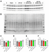
Immunoblotting was performed to evaluate the protein levels of the mitochondrial respiratory complexes in kidneys on the 3rd day of reperfusion (AKI) in animals treated with the NOX4 inhibitor (NOX4i) (N = 4/group). (A) Protein expression levels using a cocktail of antibodies targeting mitochondrial complexes. (B) Protein staining of the membrane used for normalization. (CF) Statistical analysis of the protein levels of CI, CII, CIII, and CV (One-Way ANOVA).
Figure 8
Figure 8
Immunoblotting was performed to evaluate protein levels of Ser40 phosphorylated Nrf2 in kidneys on the 3rd day of reperfusion (AKI) in animals treated with NOX4 inhibitor (NOX4i) (Sham N = 5, AKI 3d N = 6, AKI 3d + NOX4i 24 h N = 5, AKI3d + NOX4i N = 6). (A) Protein expression using antibodies targeting phosphorylated Nrf2 (Ser40), Nrf2, and membranes stained for protein used for normalization. Corrections between blots were made using a control sample denoted *. (B) Statistical analysis of the levels of phosphorylated Nrf2 related to Nrf2 and (C) total protein. (D) Nrf2 normalized to total protein. One-Way ANOVA was used for statistical analysis. * for p ≤ 0.05, ** for p ≤ 0.01, and *** for p ≤ 0.001. Non-significant variations are indicated as ns.
Figure 9
Figure 9
Mitochondrial ROS production was measured using MitoSOX™ in HK-2 cells exposed to hypoxia (0.2% O2) for 3 h and reoxygenated for 24 h (H/R) in the presence of either 0.5 or 2 µM NOX4i and subsequently analyzed under fluorescent microscopy. (A) Representative photos of MitoSOX-treated cells. (B) Statistical analysis of mitochondrial ROS production. (C) Trypan blue was used to evaluate the fraction of viable cells. * for p ≤ 0.05, and *** for p ≤ 0.001. Non-significant variations are indicated as ns.
Figure 10
Figure 10
(A) Immunoblotting by targeting NOX4 was performed on HK-2 cells exposed to hypoxia (2% O2) for 3 h followed by reoxygenation for 24 h (H/R). (B) Statistical analysis of the NOX4 protein levels normalized to α-tubulin by using Student’s t-test. * for p ≤ 0.05.
Figure 11
Figure 11
Cells were exposed to hypoxia (0.2% O2) for 3 h followed by reoxygenation for 12 h (H/R). (A) Superoxide production by NADPH oxidase was evaluated luminometrically in whole cells using lucigenin as a superoxide probe after supplementing with NADPH. (B) H2O2 production by NADPH oxidase was measured in sonicated cells by using luminol as a H2O2 probe after supplementing with NADPH in presence of HRP. One–Way ANOVA was used for statistical analysis. ** for p ≤ 0.01, *** for p ≤ 0.001, and **** for p ≤ 0.0001. Non-significant variations are indicated as ns.

Similar articles

Cited by

References

    1. Lewington A.J., Cerda J., Mehta R.L. Raising awareness of acute kidney injury: A global perspective of a silent killer. Kidney Int. 2013;84:457–467. doi: 10.1038/ki.2013.153. - DOI - PMC - PubMed
    1. Kellum J.A., Romagnani P., Ashuntantang G., Ronco C., Zarbock A., Anders H.-J. Acute kidney injury. Nat. Rev. Dis. Primers. 2021;7:52. doi: 10.1038/s41572-021-00284-z. - DOI - PubMed
    1. Susantitaphong P., Cruz D.N., Cerda J., Abulfaraj M., Alqahtani F., Koulouridis I., Jaber B.L. World incidence of AKI: A meta-analysis. Clin. J. Am. Soc. Nephrol. 2013;8:1482–1493. doi: 10.2215/CJN.00710113. - DOI - PMC - PubMed
    1. Martin-Cleary C., Molinero-Casares L.M., Ortiz A., Arce-Obieta J.M. Development and internal validation of a prediction model for hospital-acquired acute kidney injury. Clin. Kidney J. 2021;14:309–316. doi: 10.1093/ckj/sfz139. - DOI - PMC - PubMed
    1. Hoste E.A.J., Kellum J.A., Selby N.M., Zarbock A., Palevsky P.M., Bagshaw S.M., Goldstein S.L., Cerda J., Chawla L.S. Global epidemiology and outcomes of acute kidney injury. Nat. Rev. Nephrol. 2018;14:607–625. doi: 10.1038/s41581-018-0052-0. - DOI - PubMed

LinkOut - more resources